

Retinal Arteriovenous Malformation (AVM)
<< Back to Cases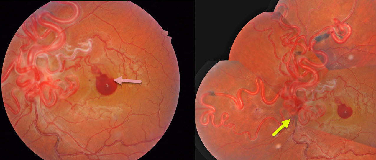
RETINAL ARTERIOVENOUS MALFORMATION
The European VitreoRetinal Society (EVRS) and Rajiv Gandhi
Originally posted on @retina.rocks April 17, 2026
This healthy 34YO man noticed sudden vision loss in his left eye following heavy lifting 2 days earlier. Vision was 20/20 in his normal OD and 20/200 OS.
Color photography shows a diffuse network of markedly dilated, tortuous vessels involving the optic disc and posterior pole, extending into the peripheral retina, with an associated small premacular hemorrhage. The vessels are sheathed superonasal to this blood. Unfortunately, our patient was lost to follow-up immediately.
Learning Points:
A retinal arteriovenous malformation (AVM) is a congenital connection between an artery and a vein without an intervening capillary network. These lesions appear along a spectrum ranging from small, isolated lesions to the large ‘bag of worms’ appearance. These more extensive AVMs are usually part of the Wyburn-Mason Syndrome, which includes ocular and midbrain vascular lesions. Although retinal AVMs are usually stable lesions, bleeding or secondary venous occlusions may occur. Our patient’s vascular sheathing may indicate prior occlusion.
RETINAL ARTERIOVENOUS MALFORMATION
Seif Anwar
Originally posted on @retina.rocks December 1, 2025
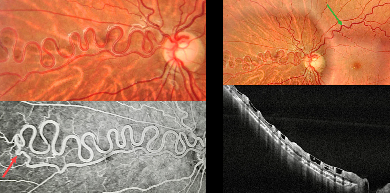
This healthy 12YO boy was referred for asymptomatic retinal findings. Vision was 20/20 in his normal OD and 20/20 OS.
Color photography shows a dilated multi-corkscrewed arteriole extending into the nasal midperiphery. The major superotemporal vein is also somewhat dilated and tortuous. On OCT, these anomalous vessels are hyperreflective and extend through the full thickness of the retina. ILM draping spans between each vessel. Angiographically, the artery directly connects to a paired vein. There is no angiographic leakage.
Learning Points:
A retinal arteriovenous malformation (AVM) is a congenital connection between an artery and a vein without an intervening capillary network. These lesions appear along a spectrum ranging from small, isolated lesions to the large ‘bag of worms’ appearance. These more extensive lesions are usually part of the Wyburn-Mason Syndrome, which includes ocular and midbrain vascular lesions. Although retinal AVMs are usually stable lesions, secondary venous occlusions may occur. Our patient has an isolated retinal AVM due to the lack of intracranial lesions on neurologic imaging.
PROLIFERATIVE DIABETIC RETINOPATHY NVD VS RETINAL ARTERIOVENOUS MALFORMATION
Ayushi Gupta and Vishal Agrawal.
Originally posted on @retina.rocks November 12, 2025
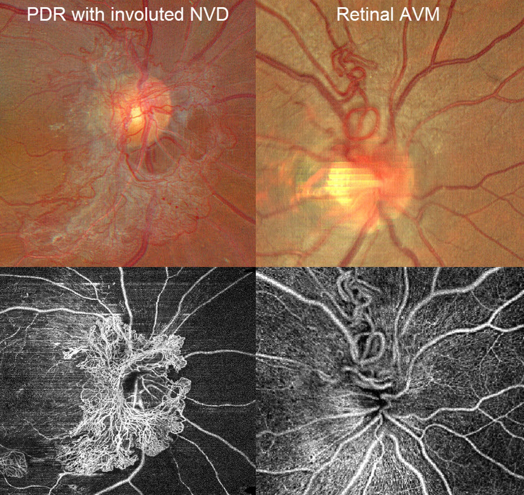
A patient with disc neovascularization (NVD) due to proliferative diabetic retinopathy (PDR) following full panretinal photocoagulation is compared with a patient with an asymptomatic retinal arteriovenous malformation (AVM).
Color photography of the PDR patient shows diffuse flat NVD with variable tight capillary networks of new vessels, admixed with larger vessels and fibrosis. OCT angiography (OCTA) confirms flow signals within these vessels, along with diffuse capillary loss superior, nasal, and inferior to the nerve. Color photography and OCTA of the retinal AVM show a tangle of arterioles and veins extending superiorly from the nerve. Fluorescein angiography, if performed, would show leakage from the NVD but not from the AVM.
RETINAL ARTERIOVENOUS MALFORMATION
Rohan Jain and Manish Nagpal
Originally posted on @retina.rocks August 12, 2025
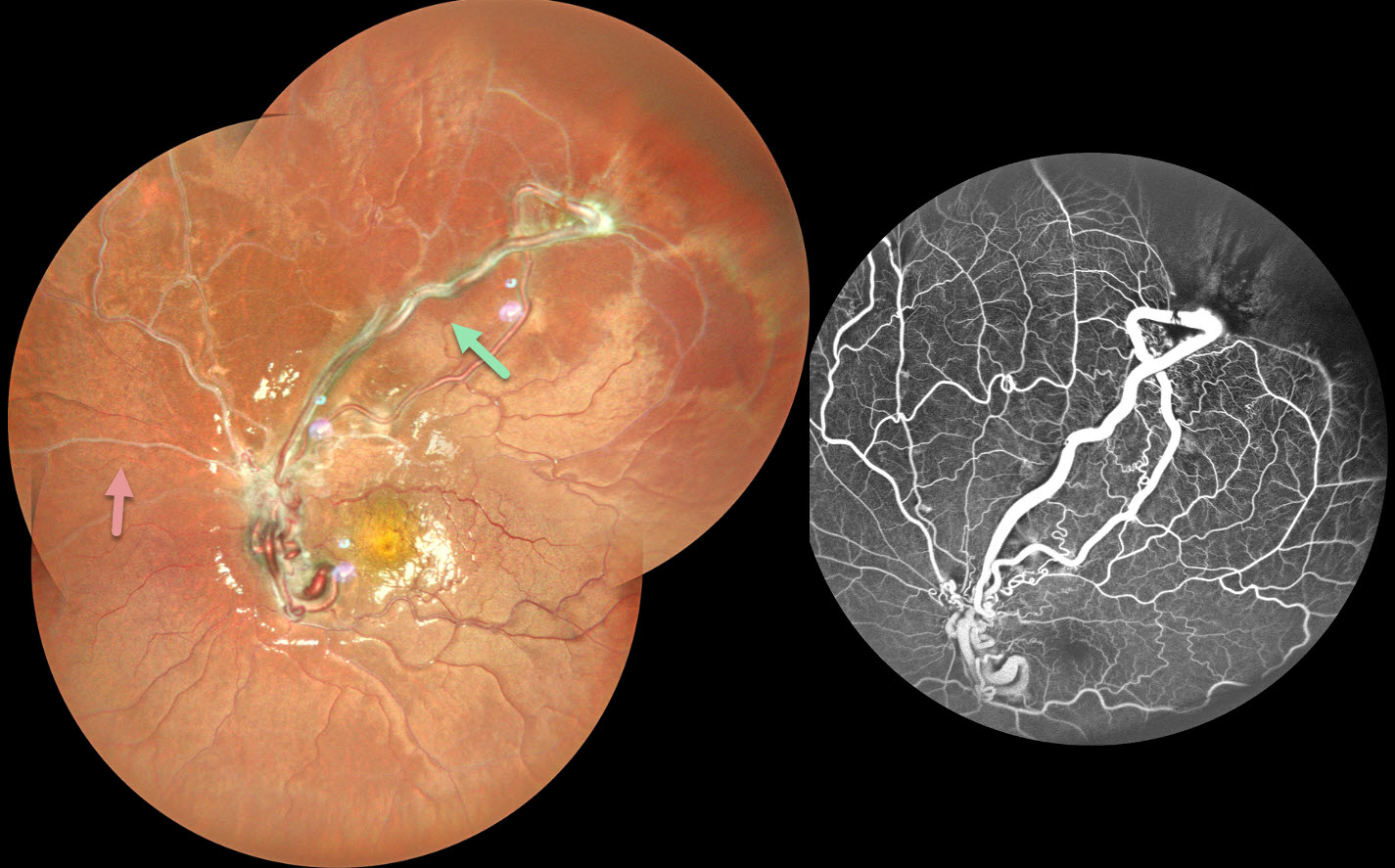
This healthy 10YO female presented with a 3-month history of decreased vision in her left eye. Vision was 20/15 in her normal OD and 20/200 OS.
Pseudocolor SLO imaging shows tangles of markedly dilated and tortuous vessels, which extend into the superotemporal midperiphery and obscure the optic disc. The retinal vessels are sheathed nasally and superiorly. Fluorescein angiography shows no leakage with ischemic retina distal to the anomalous vessels. Neurologic imaging was negative.
Learning Points:
A retinal arteriovenous malformation (AVM) is a congenital connection between an artery and a vein without an intervening capillary network. These lesions appear along a spectrum ranging from small, isolated lesions to the large ‘bag of worms’ appearance as seen in our patient. These more extensive lesions are usually part of the Wyburn-Mason Syndrome, which includes vascular lesions in the eyes and midbrain.
Retinal venous malformations, also known as retinal macrovessels, are now thought to represent an AVM (Pichi et al., JAMA Ophthalmol 2018;136:372-379). These lesions can rarely bleed or cause retinal venous occlusions. The diffuse vascular sheathing may represent such a prior occlusion.
RETINAL ARTERIOVENOUS MALFORMATION
Mattie Adams
Originally posted on @retina.rocks April 15, 2025
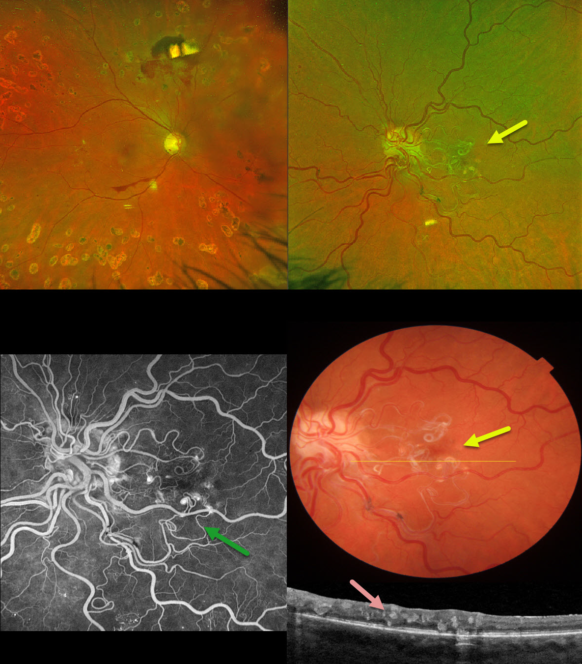
This 53YO female with a history of type 2 diabetes was referred for a symptomatic vitreous hemorrhage in her right eye from proliferative diabetic retinopathy. She received prior panretinal photocoagulation (PRP) in this eye elsewhere. Vision was 20/60 OD and 20/70 OS.
Optos color RG imaging OD shows preretinal blood in the inferior macula, and PRP scarring is noted superior to the nerve. The left eye shows a tangle of dilated and partially sheathed vessels extending from the nerve into the central macula. The more peripherally located vessels are dilated and tortuous.
On B-scan OCT, these vessels are noted in all retinal layers. These vessels did not leak on fluorescein angiography. Neurologic imaging was negative.
Learning Points:
A retinal arteriovenous malformation (AVM) is a congenital connection between an artery and a vein without an intervening capillary network. These lesions appear along a spectrum ranging from small, isolated lesions to the large ‘bag of worms’ appearance as seen in our patient. These more extensive lesions are usually part of the Wyburn-Mason Syndrome, which includes ocular and midbrain vascular lesions. Our patient has an isolated retinal AVM, as there are no intracranial lesions.
Unlike diabetic neovascularization that proliferates from the retinal surface into the cortical vitreous, the vessels in an AVM are intraretinal. Even though the AVM somewhat mimics diabetic disc neovascularization, its presence in our diabetic patient is purely coincidental. Although retinal AVMs are usually stable lesions, retinal hemorrhages, vitreous hemorrhage, and venous occlusions may occur.
RETINAL ARTERIOVENOUS MALFORMATION
Evgenii Chernov
Originally posted on @retina.rocks February 17, 2025
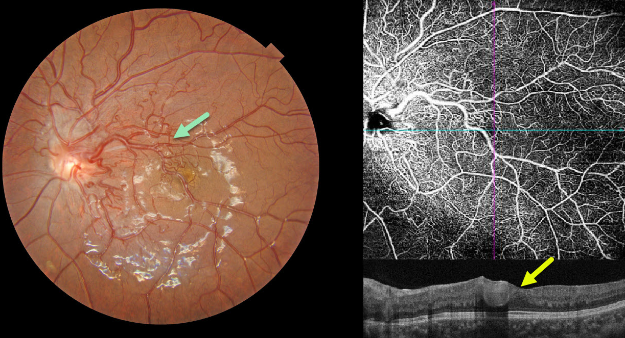
This 10YO girl presented with asymptomatic retinal findings in her left eye. Vision was 20/25 in her normal OD and 20/30 OS.
Color photography shows a retinal arteriovenous malformation (AVM) composed of dilated, tortuous vessels, mostly in the macular area. The foveal pit is crossed by a large tortuous vessel. OCT angiography highlights the profoundly abnormal macular vasculature, and an OCT B-scan shows the markedly dilated inner foveal vessel. MRI scanning of the brain and orbits was normal.
Learning Points:
A retinal AVM is a congenital connection between an artery and a vein without an intervening capillary network. An AVM appears along a spectrum ranging from small, isolated lesions to the large ‘bag of worms’ appearance. These more extensive lesions are usually part of the Wyburn-Mason Syndrome, which includes ocular and midbrain vascular lesions. Our patient has an isolated retinal AVM, as there are no intracranial lesions.
RETINAL ARTERIOVENOUS MALFORMATION
Mattie Adams and Jonathan Bunnell
Originally posted on @retina.rocks November 28, 2024
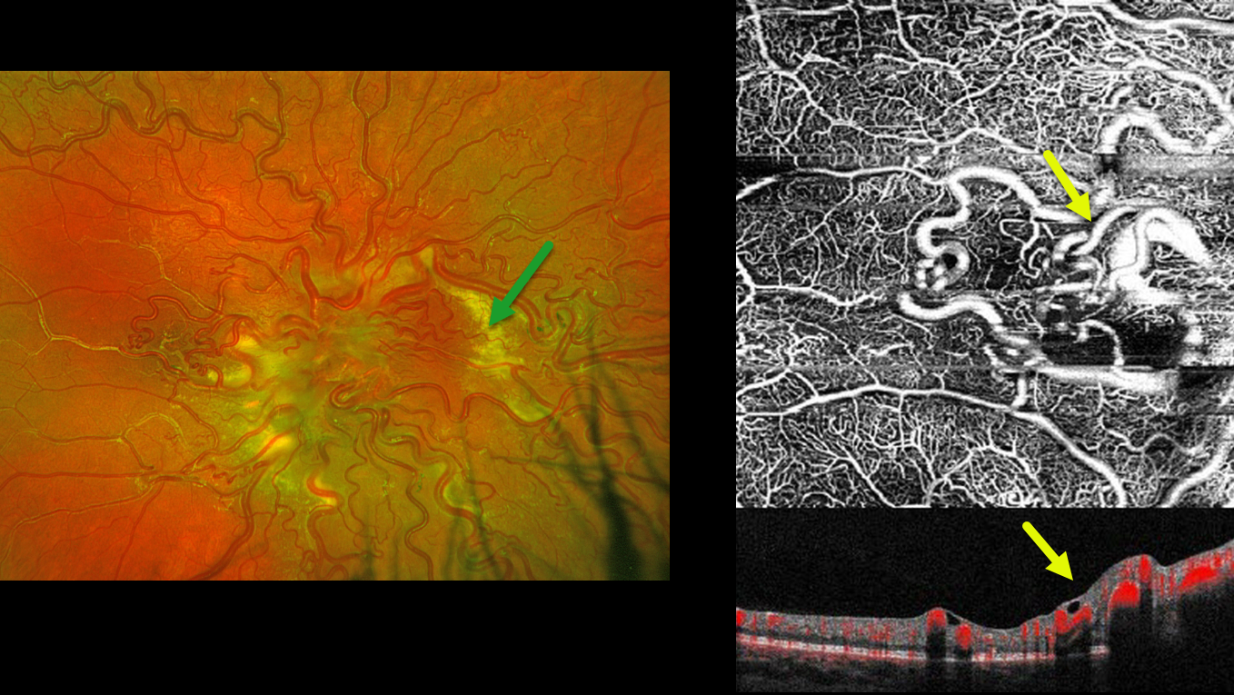
This 10YO boy recently moved to the area with a prior diagnosis of Wyburn-Mason syndrome. Prior neurologic imaging was negative, and he was otherwise healthy. Vision was 20/400 OD and 20/20 in his normal left eye.
Optos color RG imaging of his right eye shows tangles of markedly dilated and tortuous vessels covering the optic nerve and posterior pole. The vessels are sheathed superiorly and temporally. Subretinal lipid surrounds the posterior pole. En face OCT angiography highlights these dilated inner retinal vessels. Observation was recommended.
Learning Points:
A retinal arteriovenous malformation (AVM) is a congenital connection between an artery and a vein without an intervening capillary network. These lesions appear along a spectrum ranging from small, isolated lesions to the large ‘bag of worms’ appearance as seen in our patient. These more extensive lesions are usually part of the Wyburn-Mason Syndrome, which includes ocular and midbrain vascular lesions. Our patient has an isolated retinal AVM, with no intracranial lesions.
Although retinal AVMs are usually stable lesions, secondary venous occlusions may occur, and the peripheral vascular sheathing in our patient likely indicates a prior occlusion. The subretinal lipid in our patient is a very atypical finding in our experience.
RETINAL ARTERIOVENOUS MALFORMATION
Şengül Özdek and Ece Özdemir Zeydanlı
Originally posted on @retina.rocks May 8, 2024
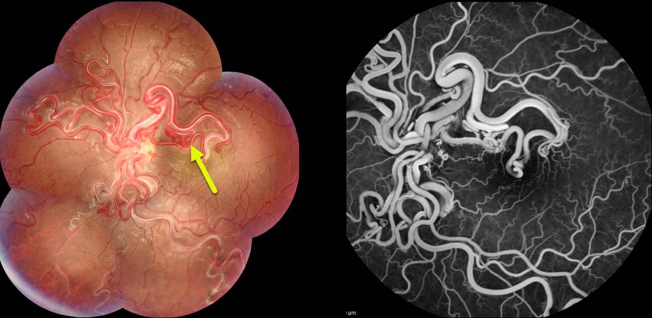
This healthy 16YO girl presented with a long history of poor vision in her left eye. Vision was 20/100 in her left eye.
Fundus photography of her left eye shows tangles of markedly dilated and tortuous vessels covering the posterior pole. Fluorescein angiography shows no leakage. Brain MRI revealed a cerebral hemangioma next to the optic chiasm.
Learning Points:
A retinal arteriovenous malformation (AVM) is a congenital connection between an artery and a vein without an intervening capillary network. These lesions appear along a spectrum ranging from small, isolated lesions to the large ‘bag of worms’ appearance as seen in our patient. These more extensive lesions are usually part of the Wyburn-Mason Syndrome, which includes vascular lesions in the eyes and midbrain. Retinal venous malformations, aka retinal macrovessels, are now thought to represent an AVM (Pichi et al JAMA Ophthalmol 2018;136:372-379).
RETINAL ARTERIOVENOUS MALFORMATION
Şengül Özdek and Ece Özdemir Zeydanl
Originally posted on @retina.rocks March 24, 2023
This healthy 11YO girl was seen for a routine ophthalmological examination. Vision was 20/20 OU.
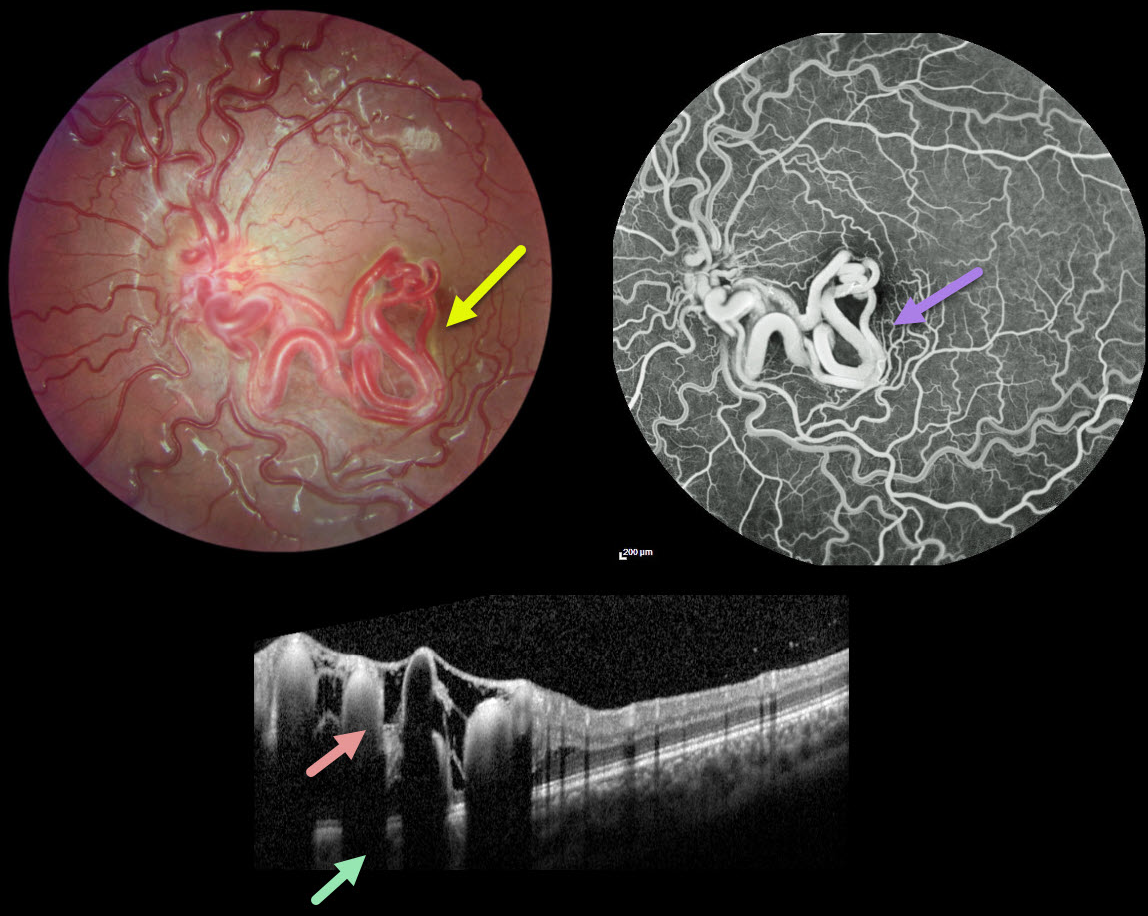
Fundus photography of her left eye shows unilateral tangles of markedly dilated and tortuous vessels extending from the disc into the inferior macula and fovea. The retinal vessels elsewhere are also somewhat dilated and tortuous. OCT scanning through the inferior macula shows these hyperreflective vessels to variably extend through the full-thickness retina with posterior shadowing.
The early-phase fluorescein angiogram shows rapid filling of the dilated artery and vein, with no intervening capillaries (not shown). The late phase shows no angiographic leakage.
Learning Points:
A retinal arteriovenous malformation (AVM) is a congenital connection between an artery and vein without an intervening capillary network. These lesions appear along a spectrum ranging from small isolated lesions to the large ‘bag of worms’ appearance as seen in our patient.
These more extensive lesions are usually part of the Wyburn-Mason Syndrome, which includes vascular lesions in the eyes and midbrain. Retinal venous malformations, also known as retinal macrovessels, are now thought to represent an AVM (Pichi et al., JAMA Ophthalmol 2018;136:372-379).
Fortunately, our patient’s MRI was normal. The AVM should likely remain stable throughout life and should not require any treatment.
RETINAL VENOUS MALFORMATION
Ryan Kern
Originally posted on @retina.rocks February 7, 2023
This healthy 24YO female presented for a general eye examination. There was no past ocular or medical history.
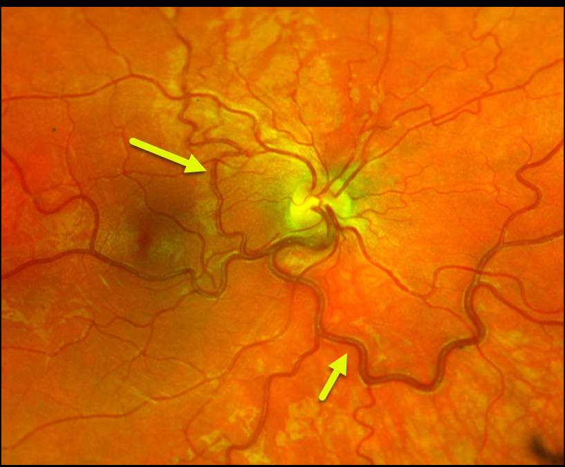
Color imaging of her right eye shows a large dilated retinal vein that drains the entire fundus except superonasally. It also crosses the temporal horizontal raphe into the superotemporal quadrant. Her left eye was normal.
Learning Points:
Retinal vessels virtually always respect the horizontal meridian. Retinal venous malformations (Pichi et al JAMA Ophthalmol 2018;136:372-379) are congenital anomalous vessels that by definition cross the horizontal.
Originally described by Gary Brown et al as congenital retinal macrovessels (Arch Ophthalmology 1982;100:1430-1436), these vessels are usually located in the macular region and are virtually always venous.
The findings in our patient are unusual, as the anomalous vessel supplies the majority of the retinal venous drainage.
These vessels are technically arterio-venous malformations (AVM) and are associated with venous anomalies in the brain (24% compared to up to 6% of the population). Brain MRI is therefore recommended to rule out any intracranial abnormalities. Neurologic imaging in our patient was fortunately normal.
RETINAL ARTERIOVENOUS MALFORMATION
Originally posted on @retina.rocks May 3, 2022
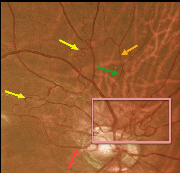
This 61YO male presented with these asymptomatic blood vessels extending superiorly from the right nerve. Vision was 20/25 OU, and the left eye was normal.
The abnormal vessels emanate from a cilioretinal artery, extend superiorly into and then above the macula, then arch nasally and descend. Note that it crosses a vein, confirming that it is still arterial (remember that arteries cross veins, veins cross arteries, but arteries never cross arteries and veins never cross veins). It then descends towards the nerve in a tangle of vessels likely consisting of both arteries and veins.
We felt that these vessels represented a low-grade arteriovenous malformation (AVM).
Learning Points:
Since about 25% of patients with AVMs can have intracranial venous anomalies, a brain MRI is usually recommended.
These vessels somewhat resemble collateral vessels noted after retinal vein occlusions. Collaterals are dilated capillaries that result from increased hydrostatic pressure from an occlusion. They are almost always venous, and when seen, are a telltale sign for a prior occlusion. Unlike retinal neovascularization, they do not leak angiographically.
RETINAL ARTERIOVENOUS MALFORMATION
Originally posted on @retina.rocks December 9, 2021
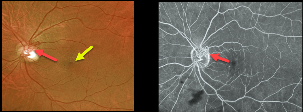
This 67YO male with type 2 diabetes presented with these asymptomatic blood vessels overlying the left nerve, which did not leak on fluorescein angiography. Vision was 20/20 OU. Mild non-proliferative retinopathy was noted bilaterally, and there was a mild asymptomatic macular pucker in the left eye.
We felt that these vessels represented an arteriovenous malformation (AVM). The other possibility would be extremely large and complex collateral vessels, but we saw no evidence of retinal vein occlusion, clinically or angiographically.
Learning Points:
Since about 25% of patients with AVMs can have intracranial venous anomalies, a brain MRI is usually recommended. Our patient was asymptomatic, and his MRI and MRA were normal.
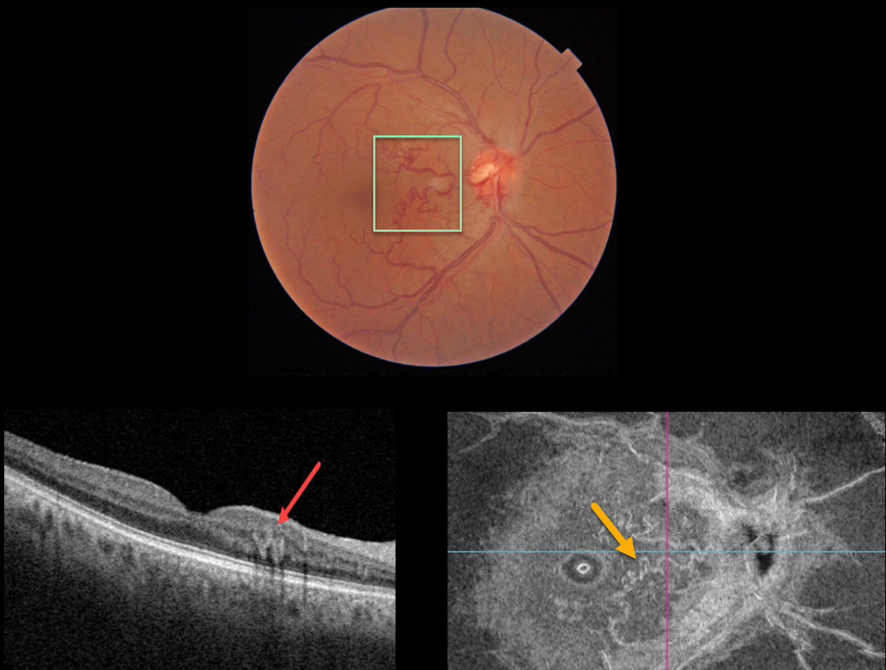
RETINAL ARTERIOVENOUS MALFORMATION
Originally posted on @retina.rocks September 3, 2021
This 66YO male presented with an asymptomatic macular arteriovenous malformation (AVM). Vision was 20/30.
Triton imaging shows an AVM involving the nasal macula. Swept-source OCT shows that the abnormal vessels lie within the deep vascular complex. En face OCT beautifully displays the abnormal vasculature.
Learning Points:
Since about 25% of patients with AVMs can have intracranial venous anomalies, a brain MRI is usually recommended.
Our patient was asymptomatic, and his MRI and MRA were normal.
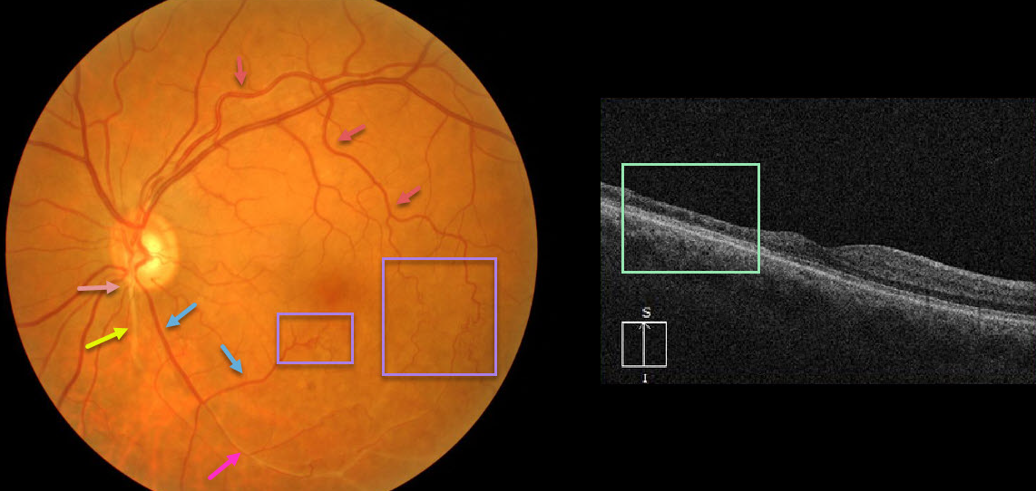
RETINAL ARTERIOVENOUS MALFORMATION
Originally posted on @retina.rocks July 28, 2020
At first or quick glance, this looks like an old inferotemporal branch retinal vein occlusion (BRVO).
On closer inspection, there is a white-sheathed vessel arising from the nerve inferiorly. Numerous collateral vessels are noted inferior and temporal to the fovea, and there is a sheathed vessel in the inferior arcade.
OCT shows marked inner retinal atrophy with preservation of the photoreceptor layers, consistent with a prior branch retinal artery occlusion. So the collateral vessels (virtually always a sign of a prior venous occlusion) indicate an old BRVO, and the atrophic and sheathed vessel indicates an old BRAO, both in the same quadrant. Right?
Tracing the large branch of the superotemporal arcade inferotemporally, we see that the collaterals appear to be arterial. Inferior to the nerve, we see an arteriole that crosses the sheathed vessel, indicating that the sheathed vessel is not an artery but a vein. Moreover, the large vessel off the nerve, inferotemporal in location, appears to be a vein that eventually connects to the collateral vessels we previously identified as arterial.
Our best guess is that this may be a low-grade arteriovenous malformation, possibly with a prior secondary BRAO/BRVO. Unfortunately, we don’t have a fluorescein angiogram, which may help better define the vascularization.
Learning Points:
Remember that arteries don’t cross arteries and veins don’t cross veins. Tracing vessels can be a helpful key to identifying retinal vascular disorders.
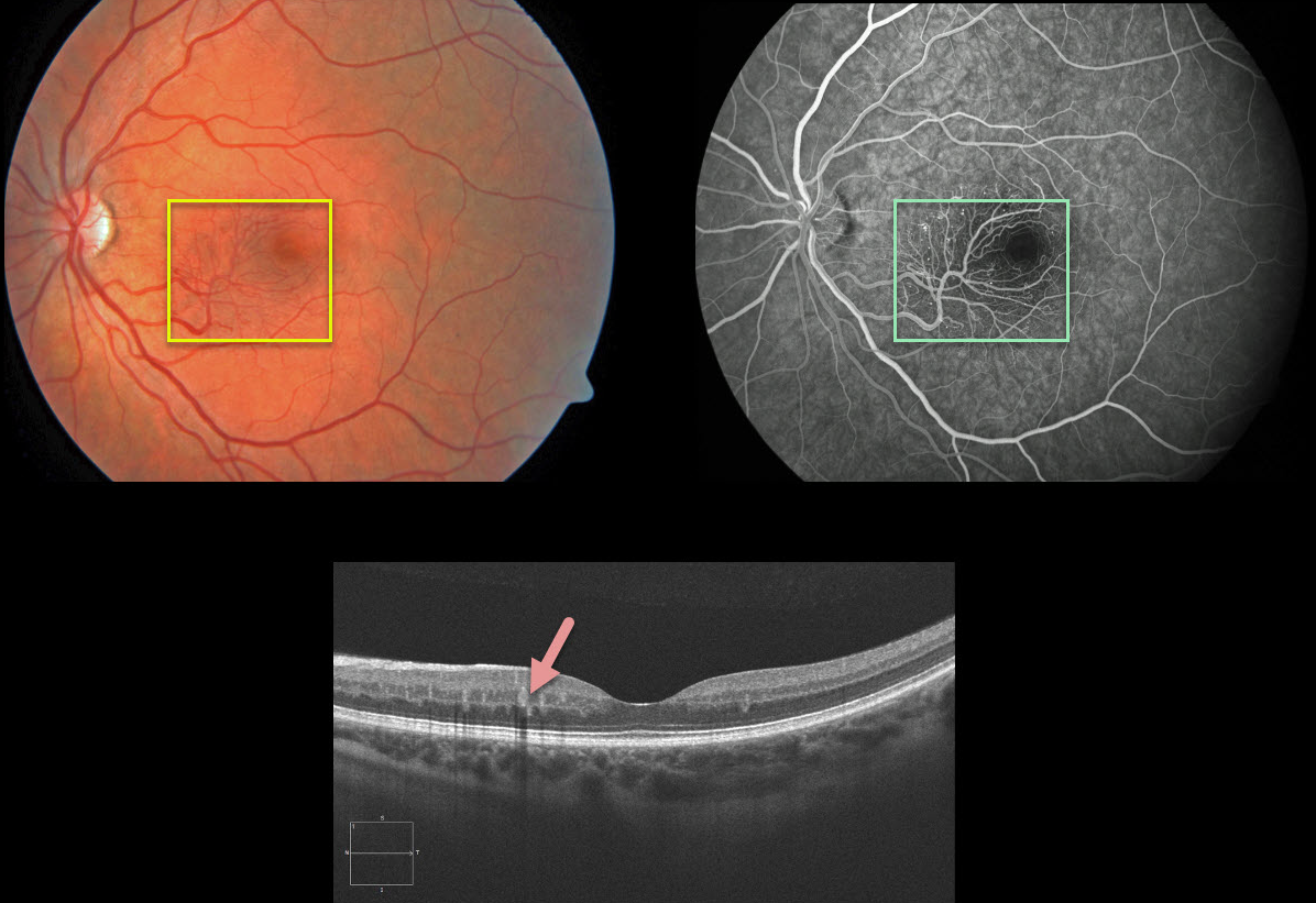
RETINAL ARTERIOVENOUS MALFORMATION
Originally posted on @retina.rocks December 19, 2019
This patient was asymptomatic and found to have a macular arteriovenous malformation (AVM). OCT B-scan shows the vascular abnormality mostly involves the deep vascular complex. Fluorescein angiography demonstrates no leakage.
The patient was asymptomatic with a normal MRI and MRA of the brain/orbits.
Learning Points:
Reinal arteriovenous malformations (AVM) are typically unilateral and sporadic.
Our patient had an isolated AVM. Wyburn-Mason Syndrome is characterized by the presence of retinal AVMs, as well as AVMs of the cerebral nervous system (CNS) and/or orbit.


