

Retinal Astrocytic Hamartoma
<< Back to Cases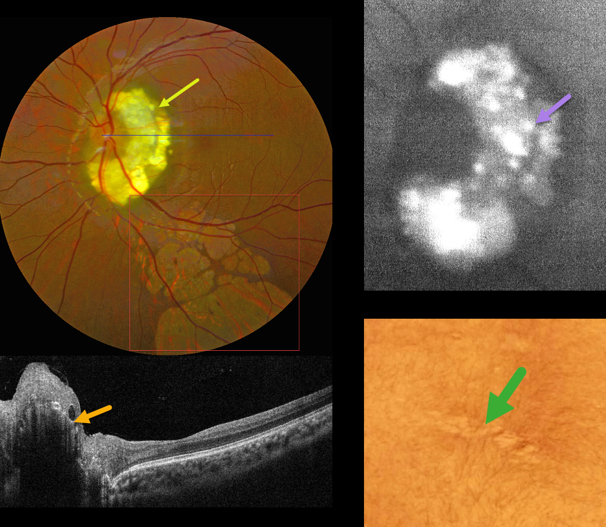
TUBEROUS SCLEROSIS WITH RETINAL ACHROMIC PATCHES AND RETINAL ASTROCYTIC HAMARTOMA
Kanwaljeet Harjot Madan
Originally posted on @retina.rocks September 4, 2026
This 17YO male presented with a known history of tuberous sclerosis, childhood seizures, and decreased vision in his left eye. Vision was 20/20 in his normal OD and 20/40 OS.
Color photography shows a glistening yellow-white inner peripapillary retinal lesion surrounding the superior, temporal, and inferior edge of the optic nerve, composed of innumerable semi-transparent spherules which hyper-autofluoresce. OCT scanning shows an elevated, hyperreflective inner retinal lesion with a hyporeflective intralesional cavity. An area of skin depigmentation (ash leaf spot) was noted on his lower back. There are numerous variably sized and variably confluent lobules of subretinal depigmentation extending from the macula inferiorly.
Learning Points:
The best-known retinal finding in tuberous sclerosis is retinal astrocytic hamartoma. Early in life, these inner retinal lesions have a fleshy white appearance and are often multifocal and bilateral. With time, as in this patient, they show a more classic “mulberry appearance” with significant calcification. The calcified lesions, as in our patient, hyper-autofluoresce while the flat, semitransparent lesions found earlier in life are hypoautofluorescent. Rarely, the vascular component can cause intraretinal exudation requiring treatment.
Retinal achromic patches, although much less described in the literature, are found in up to 39% of tuberous sclerosis patients (Rowley et al, Br J Ophthalmology 2001;85:420-423). They are thought to be analogous to the hypopigmented skin lesions and can appear in one or both eyes, singly or in multiples, and are typically located in the retinal periphery. Unliked astrocytomas, these lesions are flat and subretinal.
RETINAL ASTROCYTIC HAMARTOMA
European VitreoRetinal Society (EVRS) Dhaivat Shah, Nida Khan and Choithram Netralaya
Originally posted on @retina.rocks January 30, 2026
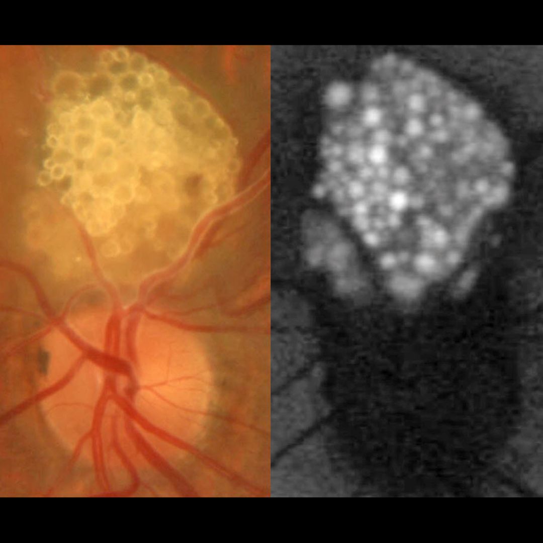
This healthy 43 YO male presented with an asymptomatic lesion in his left eye. Family history was negative. Vision was 20/20 in his normal OD and 20/20 OS.
Color photography shows a glistening yellow-white inner retinal lesion just superior to the optic nerve composed of innumerable semi-transparent spherules which hyper-autofluoresce.
Learning Points:
A retinal astrocytic hamartoma is a benign tumor composed of spindle-shaped fibrous astrocytes located in the nerve fiber layer, most commonly associated with tuberous sclerosis. Tuberous sclerosis includes the classic triad of seizures, mental deficiency and fibroangiomas, but patients can have lesions throughout the body including the eyes, skin (ash-leaf spots), bone, nails, teeth, brain, heart, lungs and kidneys. Early in life the inner retinal lesions have a fleshy white appearance and are often multifocal and bilateral. With time, as in this patient, they show a more classic “mulberry appearance” with significant calcification. Rarely, the vascular component can cause intraretinal exudation requiring treatment.
Clinically retinocytomas can resemble astrocytic hamartomas. However, retinocytomas often have underlying RPE changes and chorioretinal scarring (lacking in our patient), which are absent in astrocytic hamartomas.
Since the patient was self-paying, neurologic imaging was not performed and observation was recommended. A neurology reference was given.
ASTROCYTIC HAMARTOMA
Yuenpang Cheung, Stephanie Choi and Brian Marr
Originally posted on @retina.rocks December 5, 2023
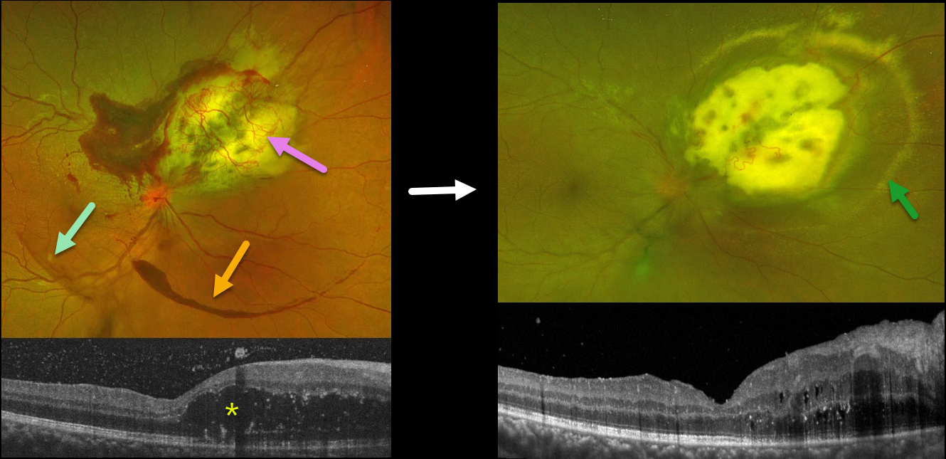
This 27YO female has been followed for an exudative astrocytic hamartoma in her right eye. Vision is 20/20 bilaterally. She was diagnosed with tuberous sclerosis at age 5 and has been followed closely by neurology.
Optos color RG imaging shows a superonasal yellow-and-white lesion with overlying neovascularization and exudates, along with areas of vitreous and subhyaloid blood.
OCT shows significant mostly outer retinal cystoid edema nasal to the fovea, as well as some vitreous blood.
She received a series of intravitreal Avastin injections with modest response and was more recently switched to Vabysmo injections. Two weeks following her third Vabysmo injection, the overlying neovascular network and blood are nearly resolved, but the surrounding subretinal lipid is increasing. The cystoid macular edema has mildly improved.
This case was submitted by Yuenpang Cheung, Stephanie Choi, and Brian Marr.
Learning Points:
Tuberous sclerosis is an autosomal dominant condition caused by mutations in the tumor suppressor genes TSC1 or TSC2. Tuberous sclerosis has multiple systemic manifestations affecting the brain (cortical tubers, subependymal nodules, epilepsy), skin (facial angiofibromas, shagreen patch, ash leaf spots), heart (cardiac rhabdomyoma), and the kidneys (angiomyolipomas).
The primary ocular manifestation is a retinal hamartoma arising from glial astrocytes. Retinal hamartomas can present as subtle gray areas in the nerve fiber layer without calcification or as prominent calcified nodules arising from the inner surface of the retina with a mulberry or fish egg-like appearance.
Although patients usually remain asymptomatic, these benign tumors can enlarge and cause macular edema, serous retinal detachment, or vitreous hemorrhage.
Exudative treatment options include anti-VEGF injections, photodynamic therapy, transpupillary thermotherapy, and radiation therapy. In cases with suboptimal response, reports of systemic mTOR inhibitors, such as Sirolimus, have shown some benefit.
RETINAL ASTROCYTIC HAMARTOMA
Originally posted on @retina.rocks January 10, 2022
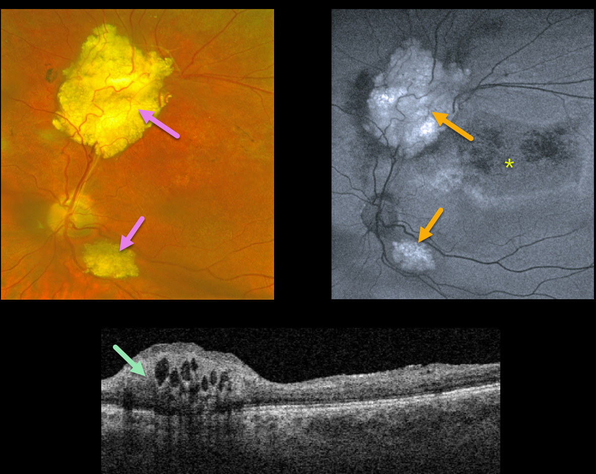
This 21YO male patient with a known history of tuberous sclerosis was referred for multiple, asymptomatic, thickened whitish inner retinal lesions.
On fundus autofluorescence (FAF), these lesions are hyper-FAF. Surrounding hypo-FAF changes that extend into the macula are a sign of prior exudative fluid.
OCT scanning shows an elevated, hyperreflective inner retinal lesion with multiple hyporeflective intralesional cavities.
Learning Points:
A retinal astrocytic hamartoma is a benign tumor composed of spindle-shaped fibrous astrocytes located in the nerve fiber layer, most commonly associated with tuberous sclerosis.
Tuberous sclerosis includes the classic triad of seizures, intellectual disability, and fibroangiomas. Still, patients can have lesions throughout the body, including the eyes, skin (ash-leaf spots), bone, nails, teeth, brain, heart, lungs, and kidneys.
Early in life, the inner retinal lesions appear fleshy white and are often multifocal and bilateral. With time, as in this patient, they show a more classic “mulberry appearance” with significant calcification. Rarely, the vascular component can cause intraretinal exudation requiring treatment.
RETINAL ASTROCYTIC HAMARTOMA
Originally posted on @retina.rocks February 18, 2021
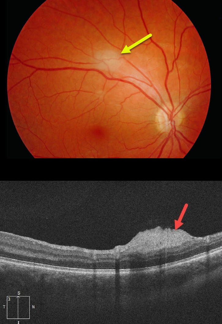
This patient with a known history of tuberous sclerosis was referred for an asymptomatic, slightly thickened whitish inner retinal lesion. OCT scanning showed an elevated, hyperreflective nerve fiber layer lesion.
Learning Points:
A retinal astrocytic hamartoma is a benign tumor composed of spindle-shaped fibrous astrocytes located in the nerve fiber layer, most commonly associated with tuberous sclerosis.
Tuberous sclerosis includes the classic triad of seizures, intellectual disability, and fibroangiomas, but patients can have lesions throughout the body, including the eyes, skin (ash-leaf spots), bones, nails, teeth, brain, heart, lungs, and kidneys.
Early in life, the inner retinal lesions appear fleshy white and are often multifocal and bilateral. Later in life, they show a more classic “mulberry appearance” with significant calcification. Rarely, the vascular component can cause intraretinal exudation requiring treatment.
RETINAL ASTROCYTIC HAMARTOMA
Originally posted on @retina.rocks April 27, 2020
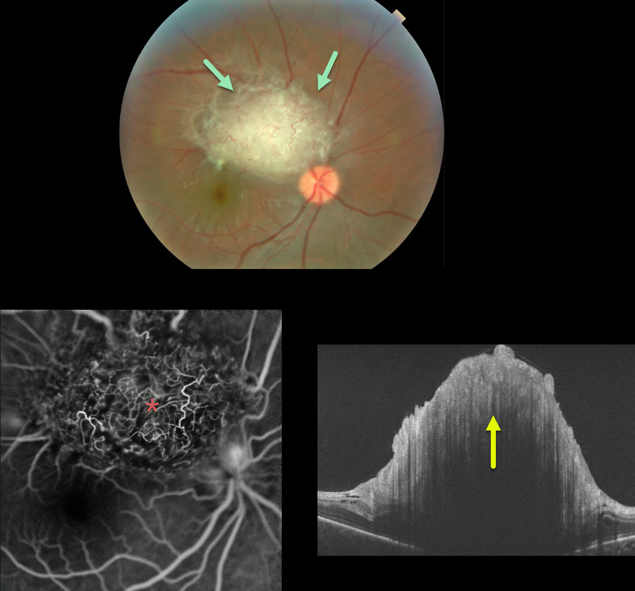
Our patient (courtesy of Tufts) has the classic mulberry appearance of a retinal astrocytic hamartoma. Early in life the inner retinal lesions have a fleshy white appearance and are often multifocal and bilateral. Later in life they develop the mulberry or fish egg-like appearance with significant calcification.
Fluorescein angiography (FA) shows the often angiomatous nature of these lesions. OCT shows the elevated, hyperreflective inner retinal lesion, with marked shadowing more posteriorly.
These tumors are generally are just observed. Rarely, the vascular component can cause intraretinal exudation requiring treatment.
Learning Points:
A retinal astrocytic hamartoma is a benign tumor composed of spindle-shaped fibrous astrocytes located in the nerve fiber layer, most commonly associated with tuberous sclerosis.
Tuberous sclerosis includes the classic triad of seizures, mental deficiency and fibroangiomas, but patients can have lesions throughout the body including the eyes, skin (ash-leaf spots), bone, nails, teeth, brain, heart, lungs and kidneys.


