

Retinal Break
<< Back to Cases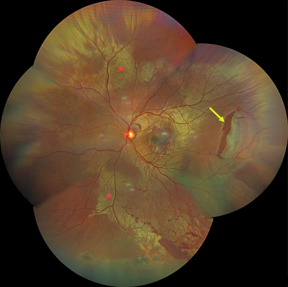
COMMOTIO RETINAE WITH TRAUMATIC RETINAL BREAK
Dibya Prabha
Originally posted on @retina.rocks August 7, 2026
This 38YO male presented with sudden vision loss in his left eye immediately following blunt trauma from a cricket bat 2 days earlier. Vision was 20/200.
Color fundus photography shows extensive commotio retinae throughout the posterior pole, along with a large retinal tear in the temporal midperiphery. Oral and topical steroids were prescribed, and the retinal break was surrounded with laser photocoagulation.
Learning Points:
Commotio retinae is caused by direct, blunt eye trauma. The outer retinal whitening is due to shearing of the photoreceptor outer segments. The term “Berlin’s edema” is a misnomer and should not be used, as the retina is not edematous. The acute white retinal changes usually resolve within several weeks. With resolution, the fundus appearance can normalize. With more severe injury, outer retinal loss can allow RPE cells to migrate into the retina, giving a pseudo-retinitis pigmentosa appearance. Patients can also develop retinal breaks and detachment.
LATTICE DEGENERATION WITH SYMPTOMATIC RETINAL TEARS
Originally posted on @retina.rocks May 14, 2026
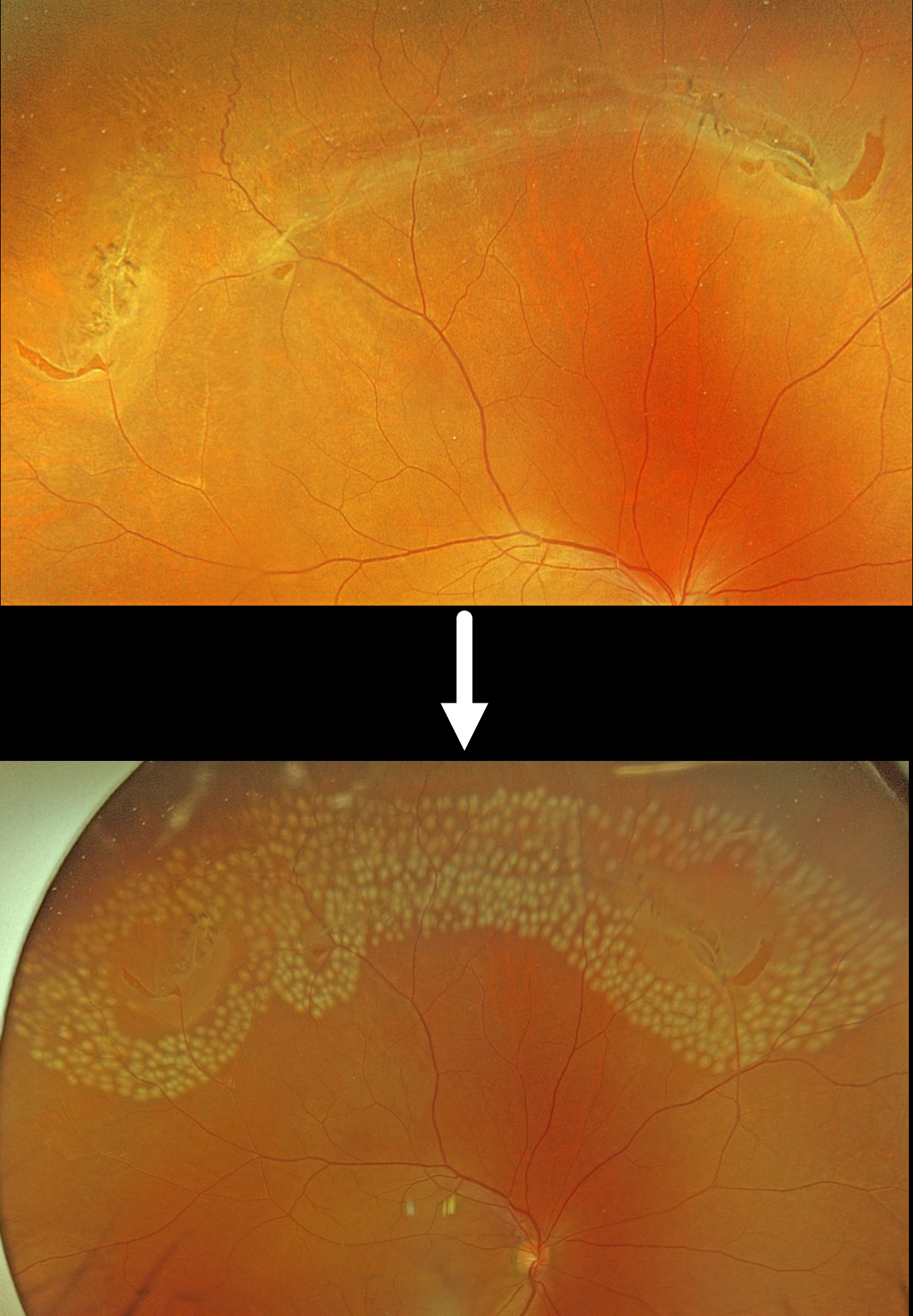
This 55YO male was referred for an acute posterior vitreous detachment with symptomatic retinal tears. Vision was 20/25.
Optos color RGB imaging shows several patches of lattice degeneration extending from the 10 to 1:30 o’clock meridians associated with multiple retinal tears. Some inferior vitreous hemorrhage was noted (not shown). Immediate prophylactic laser photocoagulation was applied to all surrounding the lattice and breaks.
Lattice lesions have a pocket of liquefied vitreous overlying thinned retina with a firm area of surrounding vitreoretinal adhesion. White, fibrosed vessels often crisscross the lesions, giving them their name, “lattice.” This firm vitreoretinal adhesion, along with the often-present atrophic holes, is what can predispose these eyes to retinal breaks and detachment. Although prophylactic treatment is rarely needed, symptomatic breaks, as in our patient, need prompt retinopexy.
SHOULD THIS LESION BE EMERGENTLY LASERED???
Originally posted on @retina.rocks March 11, 2026
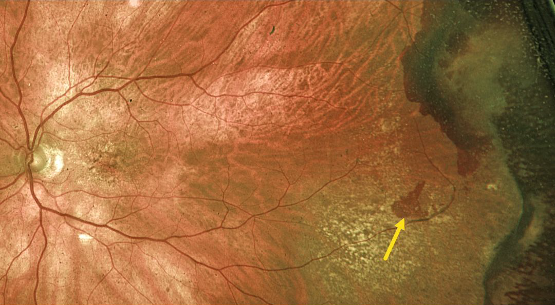
This 89YO male was referred for an asymptomatic retinal tear with possible retinal detachment in his left eye. Vision was 20/60.
Optos color RG imaging shows a red horseshoe-shaped lesion in the temporal midperiphery. Would you recommend immediate thermal laser demarcation of this retinal tear?
We hope not! This patient has asymptomatic and chronic ectopic choroidal neovascularization (CNV), which is evidenced by the more peripheral subretinal scarring and superotemporal dark subretinal blood. Ectopic CNV can often masquerade as a choroidal mass or uveal melanoma. It can also cause extensive choroidal/suprachoroidal, subretinal and vitreous hemorrhage, more commonly in patients who are on blood thinners. The subretinal blood often spontaneously regresses without treatment, although anti-VEGF injections can be used for symptomatic or increasing exudation.
We recommended observation and expect this lesion to remain relatively stable.
RETINAL TEAR
Originally posted on @retina.rocks October 22, 2025
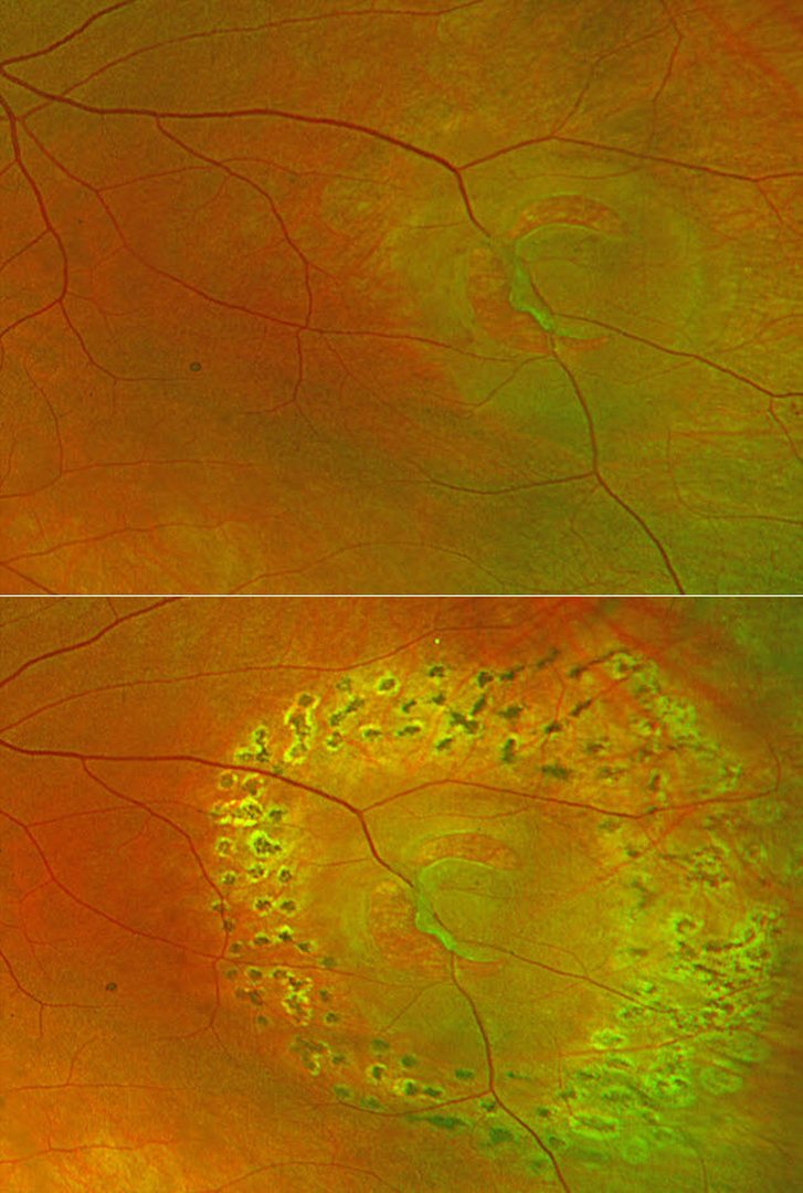
This 60YO female presented with a symptomatic retinal tear in her left eye. Optos color RG imaging shows the break with localized surrounding subretinal fluid and a bridging vessel. The break was immediately surrounded by laser. When examined 1 month later, the tear and fluid were well demarcated, and no new breaks were found.
Learning Points:
Symptomatic retinal tears in the presence of an acute posterior vitreous detachment have a high risk of causing a rhegmatogenous retinal detachment. Prophylactic retinopexy is therefore indicated. Treatment must completely surround the tear and, ideally, extend to the ora serrata. Both thermal laser photocoagulation and cryotherapy are equally effective in creating a vitreoretinal adhesion, which takes at least several weeks to form. The treatment must also be inspected during the postoperative period to ensure adequate scarring surrounds the break.
TRAUMATIC RETINAL HOLES
Fatma Shakarchi and Ahmed Sallam
Originally posted on @retina.rocks October 6, 2025
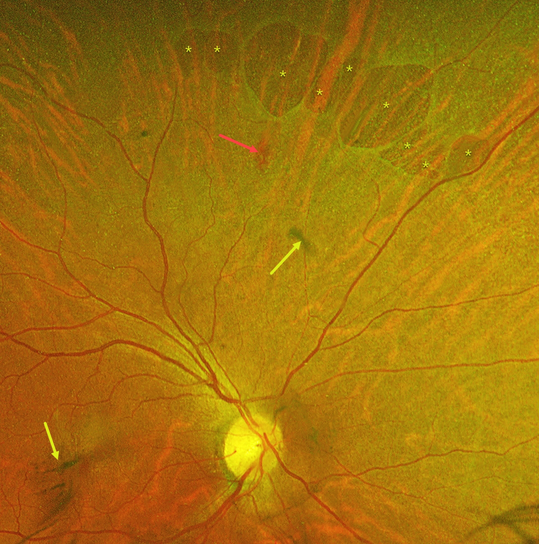
This 55YO female was referred with mildly decreased vision in her right eye two days following blunt ocular trauma. Vision was 20/40 OD and 20/20 in her normal OS.
Optos color RG imaging reveals numerous irregularly oval superonasal retinal holes, a retinal hemorrhage, and some clumps of vitreous blood. There was no vitreous detachment clinically.
Learning Points:
Retinal breaks following blunt trauma include retinal dialysis, horseshoe tears, and giant retinal tears. Breaks can also develop from concussive injury at the impact site or from a coup-contrecoup injury, so-called impact or necrotic breaks, which may be associated with varying degrees of commotio retinae acutely (Cox et al, Arch Ophthalmol 1966;76:678-685) or following its resolution (Longstaff et al, Br J Ophthalmol 1987;71:375-376).
In this case, the breaks identified 2 days after the injury were not associated with commotio. However, their appearance and the adjacent hemorrhage support an acute traumatic etiology. Therefore, prophylactic laser retinopexy was promptly applied around all breaks.
COMMOTIO RETINAE WITH RETINAL BREAKS AND CONTUSIVE SEROUS MACULAR DETACHMENT
Tejaswita Verma and Manish Nagpal
Originally posted on @retina.rocks September 24, 2025
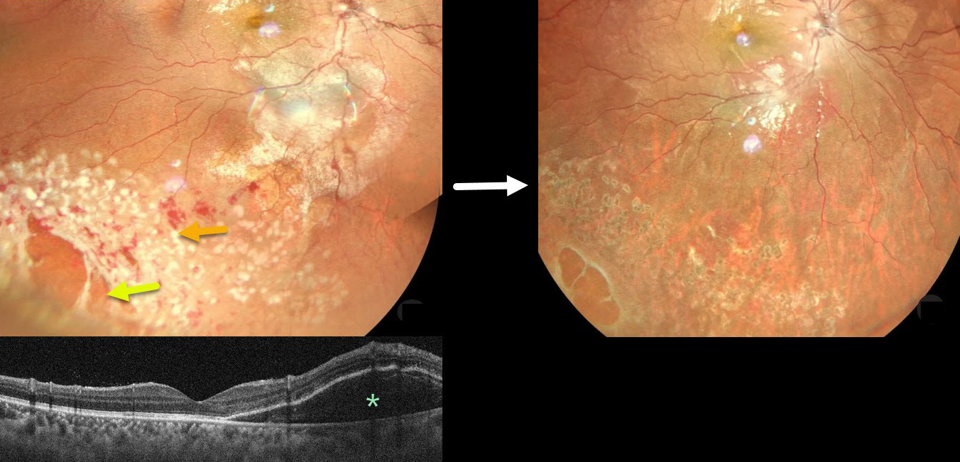
This 13YO male was hit in his right eye with a cricket ball. When he presented later that day vision was 20/30.
Color photography shows a large area of commotio retinae with scattered retinal hemorrhages involving the inferotemporal fundus. There is a poor foveal reflex with possible nasal macular subretinal fluid, which is confirmed on OCT. Several atrophic retinal breaks are noted peripherally. Immediate prophylactic laser was applied around all breaks. Seven weeks later, the commotio and hemorrhages are resolved, and all breaks are well-surrounded with laser scarring. Vision was 20/20.
Although blunt trauma is a common cause of retinal breaks, acute or subsequent dissolution of the retina within areas of commotio with secondary holes is quite rare in our experience (Cox et al Arch Ophthalmol 1966;76:678-685 and Longstaff et al Br J Ophthalmology 1987;71:375-376). The acute white retinal changes usually resolve within several weeks. With resolution, the fundus appearance can normalize. With more severe injury, outer retinal loss can allow RPE cells to migrate into the retina, giving a pseudo-retinitis pigmentosa appearance.
Transient subretinal fluid is an uncommon finding with blunt trauma. Injury to the choroid and RPE allows fluid to accumulate in the subretinal space and outer retina (Mishra et al, Journal of Vitreoretinal Diseases 2021;5:165-169).
RETINAL TEAR WITH BRIDGING VESSEL
Originally posted on @retina.rocks January 9, 2025
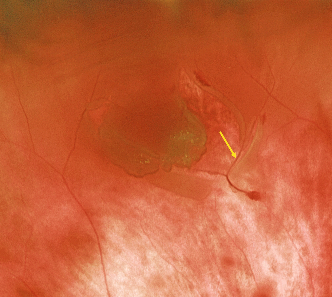
This patient presented with a macula-on rhegmatogenous retinal detachment (RRD). Optos color RG imaging shows the causative retinal tear with a bridging vessel. The RRD was successfully repaired with vitrectomy.
Learning Points:
The overall risk of an acute PVD causing a retinal break or detachment is about 5% (Seider et al, Ophthalmology 2022;129:67-72), and this risk is increased in the presence of vitreous pigment or blood. The reason for this is that, as in our patient, the retina often tears along a blood vessel. An occult retinal break is often the cause of an unexplained vitreous hemorrhage, which is why early vitrectomy is often recommended for these eyes, even when a break cannot be found on office examination (Flores-Sanchez et al, Eye 2023;37:3191-3196).
The bridging vessels in retinal breaks can also cause recurrent vitreous hemorrhage, a condition known as the avulsed retinal vessel syndrome (Robertson et al, Arch Ophthalmology 1971;85:669-672). The traction can spontaneously release, or vitrectomy may be required for sputtering hemorrhages or non-clearing blood.
RETINAL BREAK
Justin Grassmeyer, Ambar Faridi, and Brittany Heckerman
Originally posted on @retina.rocks June 5, 2024
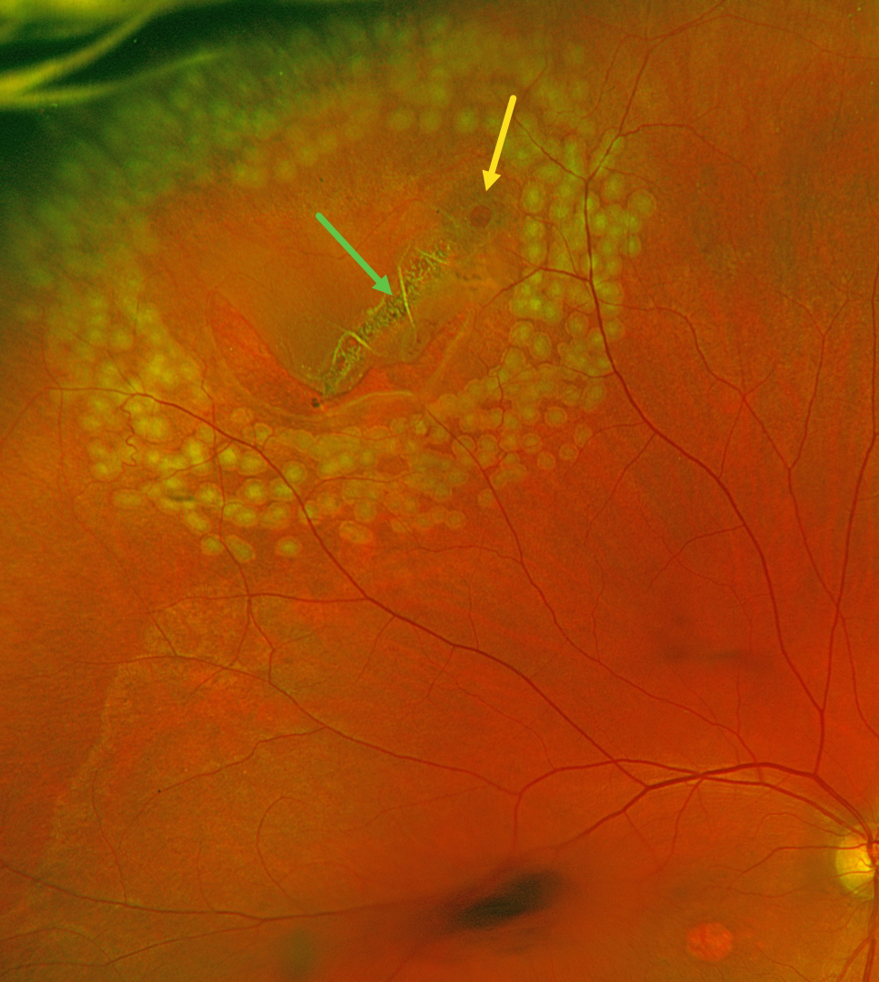
This 63YO male with history of idiopathic central serous chorioretinopathy (ICSC) with secondary macular neovascularization OD and lattice degeneration OU presented with new floaters in his right eye. He was scheduled for an anti-VEGF injection for the right eye on a treat-and-extend protocol the same day. Vision was stable at 20/100 OD and 20/20 OS. An acute, related retinal tear was found and immediately demarcated with thermal laser photocoagulation.
Optos color RG imaging immediately following the laser procedure shows the retinal break well surrounded by multiple nearly confluent rows of laser burns. The flap of the tear consists of an entire lattice lesion along with a chronic atrophic hole with surrounding pigment. There is an area of central macular atrophy from the ICSC.
Learning Points:
Lattice lesions have a pocket of liquefied vitreous overlying thinned retina with a firm area of surrounding vitreoretinal adhesion. White, fibrosed vessels often crisscross the lesions, which gives the lesions their name, “lattice.” This firm vitreoretinal adhesion, along with the often-present atrophic holes, is what can predispose these eyes to retinal breaks and detachment. Although prophylactic treatment is rarely needed, symptomatic breaks, as in our patient, need prompt retinopexy.
RETINAL TEAR
Originally posted on @retina.rocks May 6, 2024
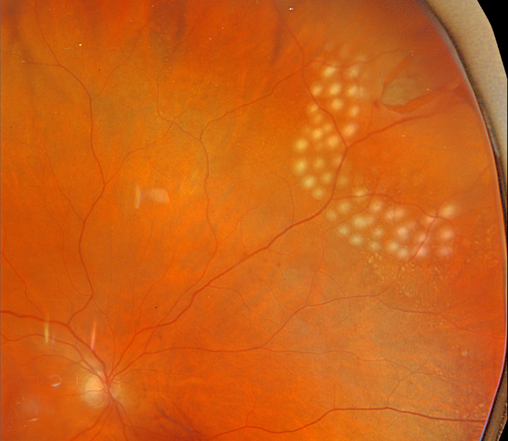
This 70YO female was examined for new flashes and floaters in her right eye and was found to have a retinal tear with localized subretinal fluid at the superonasal equator.
Several nearly confluent rows of laser photocoagulation were applied along its posterior and lateral edges, but we could not extend treatment around its anterior margins with the available slit lamp delivery.
The patient returned the following day to another office where we have our cryotherapy device, where the remainder of the break and fluid were administered.
Learning Points:
Symptomatic retinal tears in the presence of an acute posterior vitreous detachment have a high risk of causing a rhegmatogenous retinal detachment. Prophylactic retinopexy is therefore indicated. Treatment must completely surround the tear and, ideally, extend to the ora serrata. Both thermal laser photocoagulation and cryotherapy are equally effective in creating a vitreoretinal adhesion, which takes at least several weeks to form.
RETINAL BREAK
Mohammad Abbas
Originally posted on @retina.rocks July 10, 2023
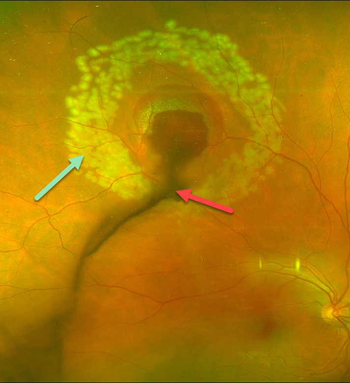
This 27 YO technician working without eye protection in a dermatology laser clinic presented with sudden, painless vision loss and floaters in his right eye. Vision was 20/40.
Optos color imaging shows vitreous blood emanating from the site of laser injury in the super midperiphery. A rim of subretinal blood surrounds the lesion. Fresh laser retinopexy encircles the hemorrhage.
A retinal break is likely obscured by the blood, and this laser should hopefully reduce the risk of a retinal detachment.
Learning Points:
Ocular damage from these lasers depends highly on the laser wavelength, power, and duration. There are several types of lasers used in dermatology, and eye protection for both patients and clinic staff should be used.
RETINAL TEAR + BRVO
Originally posted on @retina.rocks June 5, 2023
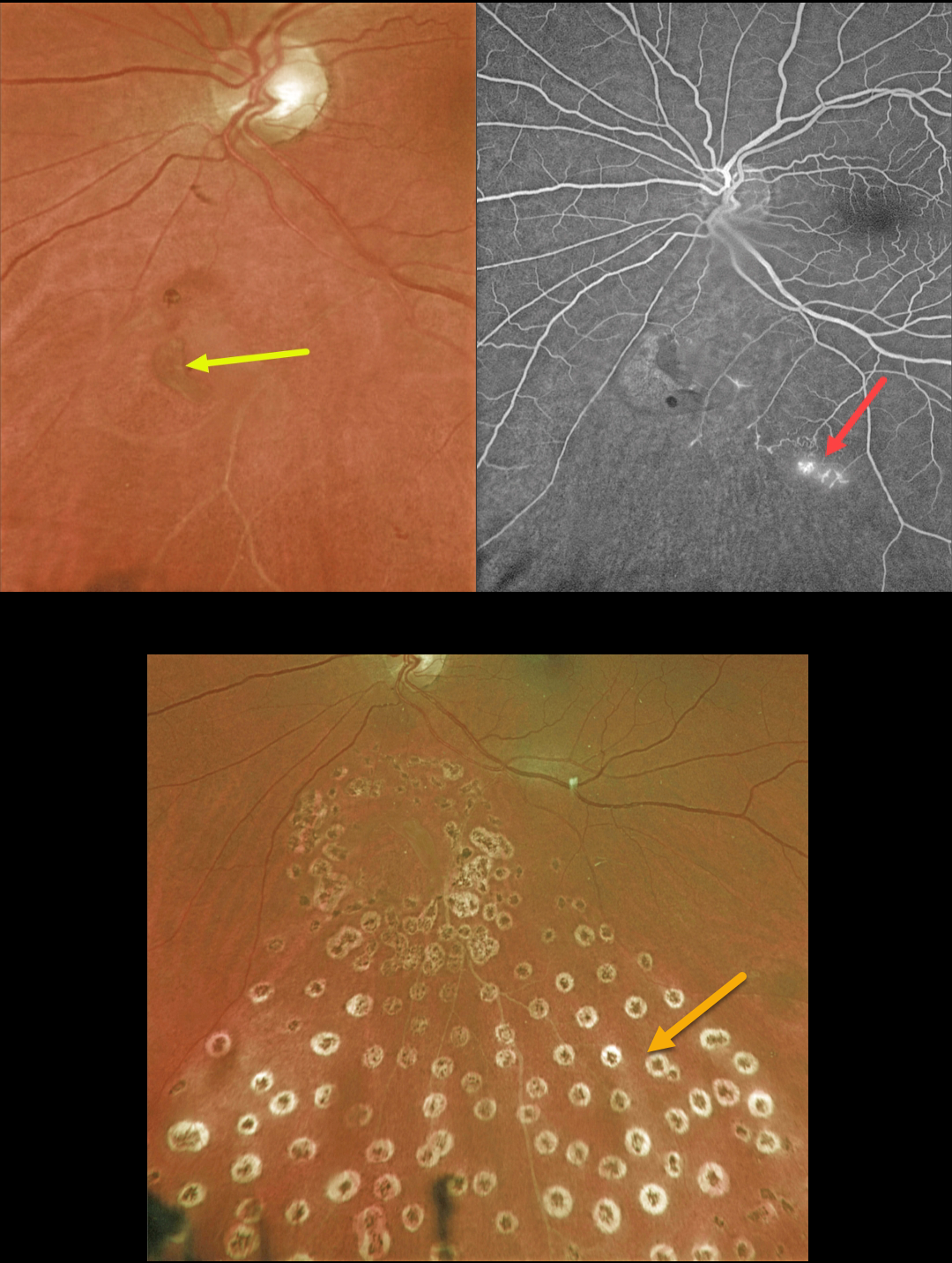
This 63YO female presented with symptomatic flashes and floaters in her left eye.
Optos color imaging shows a retinal tear just inferior to the nerve. An ischemic branch retinal vein occlusion (BRVO) is noted inferiorly. A posterior vitreous detachment (PVD) was noted clinically.
Fluorescein angiography confirmed the inferior ischemic BRVO with some small areas of retinal neovascularization. The tear appeared to be completely independent of the venous occlusion.
The retinal tear was surrounded by a few nearly confluent rows of laser photocoagulation burns, and inferior scatter laser was applied into the distribution of the BRVO. Fundus photography, a few months later, shows the laser scarring.
Learning Points:
Retinal tears are virtually always seen in the retinal periphery and are caused by an adherent vitreous pulling the retina anteriorly towards the vitreous base, most commonly during an acute PVD.
Our patient’s tear is unusual due to its quite posterior location and the flap occurring along its posterior edge. This implies that the vitreous was tugging towards the optic nerve. The optic nerve is usually the last site for the vitreous to release from during an acute PVD (Johnson, AJO 2010;149:371-382).
We hypothesize that the vitreous was completely detached except at the optic nerve and just inferiorly, and the tear was created from anterior to posterior traction when the vitreous separated from the nerve.
LATTICE DEGENERATION WITH RETINAL TEAR
Originally posted on @retina.rocks December 13, 2022
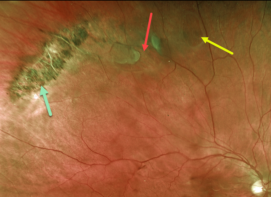
This 64YO male presented with acute flashes and floaters in his right eye. Optos imaging shows a patch of pigmented lattice superotemporally, associated with a retinal tear and atrophic hole. The breaks were surrounded with laser photocoagulation.
Learning Points:
Retinal detachment occurs in 1 in 10,000 people per year. Lattice is found in 10% of the general population, especially in high myopes. Since about one-third of patients with retinal detachment have lattice as the cause, the question always arises: Should prophylactic retinopexy be performed?
The short answer is that asymptomatic lattice generally should not be treated. However, an eye with symptomatic lattice breaks, as in our patient, is at high risk for developing rhegmatogenous detachment and should always be treated.
Excellent references on this controversial topic include: Byer Ophthalmology 1989;96:1396-1402, Folk et al Ophthalmology 1989;96:72-79 and Folk et al Retina 1990;10:165-169.
RETINAL BREAK
Originally posted on @retina.rocks December 7, 2022
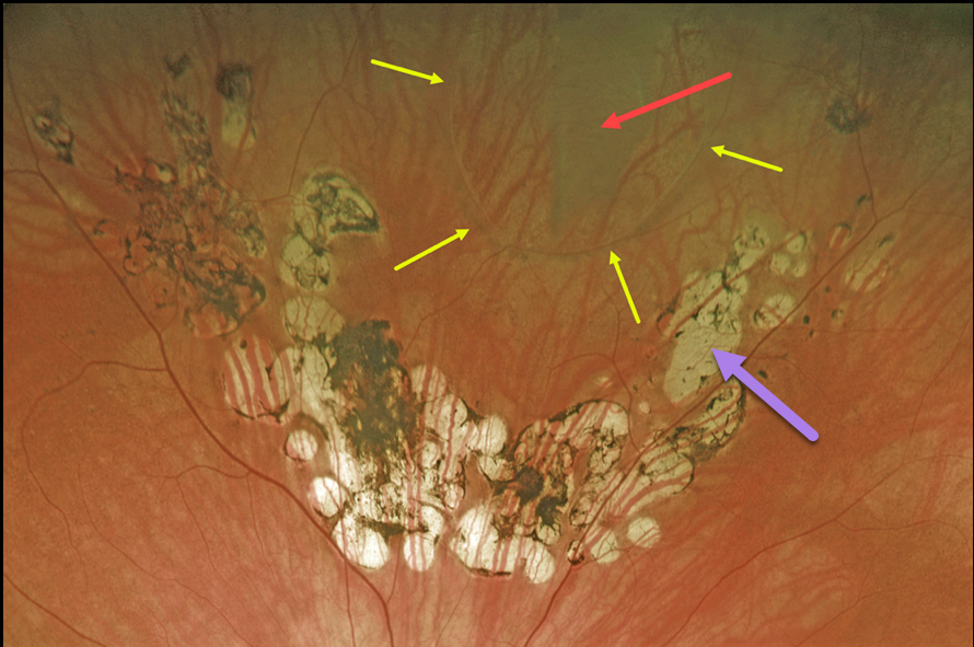
This 67YO female presented with a symptomatic vitreous floater that started about 6 months earlier when she developed a symptomatic retinal tear.
The break was treated elsewhere with laser photocoagulation, and on subsequent visits, she was told the treatment was successful and looked perfect.
However, Optos imaging in our office shows otherwise. A large tear is visualized superiorly. Laser scars are seen posterior to the tear, but barely extend up its lateral margins and completely miss the anterior edge. Even where present, the scarring doesn’t extend to the edge of the break.
Elective vitrectomy surgery will be performed in the near future for her symptomatic vitreous floaters.
We will complete the prophylactic retinopexy at the time of surgery.
Learning Points:
Symptomatic retinal tears in the presence of an acute posterior vitreous detachment have a high risk of causing a rhegmatogenous retinal detachment. Prophylactic retinopexy is therefore indicated.
Treatment must completely surround the tear and, ideally, extend to the ora serrata. Both thermal laser photocoagulation and cryotherapy are equally effective in creating a vitreoretinal adhesion, which takes at least several weeks to form.
RETINAL TEAR
Originally posted on @retina.rocks November 17, 2022
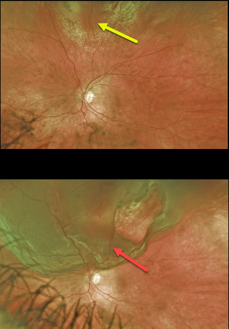
This 37YO female presented with a symptomatic superior retinal tear in her left eye. The posterior and lateral edges of the break were surrounded with thermal laser. The anterior edges could not be reached with the laser, so these were surrounded with cryotherapy. One week later, pigmented scarring surrounds the treated tear.
Three weeks later, she presented with a few days of inferior visual field loss. She now has a superior macula-on rhegmatogenous retinal detachment (RRD) due to lifting off the originally treated tear. Vitrectomy surgery was performed the following day.
Learning Points:
Untreated symptomatic retinal tears have a high risk for RRD. Retinopexy, either with thermal laser or cryotherapy, must completely surround the break and, for more anterior lesions, ideally extend to the ora.
Patients must be followed closely, especially during the first month, due to the risk of additional breaks or retinal detachment. A firm chorioretinal adhesion takes several weeks to form (Yoon and Marmor, Ophthalmology 1988;95:1385-1388). Patients must always be cautioned to return immediately for new flashes, floaters, or vision loss.
CHRPE + RETINAL BREAK
Originally posted on @retina.rocks November 15, 2022
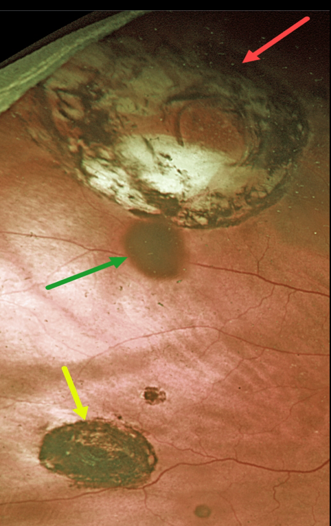
This 87YOM received thermal laser around a retinal break 23 years earlier. He presented to us without symptoms and 20/30 vision.
Optos imaging shows a small inferior congenital hypertrophy of the RPE (CHRPE) with a few tiny additional CHRPE lesions.
Confluent variably pigmented laser scarring around a superotemporal retinal break simulates an atrophic CHRPE lesion.
The operculum casts a shadow on the underlying retina, somewhat giving the impression of yet another pigmented lesion.
RETINAL TEAR
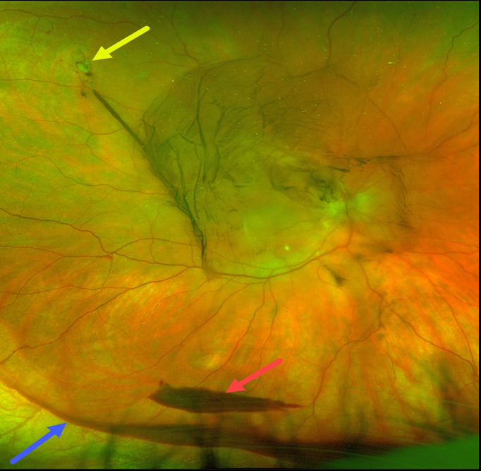
Originally posted on @retina.rocks October 10, 2022
This 59YO male presented with sudden flashes and large floaters in his right eye. Optos imaging shows a small, superotemporal causative retinal tear. There is a moderate central vitreous hemorrhage, a layered inferior pre-retinal hemorrhage, and more inferior blood collecting along the attachment of the vitreous base.
The vitreous has multiple attachments, the strongest of which is within the vitreous base. The sharp inferior border of this patient’s inferior vitreous blood beautifully highlights this anatomy.
RETINAL BREAK
Originally posted on @retina.rocks August 22, 2022
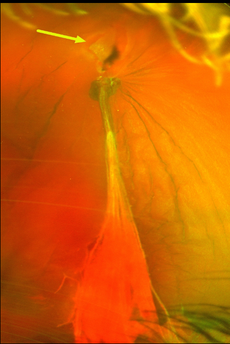
This 50YO female presented with sudden vision loss and floaters in her left eye. Despite the presence of the vitreous hemorrhage, we were able to adequately visualize the causative break and surround it with cryotherapy (not pictured).
A week later, the vitreous hemorrhage increased, and vitrectomy was performed. At the time of surgery, the vitreoretinal traction on the causative tear and blood vessel was relieved. The retina was attached without other breaks. Vision improved to 20/20 five days postoperatively.
RETINAL TEAR
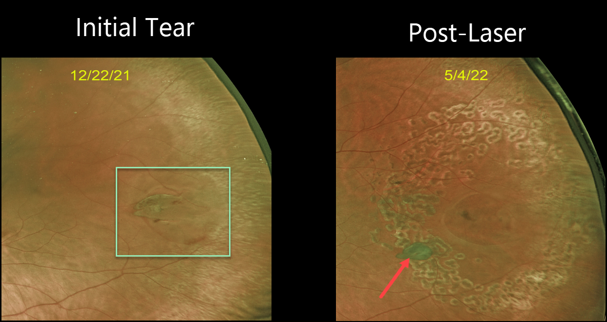
Originally posted on @retina.rocks May 19, 2022
This patient presented with new flashes and floaters in their left eye. Optos imaging shows a tractional retinal tear with a few dots of blood and localized subretinal fluid.
Barrier laser was performed to reduce the risk of retinal detachment. Six months later, good laser scarring surrounds the break, which has operculated.
In our experience, many of these tears will operculate following laser photocoagulation.
DRAGON RETINOPATHY
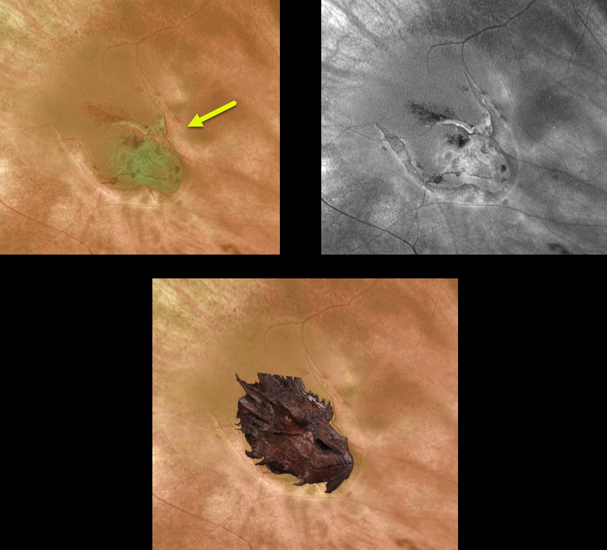
Originally posted on @retina.rocks February 1, 2021
The fiery dragon decided to make an appearance in the back of this patient’s eye. This dangerous predator caused its human prey to experience large black floaters and flashing lights.
Fortunately, the beast, aka retinal tear, was vanquished with an in-office barrier laser.
RETINAL BREAK
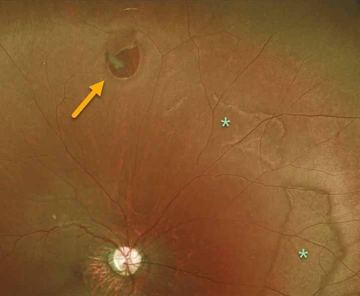
Originally posted on @retina.rocks January 13, 2021
This is a 19YO patient who presented with an asymptomatic retinal break with prominent posterior white without pressure.
Learning Points:
A retinal break is a full-thickness retinal defect. Breaks are classified as atrophic holes, operculated holes, tractional (horseshoe) tears, and retinal dialysis
(break at the ora serrata).
In general, asymptomatic retinal breaks are observed since they are at low risk of causing retinal detachment. Symptomatic retinal tears and retinal dialysis are at the highest risk for causing a detachment and are virtually always treated.
DOGGONE HOLE
Originally posted on @retina.rocks September 19, 2020
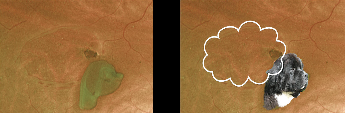
What an interesting operculated retinal break. What do you think this dog is thinking?
RETINAL BREAK
Originally posted on @retina.rocks January 14, 2020
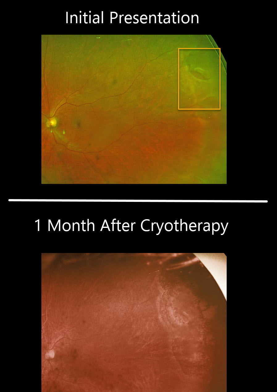
This patient had a symptomatic retinal tear superotemporally in the left eye. The tear was treat with cryotherapy and fundus image shows the appearance of the retina one month later.
Learning Points:
Untreated retinal tears have a very high risk for progressing to retinal detachment. Urgent laser photocoagulation or cryotherapy prevents liquid vitreous from leaking thru the break. Patients need to be followed closely postoperatively to be sure no new breaks develop and that chorioretinal scarring surrounds the break. Unlike thermal laser demarcation, the cryotherapy applicatiohns need to surround the break with confluent scarring in order to adequately prophylax against retinal detachment.


