

Retinal Capillary Hemangioma
<< Back to Cases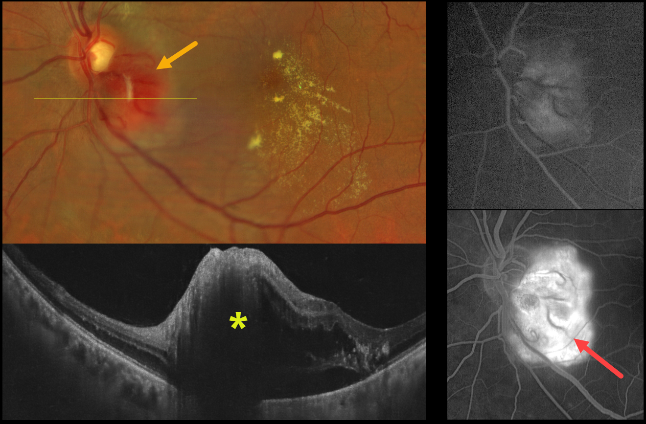
RETINAL CAPILLARY HEMANGIOMA
Ayushi Gupta and Vishal Agrawal
Originally posted on @retina.rocks June 29, 2026
This healthy 51YO male presented with one month of vision loss in his left eye. Vision was 20/20 in his normal OD and 20/200 OS.
Color fundus photography shows an elevated orange-red juxtapapillary tumor along the inferotemporal edge of the optic nerve with associated macular lipid. OCT scanning reveals the lesion to be hyperreflective with associated outer retinal and subretinal fluid. The lesion leaked on fluorescein angiography.
Family history was negative for von Hippel–Lindau disease, and systemic evaluation, including brain MRI and renal ultrasonography, was unremarkable. Anti-VEGF therapy was recommended.
Learning Points:
Retinal capillary hemangiomas appear clinically as orange-red lesions, most commonly peripherally, and are associated with prominent, dilated, tortuous paired feeding and draining vessels. They can also appear on or around the optic nerve lesions, either on the surface (endophytic) as in our patient, or buried within the substance of the nerve (exophytic). Unlike peripheral lesions, juxtapapillary tumors often lack prominent feeder vessels. These lesions can cause severe vision loss or blindness from exudative retinal detachment and thus usually require aggressive treatment. Treatment options include thermal laser, photodynamic therapy, cryotherapy, and anti-VEGF injections.
More recently, oral belzutifan, an inhibitor of hypoxia-inducible factor 2 alpha, has emerged as a highly effective treatment for these lesions. In the LITESPARK-004, a Phase 2 trial, all 16 eyes improved without new lesions after a mean follow-up of 37 months (Wiley et al., Ophthalmology 2024;131;1324-1332).
Retinal capillary hemangioma is also a predominant feature of von Hippel-Lindau disease (VHL), an autosomal dominant condition caused by mutations in the VHL tumor suppressor gene. Patients develop benign and malignant tumors, including central nervous system hemangioblastoma, pheochromocytoma, and renal cell carcinoma. Rarely, sporadic, non-hereditary tumors may be found, as in our patient.
RETINAL CAPILLARY HEMANGIOMA
The European VitreoRetina Society (EVRS), Ece Ozdemir Zeydanli and Mehmet Onen
Originally posted on @retina.rocks August 22, 2025
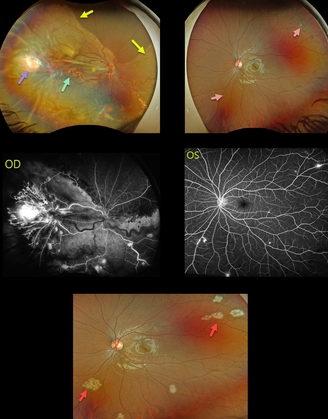
This 17YO female presented with a 3-month history of vision loss in her right eye. Vision was hand motion OD and 20/20 OS.
Optos color RGB imaging OD shows a large yellow-red angioma in the temporal periphery, associated with large dilated and draining vessels. There is another large angioma nasally, near the ora serrata, which is not well imaged. Multiple smaller angiomas are scattered throughout the temporal and inferior retina. A near-total exudative retinal detachment (RD) spares the superonasal retina. A few small peripheral angiomas are noted in her left eye. All tumors leaked angiographically.
The angiomas in the left eye were lasered during her initial examination. She then underwent lens-sparing vitrectomy in the right eye with membrane removal, endophotocoagulation, feeder vessel endodiathermy, and endoresection of the large temporal and nasal tumors. Mitomycin C was applied outside the posterior pole to reduce the risk of PVR, and silicone oil tamponade was used.
Our patient’s family history was negative. Genetic testing was subsequently positive for the von Hippel-Lindau (VHL) gene, which likely represents a de novo mutation.
Learning Points:
Retinal capillary hemangiomas are orange-red lesions associated with a prominent paired feeding and draining vessel. These benign vascular tumors need to be aggressively treated since they can cause severe vision loss or blindness from exudative RD, as in our patient. Patients require lifelong screening examinations for the development of new tumors even after successful treatment. Ultrawidefield imaging is helpful in detecting and monitoring these lesions.
Retinal capillary hemangioma is a predominant feature of VHL, an autosomal dominant condition caused by mutations in the VHL tumor suppressor gene. Patients develop benign and malignant tumors, including central nervous system hemangioblastoma, pheochromocytoma, and renal cell carcinoma.
RETINAL CAPILLARY HEMANGIOMA
Rohan Jain and Manish Nagpal
Originally posted on @retina.rocks January 20, 2025
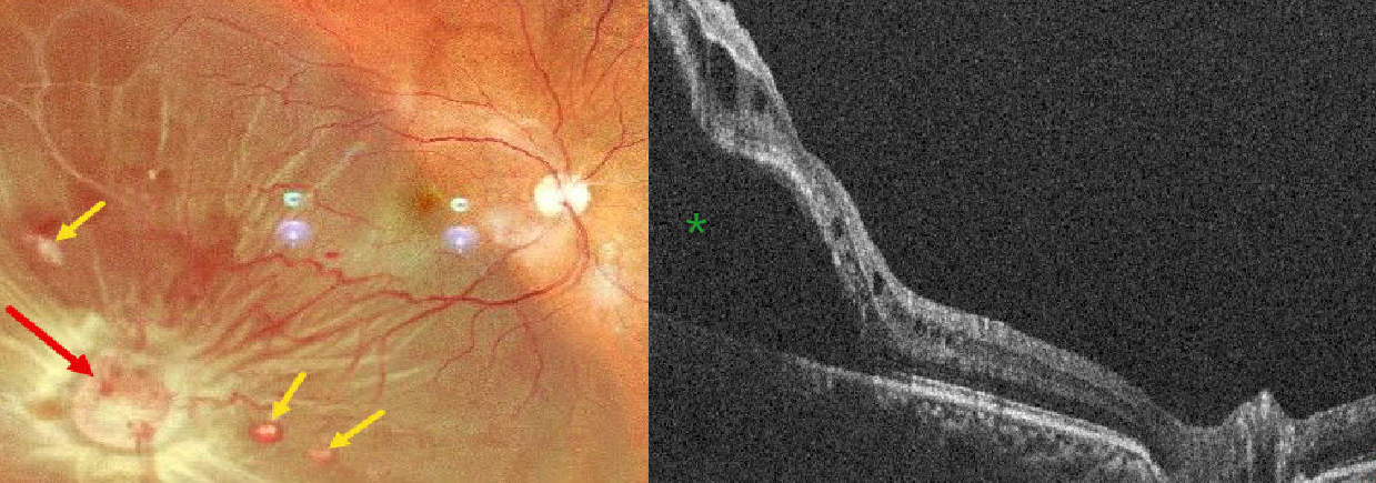
This 52YO male presented with 5 days of vision loss OD. There was a history of bilateral vision loss following facial trauma from a motor vehicle accident 20 years earlier. He denied any family history of eye disease or cancer. Vision was counting fingers OD and 20/200 OS.
Pseudocolor SLO imaging shows an orange-yellow mass consistent with a retinal capillary hemangioma in the inferotemporal midperiphery, causing an exudative retinal detachment. Several smaller hemangiomas are noted. The macula-off detachment is confirmed on OCT scanning.
A recent MRI showed bilateral optic atrophy, a partially empty sella, and an old, healed fracture of the right lateral wall of the orbit. Although emergent vitrectomy surgery was recommended, he was immediately lost to follow-up.
Learning Points:
Retinal capillary hemangiomas are orange-red lesions associated with a prominent paired feeding and draining vessel. These benign vascular tumors need to be aggressively treated since they can cause severe vision loss or blindness from exudative retinal detachment, as in our patient. The presence of hydration outer retinal folds is one of the key findings that help differentiate rhegmatogenous from exudative and traction retinal detachments (Oquendo et al, AJO 2024;268:212-221). The absence of these folds in our patient is typical of an exudative detachment. Common treatment options include thermal laser, cryotherapy, and anti-VEGF injections.
Retinal capillary hemangioma is a predominant feature of VHL, an autosomal dominant condition caused by mutations in the VHL tumor suppressor gene. Patients develop benign and malignant tumors, including central nervous system hemangioblastoma, pheochromocytoma, and renal cell carcinoma.
RETINAL CAPILLARY HEMANGIOMA
Originally posted on @retina.rocks October 1, 2024
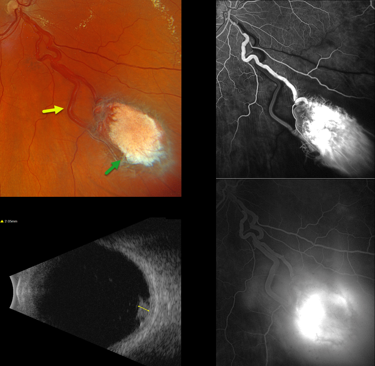
This healthy 42YO male was referred for an asymptomatic retinal lesion in his right eye. There was no family history of cancer or eye disease, although he had no knowledge about his birth father. Vision was 20/30 OD and 20/20 in his normal left eye.
Optos color RGB imaging shows an active endophytic retinal capillary hemangioma (RCH) in the inferonasal midperiphery. The tumor is supplied and drained by dilated, irregular vessels. The lesion shows profound leakage on fluorescein angiography. It measured about 2mm thick on B-scan ultrasonography. Genetic testing for the von Hippel-Lindau syndrome (VHL) gene was negative, as was MRI scanning of the brain, abdomen, and pelvis. Monthly intravitreal Avastin injections are planned to shrink the tumor, followed by thermal laser photocoagulation.
Learning Points:
Retinal capillary hemangiomas are orange-red lesions associated with a prominent paired feeding and draining vessel and can cause severe vision loss or blindness from exudative retinal detachment. These benign vascular tumors need to be aggressively treated. Common treatment options include thermal laser, cryotherapy, and anti-VEGF injections.
They are a predominant feature of VHL, an autosomal dominant condition caused by mutations in the VHL tumor suppressor gene. This prevents the degradation of hypoxia-inducible factor 1a (HIF-1a), which, in turn, causes the production of numerous growth factors, including VEGF (Kaelin, Drug Discoveries Today Disease Mechanisms 2005;2:225-231). Patients develop benign and malignant tumors, including central nervous system hemangioblastoma, pheochromocytoma, and renal cell carcinoma. Sporadic RCHs in the absence of the VHL syndrome are a rare occurrence (Singh et al, Ophthalmology 2001;108:1907-1911). The negative genetic testing fortunately ruled out VHL in our patient. Our patient’s lesion most likely resulted from a localized double-hit retinal VHL mutation.
RETINAL CAPILLARY HEMANGIOMA
Originally posted on @retina.rocks August 19, 2024
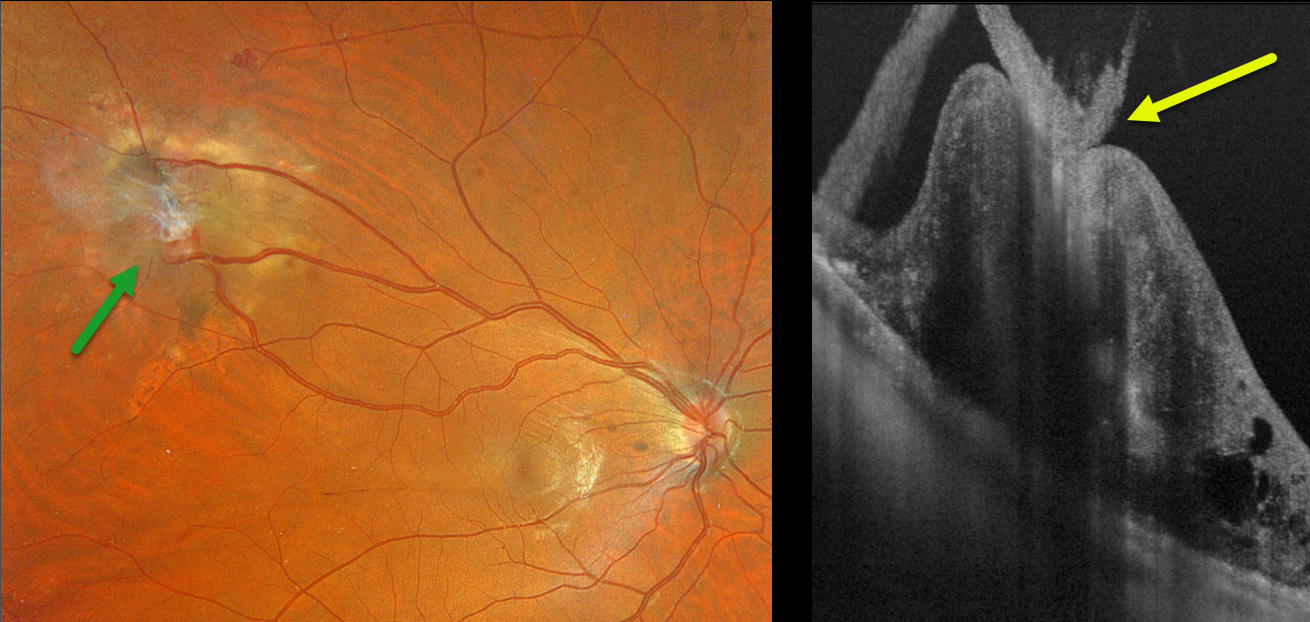
This 28YO male with a known history of Von Hippel-Lindau (VHL) recently moved to our area. He received prior anti-VEGF injections and thermal laser for a retinal capillary hemangioma in his right eye.
Optos color RGB imaging shows a regressed endophytic capillary hemangioma in the superotemporal midperiphery. The tumor is supplied and drained by dilated, irregular vessels. On Silverstone swept-source OCT, the angioma appears as a thickened, hyperreflective lesion with overlying vitreous traction.
Learning Points:
Retinal capillary hemangiomas are orange-red lesions associated with a prominent paired feeding and draining vessel and can cause severe vision loss or blindness from exudative retinal detachment. These benign vascular tumors need to be aggressively treated. Common treatment options include thermal laser, cryotherapy, and anti-VEGF injections.
Retinal capillary hemangioma is a predominant feature of VHL, an autosomal dominant condition caused by mutations in the VHL tumor suppressor gene. Patients develop benign and malignant tumors, including central nervous system hemangioblastoma, pheochromocytoma, and renal cell carcinoma.
We plan on following our patient several times per year to monitor for new lesions or recurrent exudation.
RETINAL CAPILLARY HEMANGIOMA
Yuenpang Cheung and Stephanie Choi
Originally posted on @retina.rocks December 19, 2023
This 23YO female was noted by her optometrist in 2019 to have vision loss from a retinal lesion. At presentation, vision was 20/400 in her left eye and 20/20 in the right eye.
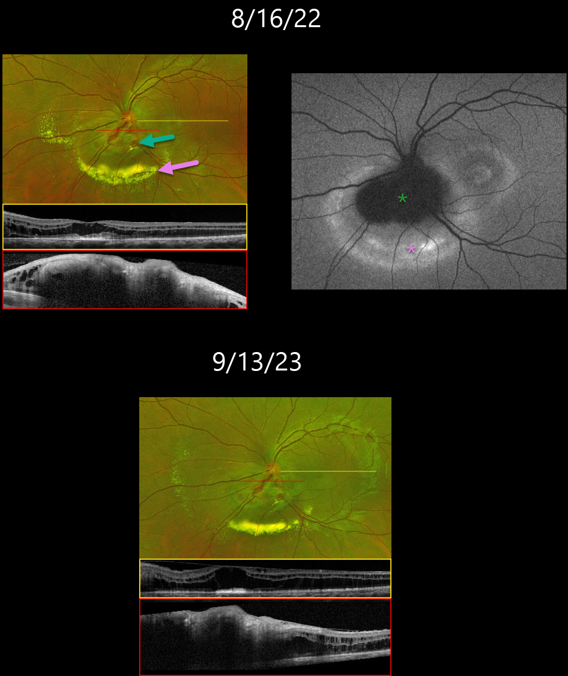
She was referred to an outside retina specialist, who gave a single anti-VEGF injection and then referred her to Columbia University. Our initial examination on 8/16/22 revealed an inferior peripapillary retinal capillary hemangioma with surrounding exudation. OCT of the macula revealed mostly outer retinal fluid. The angioma was diffusely thickened and hyperreflective.
On fundus autofluorescence (FAF), the angioma was hypo-FAF with a surrounding rim of hyper-FAF. Genetic testing for the VHL gene was negative, and thus further systemic work-up of the brain and kidneys was deferred.
She underwent subsequent monthly anti-VEGF injections with resolving exudates. On 9/13/23, the lipid decreased, but she continues to have persistent fluid and thickening on OCT.
Learning Points:
Retinal capillary hemangiomas appear clinically as orange-red lesions, most commonly peripherally, and are associated with prominent, dilated, tortuous paired feeding and draining vessels.
They can also appear on or around the optic nerve, either on the surface (endophytic), as in our patient, or buried within the substance of the nerve (exophytic).
These lesions can cause severe vision loss or blindness from exudative retinal detachment and thus usually require aggressive treatment. Treatment options include thermal laser, photodynamic therapy, cryotherapy, and anti-VEGF injections.
Retinal capillary hemangioma is also a predominant feature of von Hippel-Lindau disease (VHL), an autosomal dominant condition caused by mutations in the VHL tumor suppressor gene. Patients develop benign and malignant tumors, including central nervous system hemangioblastoma, pheochromocytoma, and renal cell carcinoma.
RETINAL CAPILLARY HEMANGIOMA
Sayali Bawankule, Prashant Bawankule, Shilpi Narnaware, and Anju Bansal
Originally posted on @retina.rocks April 6, 2023
This 31YO healthy female presented with a 3-day history of sudden vision loss in her right eye. Other than a recent history of hypertension, past medical and family history were negative. Her twins both died at 6 months of age from complications related to prematurity. Vision was counting fingers OD and 20/20 OS.
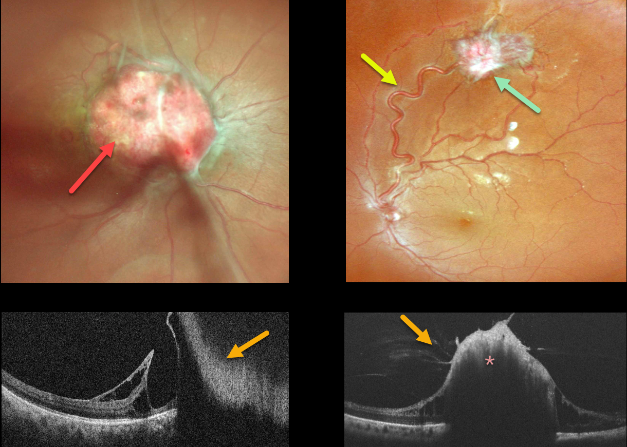
There was a moderate diffuse vitreous hemorrhage in her right eye. Color imaging shows an endophytic capillary hemangioma overlying the right optic nerve. Color imaging of her left eye shows a superotemporal peripheral variably fibrosed retinal capillary hemangioma fed and drained by dilated and irregular vessels.
On OCT, the angiomas appear as thickened, hyperreflective lesions with overlying vitreous traction.
A provisional diagnosis of Von Hippel-Lindau (VHL) disease was made, presumably from a new mutation, and she was referred to neurology and nephrology for further systemic evaluation. Unfortunately, she was subsequently lost to follow-up.
Learning Points:
Retinal capillary hemangiomas are orange-red lesions associated with a prominent pair of feeding and draining vessels and can cause severe vision loss or blindness due to exudative retinal detachment. These benign vascular tumors need to be aggressively treated. Common treatment options include thermal laser, cryotherapy, and anti-VEGF injections.
Retinal capillary hemangioma is a predominant feature of VHL, an autosomal dominant condition caused by mutations in the VHL tumor suppressor gene. Patients develop benign and malignant tumors, including central nervous system hemangioblastoma, pheochromocytoma, and renal cell carcinoma.
RETINAL CAPILLARY HEMANGIOMA
Originally posted on @retina.rocks February 1, 2023
This 39YO healthy male was referred for an asymptomatic lesion in his right eye.
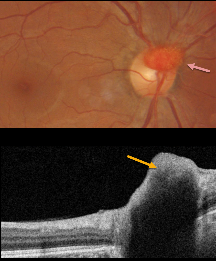
Fundus photography shows an elevated, vascularized lesion covering the superior optic nerve. The lesion was markedly hyperreflective on OCT.
On further questioning, there was a strong family history of Von Hippel-Lindau (VHL) disease, including his sister and maternal nephew. Genetic testing was ordered.
Learning Points:
Retinal capillary hemangioma is a predominant feature of VHL, an autosomal dominant condition caused by mutations in the VHL tumor suppressor gene. Patients develop benign and malignant tumors including central nervous system hemangioblastoma, pheochromocytoma and renal cell carcinoma.
Retinal capillary hemangiomas appear clinically as orange-red lesions, most commonly seen peripherally associated with a prominent paired feeding and draining vessel. They can also appear on or around the optic nerve lesions, either on the surface (endophytic) as in our patient, or buried within the substance of the nerve (exophytic).
These lesions can cause severe vision loss or blindness from exudative retinal detachment and thus usually require aggressive treatment. Treatment options include thermal laser, photodynamic therapy, cryotherapy, and anti-VEGF injections.
Since our patient is completely asymptomatic with no exudation, we will observe without treatment for now.
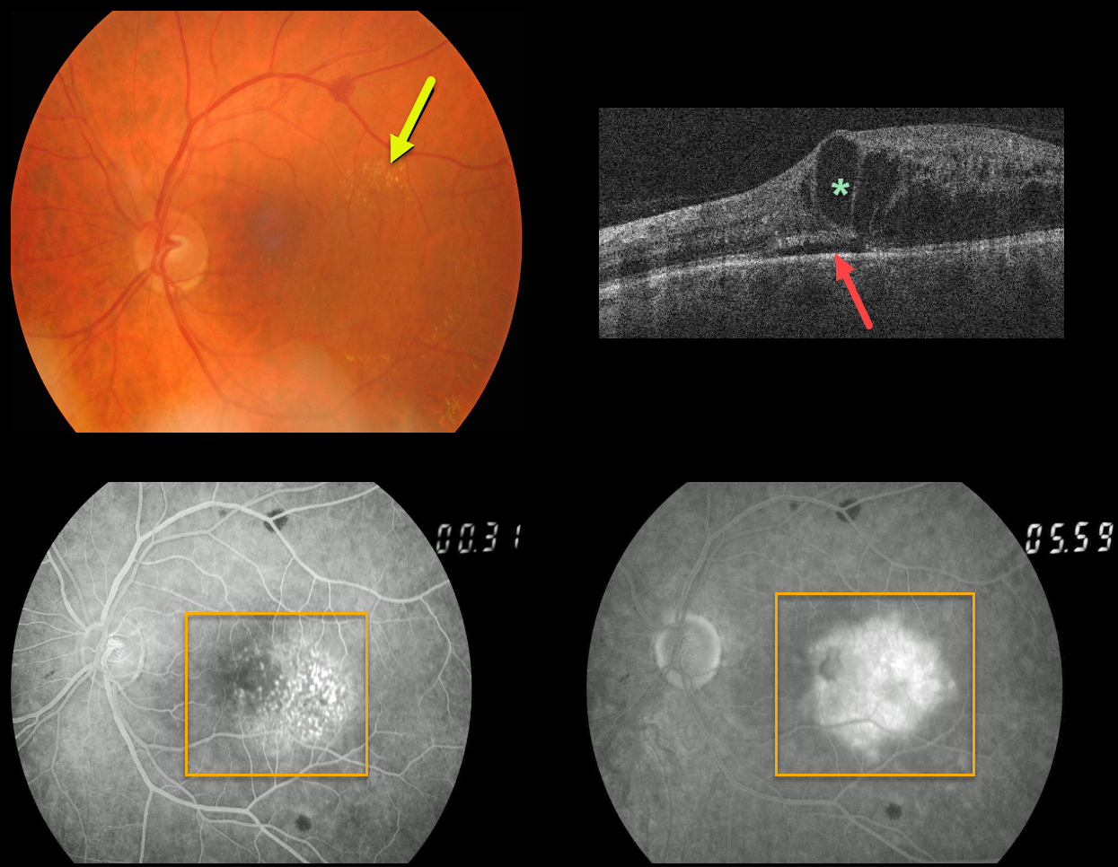
RETINAL CAPILLARY HAMARTOMA
Originally posted on @retina.rocks July 7, 2021
This patient presented with very subtle retinal vascular telangiectasia in the temporal macula, some scattered temporal macular lipid, and moderate cystic retinal edema with trace subretinal fluid on OCT. There were also a few larger scattered deep retinal hemorrhages.
Fluorescein angiography shows much more dramatic intraretinal vascular abnormalities centered in the temporal macula with moderate leakage.
Learning Points:
We believe this patient has a retinal capillary hamartoma. This is an extremely rare finding; to the best of our knowledge, only been previously described in a strikingly similar case in the 5th edition of Gass’ classic atlas (Gass’ Atlas of Macular Diseases 5th Edition, Agarwal A, Elsevier 2011, pages 446-447).
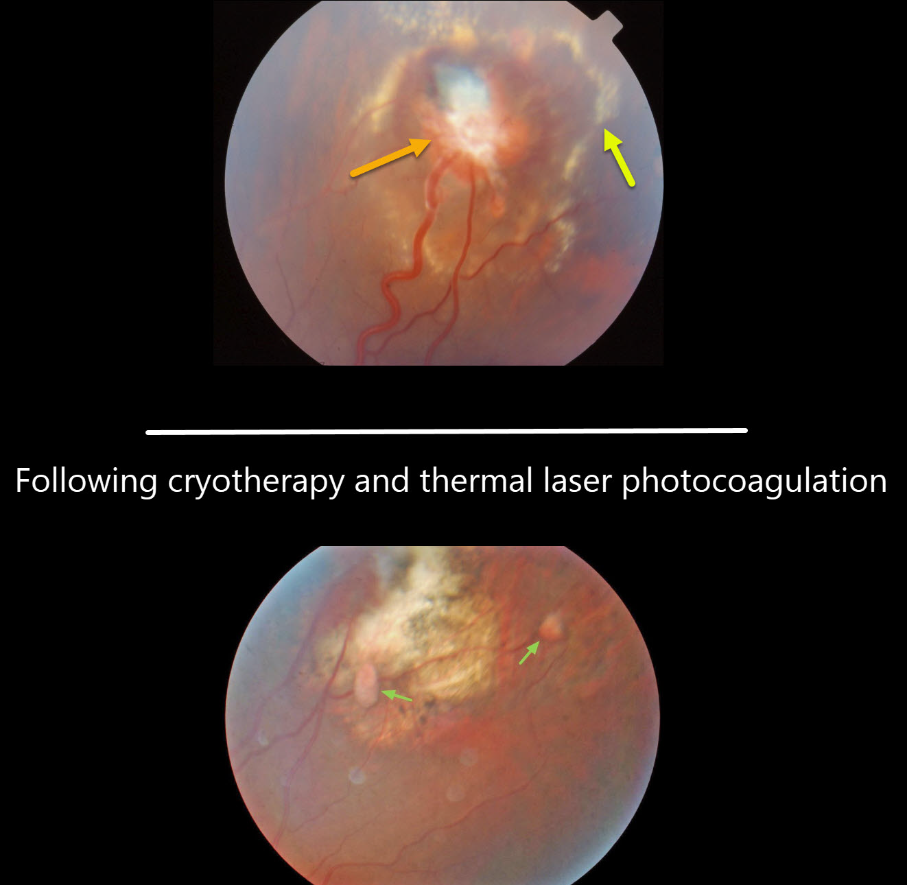
RETINAL CAPILLARY HEMANGIOMA
Originally posted on @retina.rocks January 20, 2021
We have been caring for this patient and his family with Von Hippel-Lindau (VHL) disease for over 20 years.
We initially treated the retinal capillary hemangioma in his left eye with cryotherapy. Although the tumor initially regressed, thermal laser photocoagulation was applied due to some new exudation. The exudation eventually resolved. A few smaller involuted angiomas are well-visualized in his most recent photo.
Our patient had a partial nephrectomy for renal cell carcinoma, and his mother died 2 years ago from renal cell carcinoma. His sister required enucleation of her right eye as a teenager for a total exudative detachment.
Learning Points:
Retinal capillary hemangioma is a predominant feature of VHL, an autosomal dominant condition caused by mutations in the VHL tumor suppressor gene. Patients develop benign and malignant tumors, including central nervous system hemangioblastoma, pheochromocytoma, and renal cell carcinoma.
Retinal capillary hemangiomas are orange-red lesions associated with prominent paired feeding and draining vessels and can cause severe vision loss or blindness due to exudative retinal detachment. These benign vascular tumors need to be aggressively treated. Common treatment options include thermal laser, cryotherapy, and anti-VEGF injections.


