

Retinal Choroidal Anastomosis
<< Back to Cases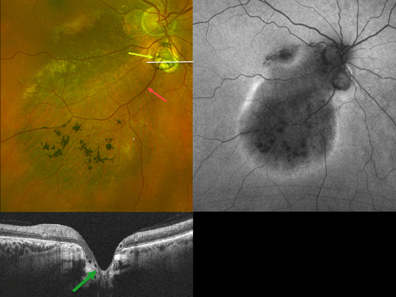
CHOROIDAL COLOBOMA WITH OPTIC NERVE PSEUDODUPLICATION
Originally posted on @retina.rocks October 8, 2025
This 60YO female was referred for asymptomatic unilateral fundus findings in her right eye. Vision was 20/30 OD.
Optos color RG imaging shows a choroidal coloboma just inferior to the disc, giving the appearance of a pseudoduplicated optic nerve. Coarse pigmentary changes with some pigment migration extend from the macula into the inferior midperiphery. An inferotemporal retinal vein drains directly into the choroid through the coloboma. Triton swept-source OCT shows that the coloboma overlies an area of ectatic sclera. Fundus autofluorescence (FAF) shows variable hypo-FAF within the area of clinical pigmentary changes, with a surrounding rim of hyper-FAF.
Learning Points:
Choroidal colobomas are most often due to failure of the optic vesicle and choroidal fissure to close during fetal development. Lesions like this may also be from other causes, including toxoplasmosis. Choroidal coloboma can present as solitary or multiple lesions. Visual prognosis depends on the location and involvement of the optic nerve and macula.
Pseudoduplication of the optic nerve occurs when a round choroidal coloboma or chorioretinal scar about the size of the optic nerve is associated with overlying radiating blood vessels (Bloom et al, Retinal Cases 2022;16:174-176). In our experience, it is most common for toxoplasmosis scars to be located near the optic nerve.
Under normal conditions, the retinal and choroidal circulations remain separate and distinct. Rarely, the two circulations connect directly through a chorioretinal anastomosis. These can occur in chorioretinal scars (typically from inactive toxoplasmosis lesions), in macular telangiectasia, and in disciform scars.
The prominent gutter of inferior pigmentary changes indicates a prior retinal detachment, most likely related to spontaneously resolved fluid associated with the coloboma (Tanaka et al, Ophthalmology Retina 2021;5:702-710). Similar pigmentary changes are seen in central serous retinopathy, although our patient’s macular OCTs showed no pachychoroid features (not shown). Finally, this may represent previously resolved pit-like macular fluid, which is rarely found in the absence of a pit or severe glaucomatous cupping (Fujimoto et al, Ophthalmology Retina 2023;7:811-818).
CHOROIDAL COLOBOMA
Evgenii Chernov
Originally posted on @retina.rocks March 18, 2025
This 32YO female has a history of congenital nystagmus and poor vision since birth. Vision was 20/130 OU.
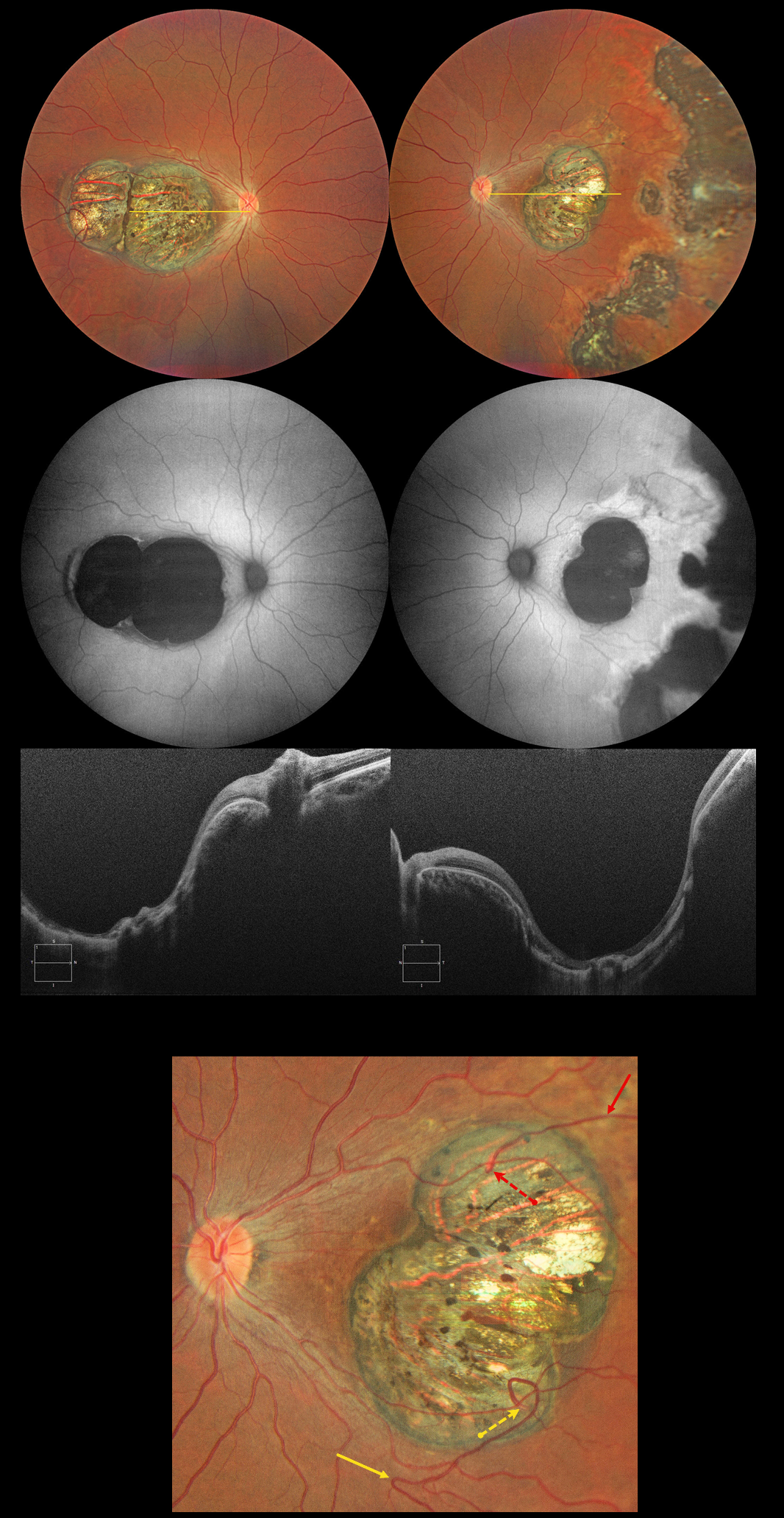
Color photography shows bilateral macular colobomas with more peripheral multifocal variably pigmented colobomatous scarring in her left eye. These lesions are hypoautofluorescent. OCT scanning through the macular lesions shows thinned hyperreflective retina draped across the posteriorly bowed sclera.
The right macular coloboma also contains several retinal choroidal anastomoses. The major inferotemporal vein acutely bends superotemporally to enter the coloboma and drain into the choroidal circulation. A superotemporal vein also enters the coloboma to drain into a visible choroidal vortex.
Learning Points:
Choroidal colobomas are most commonly caused by failure of the optic vesicle and choroidal fissure to close during embryonic development and are usually found inferonasally. They can present as solitary or multiple lesions. The differential diagnosis for congenital macular coloboma also includes post inflammatory lesions (toxoplasmosis, cytomegalovirus, and zika) and retinal dystrophies including North Carolina macular dystrophy. The retina within the coloboma is variably thinned, which increases the risk for rhegmatogenous retinal detachment.
Under normal conditions the retinal and choroidal circulations remain separate and distinct. Rarely, the two circulations directly connect with each other through a chorioretinal anastomosis. These can occur in chorioretinal scars (typically from inactive toxoplasmosis lesions), in macular telangiectasia, and in disciform scars.
TOXOPLASMOSIS WITH RETINAL CHOROIDAL ANASTOMOSIS
Originally posted on @retina.rocks October 21, 2024
This 60YO female was referred for asymptomatic scarring in her left eye. Vision was 20/50 OD and 20/40 OS due to early bilateral cataracts. Small macular drusen were noted bilaterally.
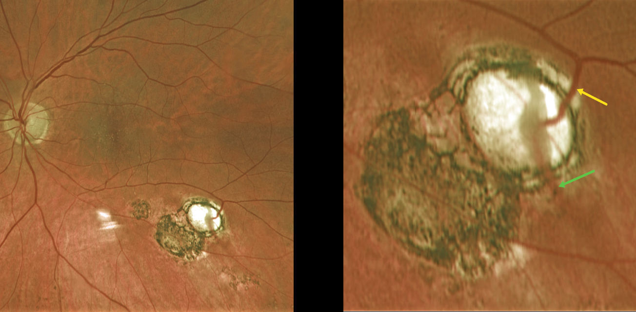
Optos color RG imaging of her left shows multifocal variably pigmented scars below the inferotemporal arcade. A magnified inferotemporal view shows a retinal vein diving into an atrophic scar and directly connecting to a choroidal vein.
Learning Points:
Under normal conditions the retinal and choroidal circulations remain separate and distinct. Rarely, the two circulations directly connect with each other through a chorioretinal anastomosis. These can occur in chorioretinal scars (typically from inactive toxoplasmosis lesions as in this patient), in macular telangiectasia, and in disciform scars. We felt our patient’s findings most consistent with inactive toxoplasmosis scarring.
TOXOPLASMOSIS WITH RETINAL CHOROIDAL ANASTOMOSIS
Originally posted on @retina.rocks September 20, 2022
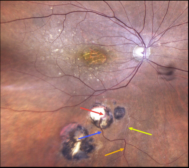
This 86YO female has a history of atrophic age-related macular degeneration with vision of 20/400 OD and 20/60 OS. Optos imaging of her right eye shows foveal atrophy with surrounding mixed drusen.
Multifocal inactive toxoplasmosis scars are noted inferiorly. An inferior retinal vein bifurcates, with a markedly attenuated branch continuing on its path towards the optic nerve. The other branch has a more normal caliber, dives into one of the toxoplasmosis scars, and drains into the choroid.
Learning Points:
Under normal conditions, the retinal and choroidal circulations remain separate and distinct. Rarely, the two circulations connect directly through a chorioretinal anastomosis.
These can occur in chorioretinal scars (typically from inactive toxoplasmosis lesions, as in this patient), in macular telangiectasia, and in disciform scars.
CHOROIDAL GRANULOMA
Originally posted on @retina.rocks December 8, 2021
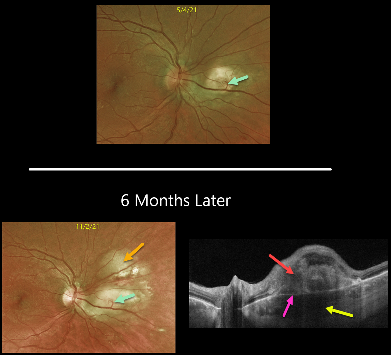
This is a follow-up from our 6/7/21 post of a choroidal granuloma, likely representing the initial manifestation of systemic sarcoidosis. The patient was sent to pulmonology to confirm the diagnosis, but he did not keep the appointment.
Six months later, the choroidal granuloma had significantly enlarged.
A retinochoroidal anastomosis is noted between the overlying retinal vein and the lesion. Triton swept-source OCT shows a thickened, hyporeflective choroidal lesion, a hyperreflective outer retinal lesion with overlying inner retinal disorganization, and visible Bruch’s membrane separating the choroidal and retinal components.
Learning Points:
Choroidal granulomas are aggregations of inflammatory cells that appear clinically as a solitary, creamy-white, round subretinal lesion. Causes include sarcoidosis, tuberculosis, and VKH.
They are hyporeflective with increased transmission on OCT imaging (see Invernizzi et al, Retina 2015;35:525-531).
NEOVASCULAR AMD + RETINAL CHOROIDAL ANASTAMOSIS
Originally posted on @retina.rocks November 25, 2021
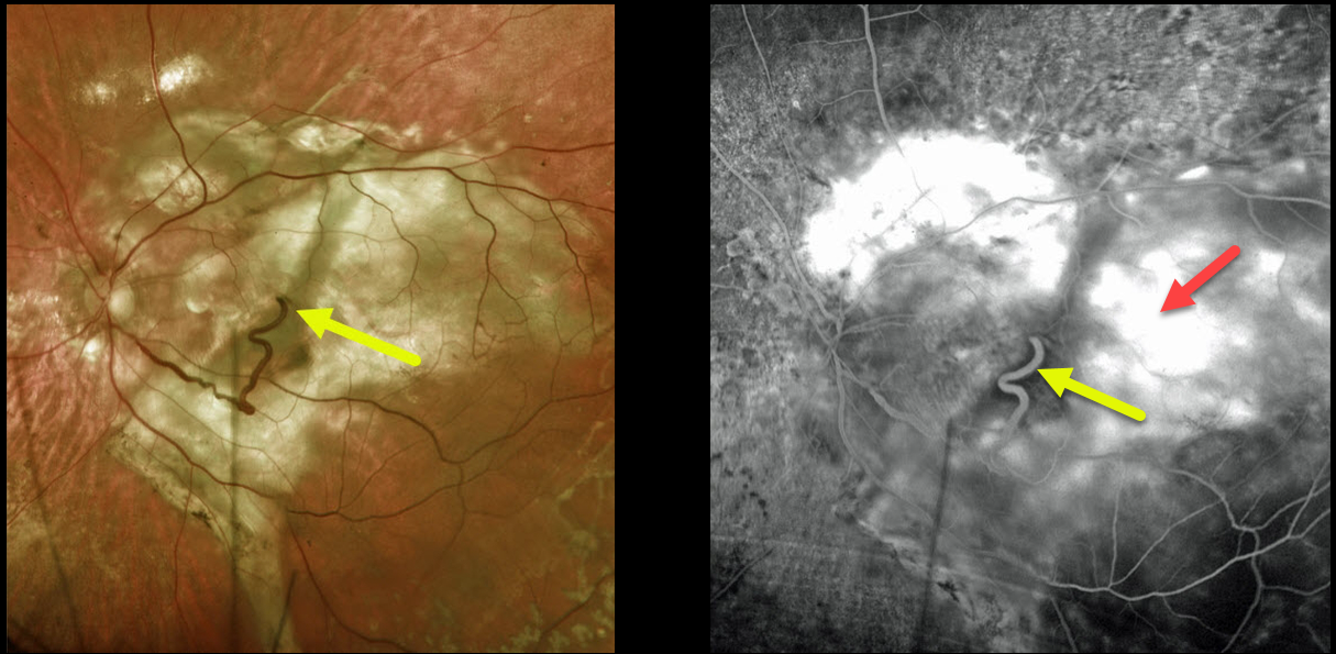
This 74YO female has a large complex disciform scar from end-stage age-related macular degeneration (AMD).
A very prominent retinal vein drains directly into the substance of the scar inferiorly, indicating a chorioretinal anastomosis. Fluorescein angiography shows diffuse staining of the subretinal scarring.
Learning Points:
Under normal conditions, the retinal and choroidal circulations remain separate and distinct. Rarely, the two circulations connect directly through a chorioretinal anastomosis.
These can occur in chorioretinal scars (typically from inactive toxoplasmosis lesions), in macular telangiectasia, and in disciform scars.
RETINAL CHOROIDAL ANASTOMOSIS
Originally posted on @retina.rocks March 24, 2020
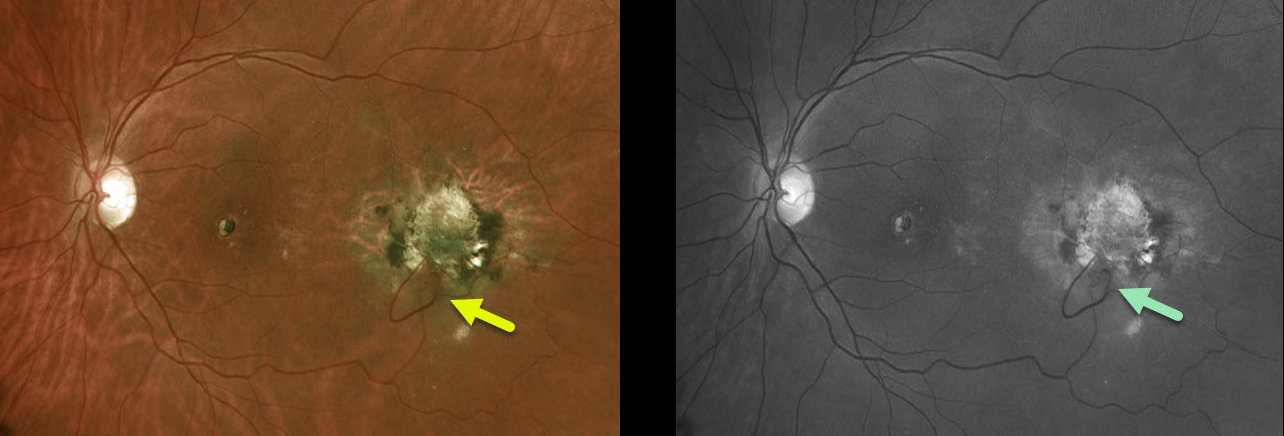
Our patient has a very prominent chorioretinal anastomosis from an idiopathic ectopic disciform scar. It is best seen in the Optos color and green-channel images.
Learning Points:
Under normal conditions the retinal and choroidal circulations remain separate and distinct. Rarely, the two circulations directly connect with each other thru a chorioretinal anastomosis.
These can occur in chorioretinal scars (typically from inactive toxoplasmosis lesions), in macular telangiectasia, and in disciform scars. Laser photocoagulation can create an iatrogenic retinal venous-choroidal anastomosis to treat macular edema in non-ischemic CRVOs, although its use has not been widely adopted.


