

Retinal Collateral Vessels
<< Back to Cases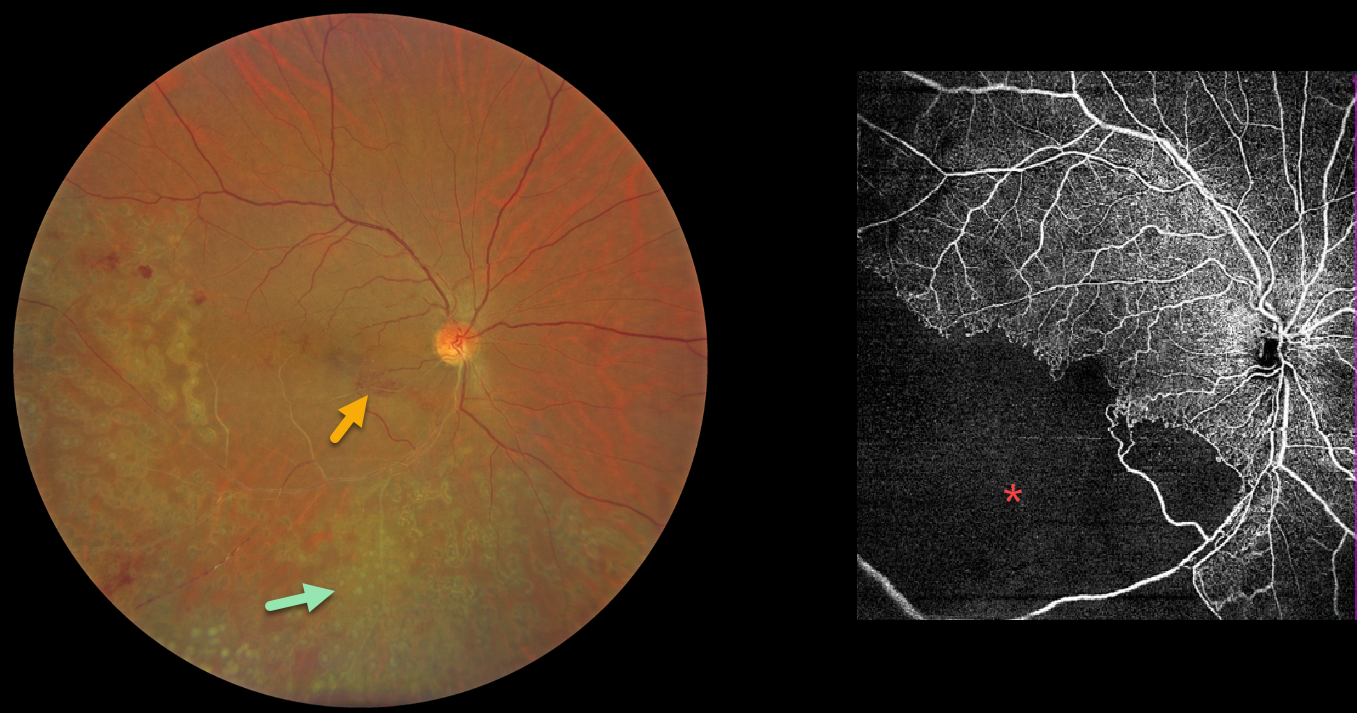
BRVO WITH RETINAL NEOVASCULARIZATION
Abhishek Karra and Ravindra Karra
Originally posted on @retina.rocks August 10, 2026
This 42YO male presented with a 3-month history of decreased vision in his right eye. He had received laser treatment in this eye elsewhere. Vision was 20/60 OD.
Color photography shows an old ischemic inferotemporal branch retinal vein occlusion (BRVO) with heavy scatter laser throughout its distribution. Collateral vessels are noted in the inferonasal macula. OCT angiography shows profound capillary loss. There was no macular edema, and observation was recommended.
Learning Points:
In 1986, the Branch Vein Occlusion Study Group reported that scatter laser decreased the risk of vitreous hemorrhage from retinal neovascularization in BRVO and suggested that laser should be applied once neovascularization develops (Arch Ophthalmol 1986;104:34-41). Thirty-five years later, scatter laser remains the gold standard, although some may prefer to stabilize the neovascularization with anti-VEGF therapy before laser.
BRVO WITH RETINAL NEOVASCULARIZATION AND COLLATERALS
Originally posted on @retina.rocks February 11, 2026
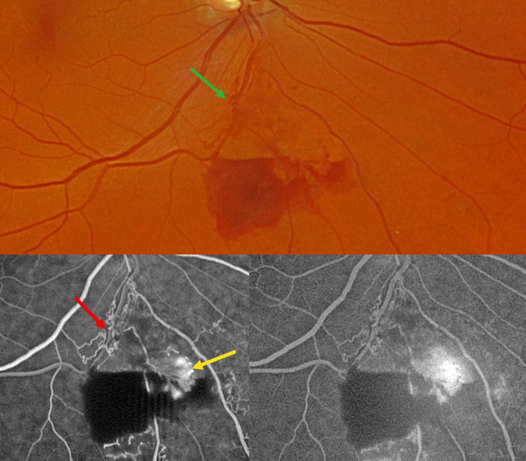
This 59YO male was referred for asymptomatic retinal findings in his right eye. Vision was 20/30.
Optos color RG imaging shows an inferior branch retinal vein occlusion with midperipheral preretinal blood. Collateral vessels are noted at the site of the occlusion. These collaterals show no leakage angiographically, although the adjacent retinal neovascularization does leak.
Learning Points:
Collateral vessels are dilated, pre-existing vessels that bypass an occlusion. These are most commonly seen in the posterior pole following retinal venous occlusions and are exceedingly rare with arterial occlusions. The endothelial tight junctions are intact, so they don’t leak angiographically. This helps differentiate them from neovascularization, which does leak. Our patient’s eye shows us a wonderful example of both.
BRVO WITH COLLATERALS AND DRIL
Malvika Singh, Akansha Sharma and Manish Nagpal
Originally posted on @retina.rocks August 19, 2025
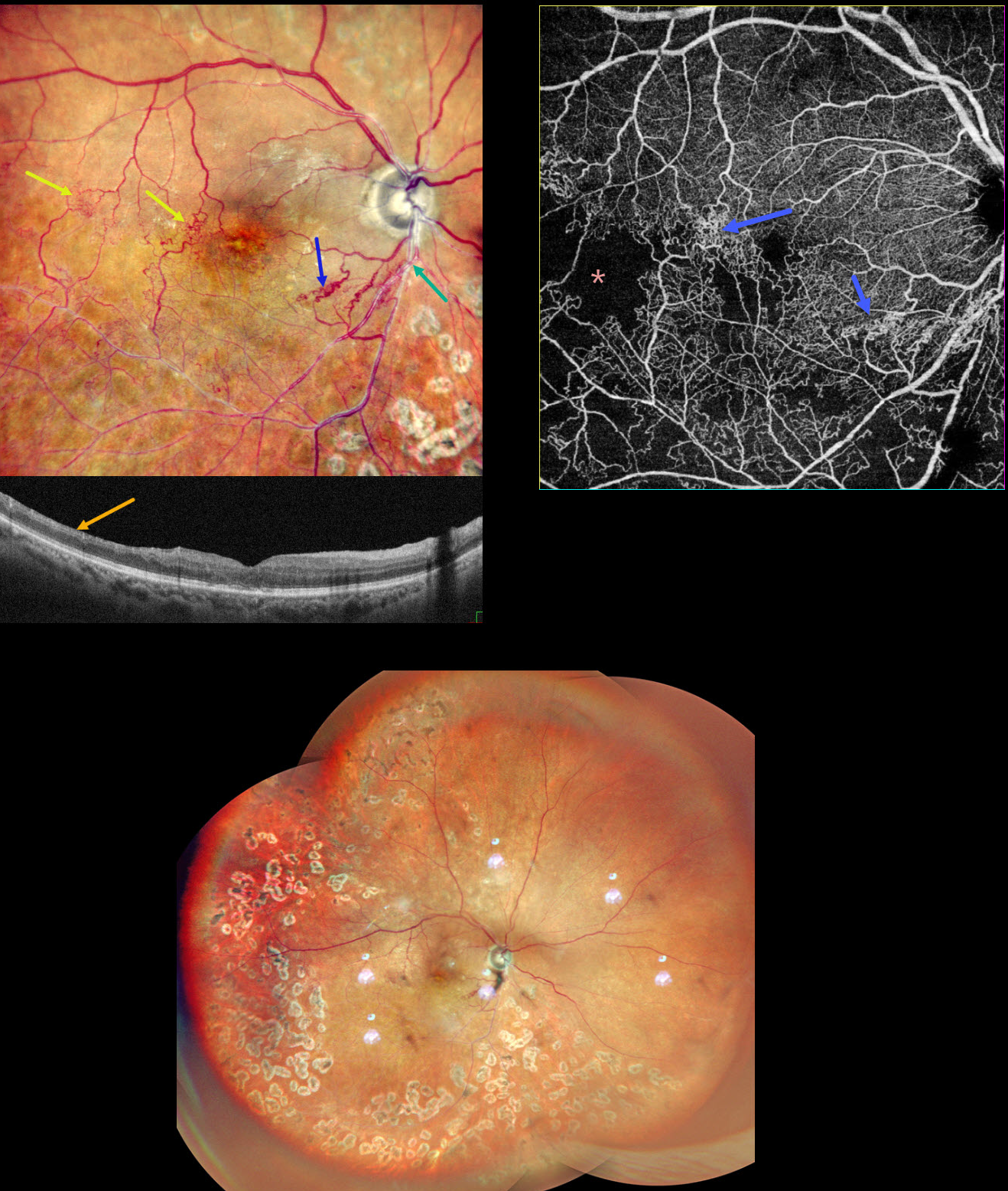
This 65YO male presented with a branch retinal vein occlusion (BRVO) in his right eye and 20/40 vision. He was treated elsewhere about a year ago with 3 intravitreal injections and scatter laser 7 years earlier.
Pseudocolor SLO shows an old major inferotemporal ischemic BRVO, which arises from an arteriovenous crossing just inferior to the disc. Extensive collateral vessels bypass the occlusion and cross the temporal horizontal raphe. OCT scanning shows thinned, featureless inner retina (disorganization of retinal inner layers, DRIL). Scatter laser is noted temporally and inferiorly. OCT angiography shows variable capillary loss throughout the BRVO distribution and highlights the collateral vessels.
Learning Points:
Collateral vessels are dilated, pre-existing vessels that bypass an occlusion. These are most commonly seen in the posterior pole following retinal venous occlusions and are exceedingly rare with arterial occlusions. The endothelial tight junctions are intact, so they don’t leak angiographically. This helps differentiate them from neovascularization, which does leak.
DRIL is seen in various retinal vascular disorders, including diabetic macular edema and retinal vein occlusions. It is generally associated with worse vision and increased anti-VEGF treatment burden (see Babiuch et al, JAMA Ophthalmol 2019;137:38-46).
CRAO
Originally posted on @retina.rocks December 23, 2024
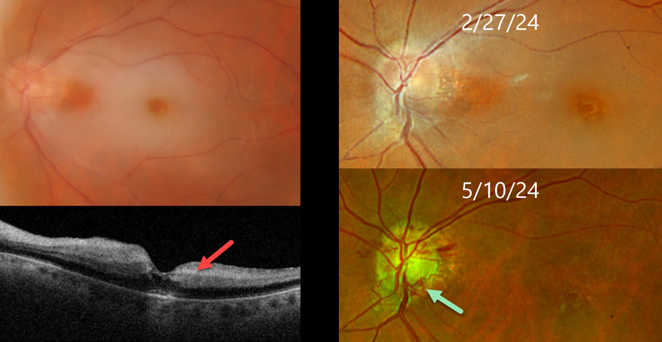
This 75YO female presented with an acute counting fingers central retinal artery occlusion. Triton color imaging shows a small area of papillomacular sparing, and swept-source OCT shows inner retinal hyperreflectivity. The retinal opacification completely resolved 10 weeks later with the development of extensive disc collaterals. Vision at counting fingers.
Learning Points:
Collateral vessels are dilated, pre-existing vessels that bypass an occlusion. These are seen in the posterior pole most commonly following retinal venous occlusions. Disc collaterals are exceedingly rare with arterial occlusions and were first described by Nettleship in 1891 (Ragge and Hoyt, Br J Ophthalmology 1992;76:186-188). The endothelial tight junctions are intact so they don’t leak angiographically. This helps to differentiate them from neovascularization which does leak.
CRAO
Originally posted on @retina.rocks July 16, 2024
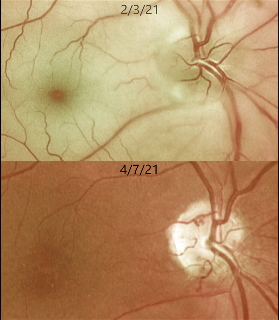
This 62YO female presented with an acute counting-fingers central retinal artery occlusion. Emergent stroke workup revealed a silent acute lacunar infarct. The retinal opacification completely resolved 2 months later. New fine collateral vessels were noted on the nerve. Vision remained at counting fingers.
Learning Points:
Collateral vessels are dilated, pre-existing vessels that bypass an occlusion. These are most commonly seen at the posterior pole following retinal venous occlusions and are exceedingly rare with arterial occlusions. The endothelial tight junctions are intact, so they don’t leak angiographically. This helps differentiate them from neovascularization, which does leak.
HRVO
Originally posted on @retina.rocks November 8, 2023
This 63YO male was referred for relatively asymptomatic retinal findings in his left eye. Vision was 20/60.
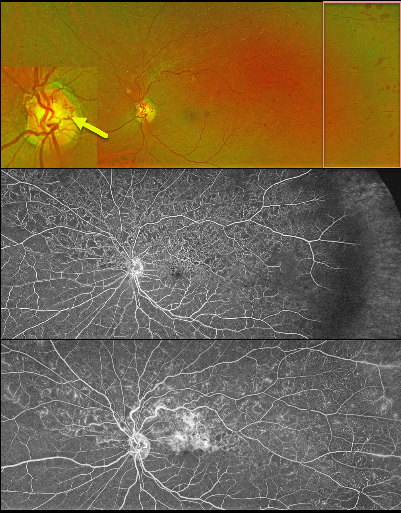
Optos color RG imaging shows a superior hemiretinal vein occlusion (HRVO) with mild hemorrhages, mostly in the temporal periphery. The occlusion is well-perfused without retinal neovascularization on fluorescein angiography. Prominent collateral vessels are noted on the optic disc superiorly. Observation was recommended.
Learning Points:
Branch retinal vein occlusions are virtually always caused by a retinal artery compressing a retinal vein within its common adventitial sheath. Some people are born with an anomalous two-trunked central retinal vein that independently drains the superior and inferior retinal hemispheres.
HRVO, similar to central retinal vein occlusion, is caused by compression of the retinal vein posterior to the lamina cribrosa (see Hayreh, Arch Ophthalmol 1980;98:1600-1609).
Collateral vessels are dilated, pre-existing vessels that bypass an occlusion. These are seen in the posterior pole most commonly following retinal venous occlusions, but can also occur on the optic disc with optic nerve tumors. The endothelial tight junctions are intact, so they don’t leak on angiography. This helps differentiate them from neovascularization, which does leak.
CRAO
Originally posted on @retina.rocks November 21, 2022
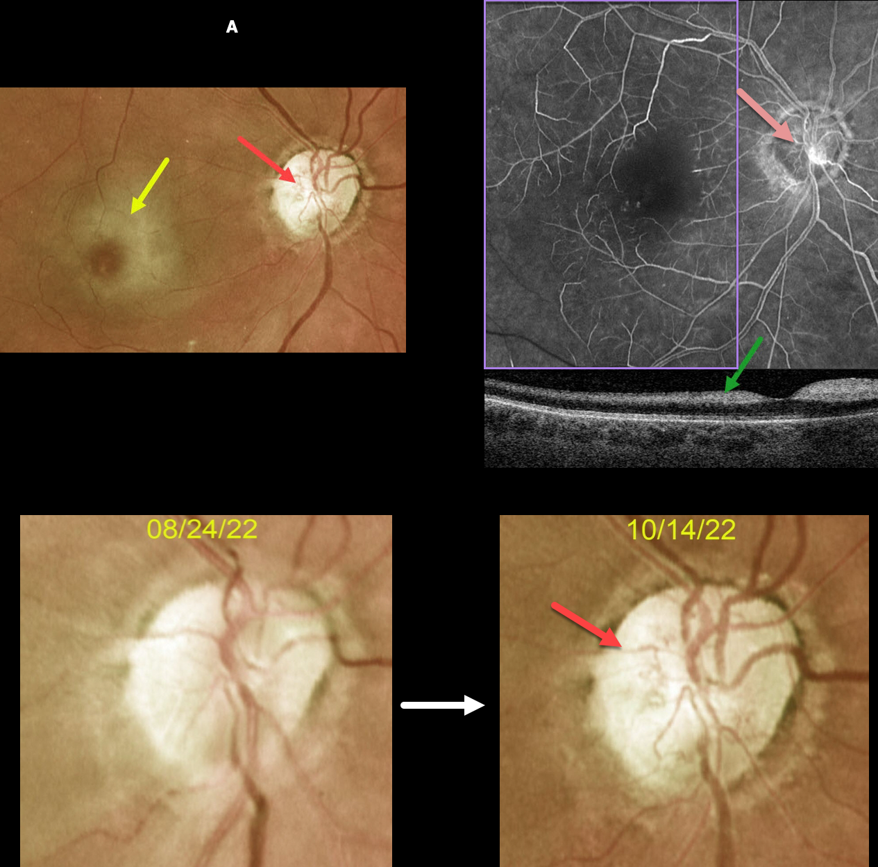
This 63YOF initially presented with an acute central retinal artery occlusion in her right eye. Emergency stroke referral revealed two prior strokes with a new asymptomatic stroke.
She returned two months later. There was subjective visual improvement, although vision remained at hand motion. Although the retinal opacification in general was improving, the perifoveal retina was more opaque with a pronounced cherry red spot.
New fine vessels were noted on the nerve. These vessels did not leak on fundus fluorescein angiography. Marked angiographic ischemia is also noted. OCT scanning shows increased reflectivity from the ischemic inner retina.
Learning Points:
Collateral vessels are dilated, pre-existing vessels that bypass an occlusion. These are seen in the posterior pole most commonly following retinal venous occlusions and are exceedingly rare with arterial occlusions.
The endothelial tight junctions are intact, so they don’t leak angiographically. This helps differentiate them from neovascularization, which does leak.
CRVO
Originally posted on @retina.rocks October 25, 2021
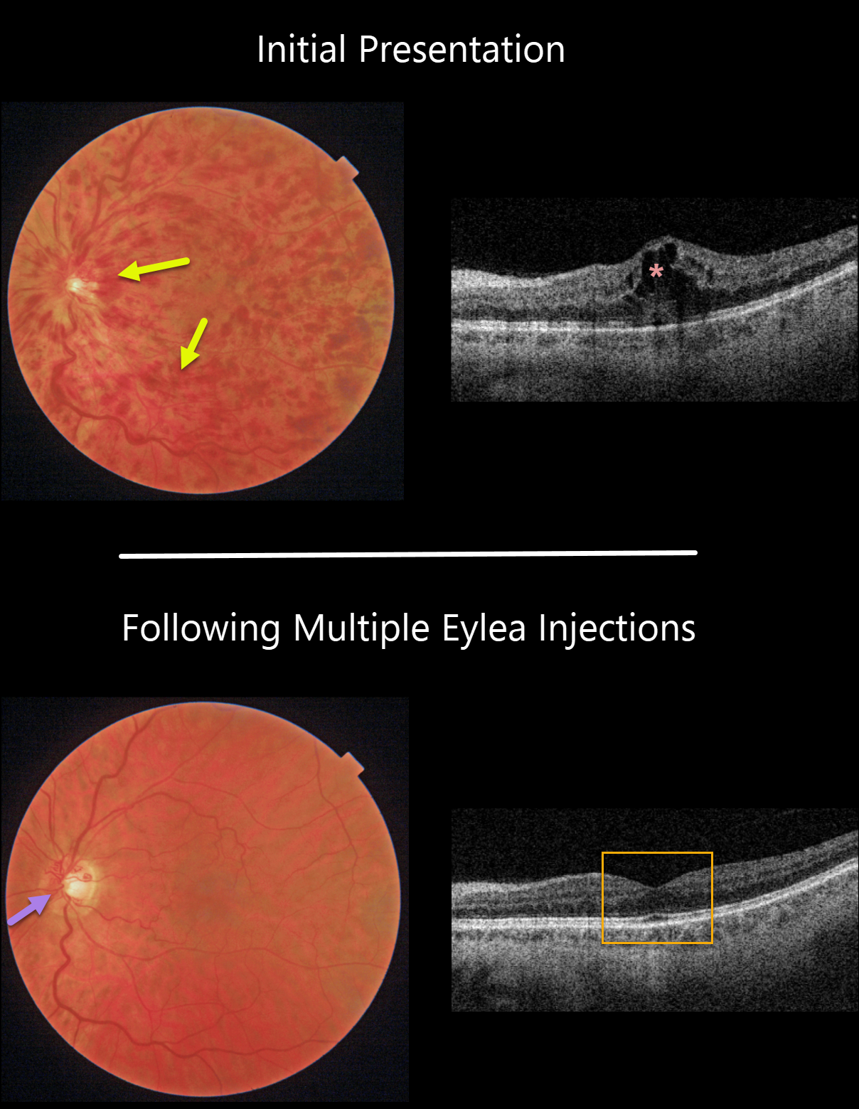
This 61YO male patient presented with a non-ischemic central retinal vein occlusion (CRVO), 20/30 vision, and macular edema.
With ongoing Eylea treat-extend protocol, the hemorrhages and macular edema completely resolved. Prominent optic nerve collaterals were noted.
Learning Points:
Before the introduction of intravitreal injections in the early 2000s, there were essentially no treatments for CRVO-related macular edema.
Anti-VEGF injections are now generally the initial treatment of choice, with about 50-60% of eyes gaining at least 3 lines of vision. Intravitreal steroids, usually Ozurdex, are more often used as secondary treatment.
Macular grid laser photocoagulation is not performed because it has been shown to confer no visual benefit in the Central Vein Occlusion Study Group (see Ophthalmology 1995;102;1425-1433).
CRVO
Originally posted on @retina.rocks April 22, 2021
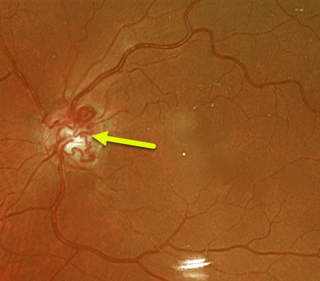
This 74YO female has large optic nerve collaterals from an old non-ischemic central retinal vein occlusion. Vision was 20/30.
Learning Points:
Collateral vessels are dilated, pre-existing vessels that bypass an occlusion. These are most commonly seen in the posterior pole, following retinal venous occlusions, often on the optic disc.
In collateral vessels, the endothelial tight junctions are intact, so they don’t leak angiographically. This helps differentiate them from neovascularization, which does leak.
CRVO
Originally posted on @retina.rocks December 21, 2020
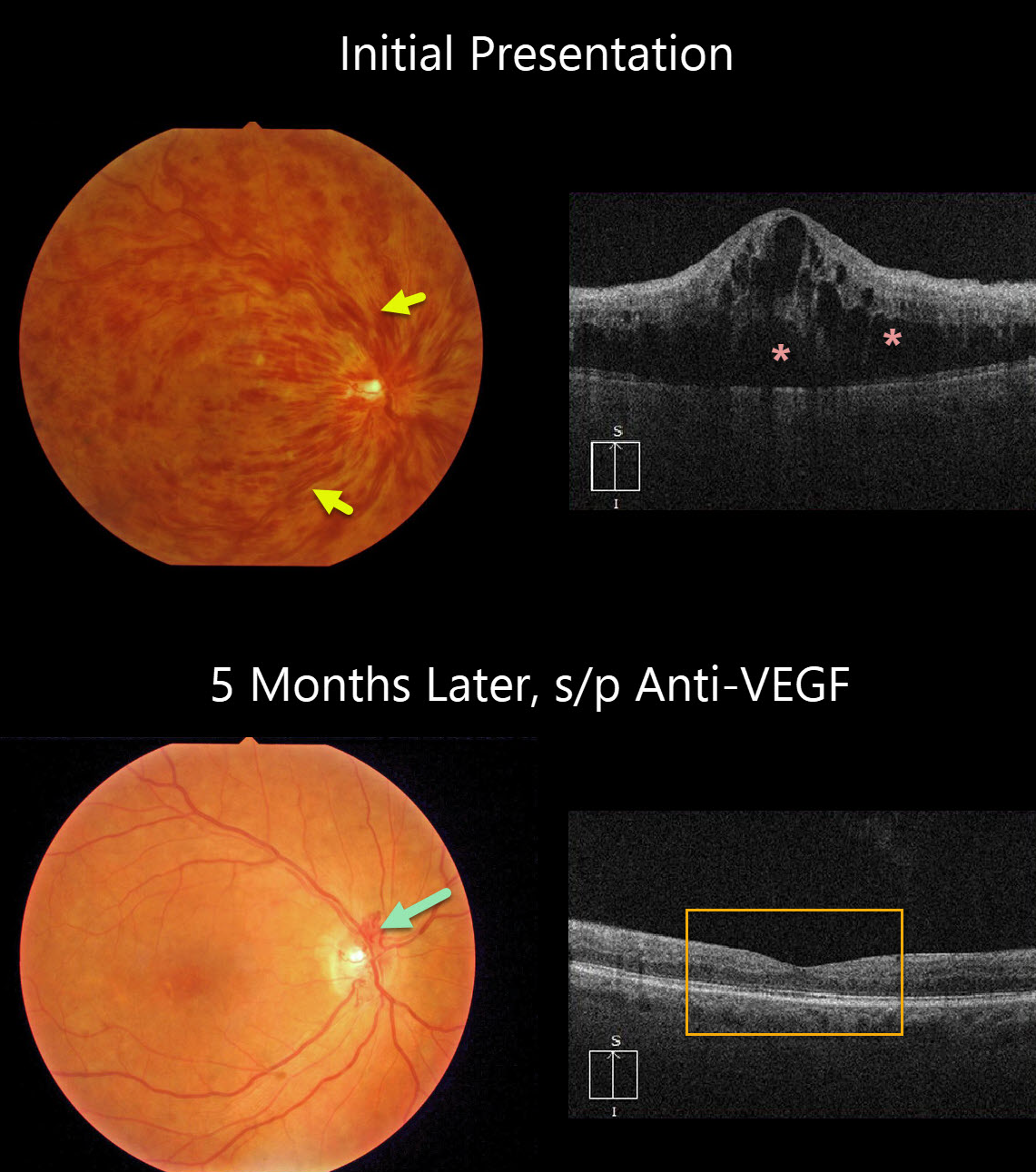
This patient presented with counting-finger vision from an acute hemorrhagic central retinal vein occlusion (CRVO) with marked macular edema. The poor vision and extensive retinal hemorrhages suggest that this is an ischemic CRVO.
After five monthly anti-VEGF injections, vision improved to 20/50, the retinal hemorrhages resolved dramatically, optic disc collaterals formed, and OCT showed complete resolution of all macular fluid. This degree of improvement is quite unusual.
Learning Points:
Patients with CRVO are at risk of vision loss due to macular edema or anterior segment neovascularization. Macular edema is common in both non-ischemic and ischemic occlusions, and this responds best to anti-VEGF therapy.
Rubeosis is found almost exclusively with ischemic occlusions, developing in about two-thirds of patients with untreated ischemic occlusions. This was common in the pre-anti-VEGF days.
However, since most patients with CRVO have macular edema and are receiving ongoing anti-VEGF therapy, neovascularization is now rarely seen unless patients present with untreated CRVO or therapy is discontinued.
BRVO
Originally posted on @retina.rocks August 31, 2020
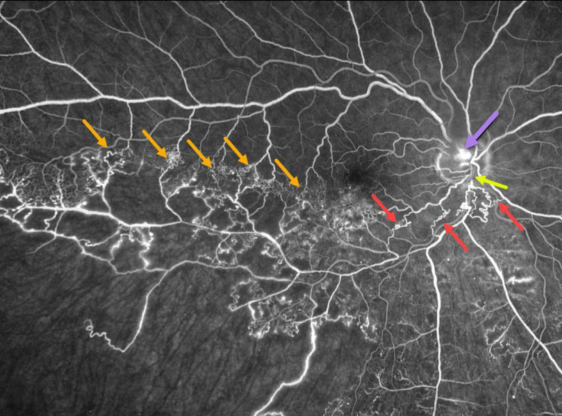
This patient has an ischemic branch retinal vein occlusion (BRVO) with extensive collateral vessels.
Occlusion occurs when an artery compresses the underlying vein where the vessels share a common adventitial sheath. Extensive collateral vessels cross the temporal horizontal raphe as well as around the occlusion more posteriorly. A small area of disc neovascularization leaks.
Learning Points:
Collateral vessels are dilated, pre-existing vessels that bypass an occlusion. These are most commonly seen in the posterior pole following retinal venous occlusions, but can also occur on the optic disc with optic nerve meningiomas.
The endothelial tight junctions are intact, so there’s no leakage on angiography. This helps differentiate them from neovascularization, which does leak.


