

Retinal Embolus
<< Back to Cases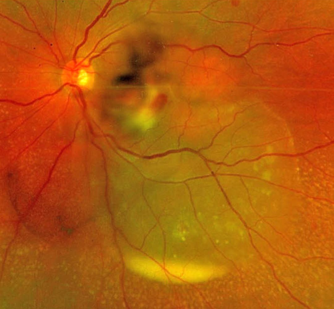
SEPTIC RETINAL EMBOLUS WITH SUBRETINAL HYPOPYON
The European VitreoRetinal Society (EVRS) and Mohit Dogra
Originally posted on @retina.rocks February 6, 2026
This previously healthy 37YO female presented to us for a second opinion regarding 4 days of decreased vision in her left eye. There was a recent history of a urinary tract infection secondary to E. coli. Vision was 20/20 in her normal OD and 20/400 OS. There was a moderate non-granulomatous uveitis with vitreous cells.
Optos color RG imaging shows opaque white retina in the inferonasal macula with overlying retinal and vitreous blood. An exudative retinal detachment extends inferotemporally with an inferior subretinal hypopyon.
She was referred back to the initially treating ophthalmologist and was subsequently lost to follow-up.
Learning Points:
Posterior segment chorioretinitis from septic embolization is a rare event, most commonly found in patients with bacterial endocarditis. Our patient presented with a constellation of findings, including retinitis, retinal and vitreous hemorrhage, and an exudative retinal detachment with an inferior subretinal hypopyon.
Subretinal hypopyon is characterized by yellowish inflammatory material settling inferiorly in the subretinal space. It has been documented in infectious (bacterial and fungal endophthalmitis, acute retinal necrosis, tuberculosis, syphilis), inflammatory (sympathetic ophthalmia), and neoplastic (leukemia, lymphoma) disorders.
RETINAL EMBOLI
Natasa Draca and Emma Oreskovic
Originally posted on @retina.rocks May 17, 2023
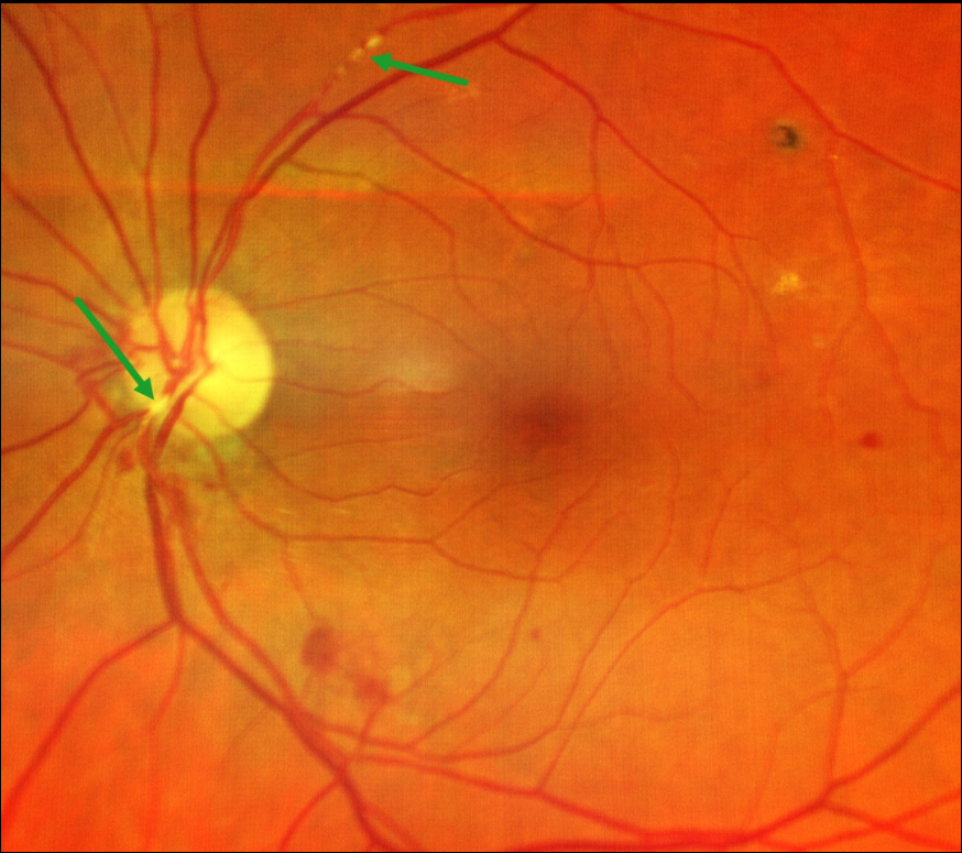
This 77YO male presented with unilateral left superior field loss in his left eye one day earlier. Vision was 18/20 bilaterally.
Color imaging shows retinal emboli along the proximal superotemporal arcade and overlying the inferonasal nerve heads. Some scattered retinal hemorrhages are also seen. The right fundus was normal. Visual field testing was normal.
He was immediately referred for a stroke evaluation, which was fortunately negative. Carotid Doppler showed bilateral 30% internal carotid artery stenosis. He was found to be in new-onset atrial fibrillation, which was the presumed source for the emboli, and was placed on oral anticoagulation therapy.
Learning Points:
Retinal emboli, which generally arise from either the heart or carotid arteries, can be calcific, platelet-fibrin, or cholesterol (Hollenhorst plaques). If large enough, they can obstruct a retinal arteriole and cause acute retinal ischemia, which is defined as transient monocular vision loss (TMVL, formerly known as amaurosis fugax), acute branch retinal artery occlusion, or acute central retinal artery occlusion.
TMVL requires an immediate referral to the nearest stroke center. Asymptomatic emboli can be more effectively evaluated for an embolic source with carotid Doppler and cardiac B-scan ultrasonography.
RETINAL EMBOLUS
Originally posted on @retina.rocks July 22, 2022
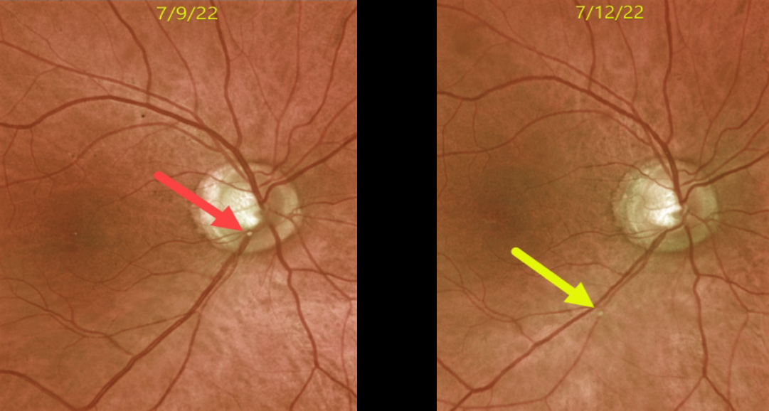
This 72YO male presented with acute superior field loss from an acute branch retinal artery occlusion (BRAO) in his right eye.
The causative calcific embolus is seen originally along the inferior optic nerve margin. Three days later, the plaque moved further downstream.
This patient had acute retinal ischemia (defined as acute BRAO, central retinal artery occlusion, or transient monocular vision loss), and was immediately referred to the nearest stroke center, which is the standard of care.
BRAO
Originally posted on @retina.rocks January 6, 2022
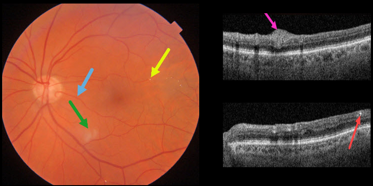
This 85YO male has 20/25 vision despite multiple retinal emboli and a resolving branch retinal artery occlusion (BRAO).
Triton swept-source OCT shows residual inner retinal edema and opacification. The embolus on Triton photography is also imaged in the OCT B-scan.
A cilioretinal artery may be responsible for the good vision.
RETINAL EMBOLUS
KMKoptometrypro
Originally posted on @retina.rocks October 14, 2020
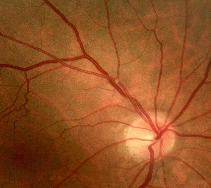
This patient had a small asymptomatic embolus. Elective carotid workup revealed a 95% ipsilateral carotid stenosis that was subsequently treated with carotid endarterectomy.
Learning Points:
Acute retinal ischemia, defined as transient monocular vision loss (or amaurosis fugax), acute branch retinal artery occlusion, or acute central retinal artery occlusion, requires an immediate referral to the nearest stroke center.
Asymptomatic emboli, as in this patient, can be more effectively evaluated for an embolic source, which should include carotid doppler and cardiac B-scan ultrasonography.


