

Retinal Vascular Loop
<< Back to Cases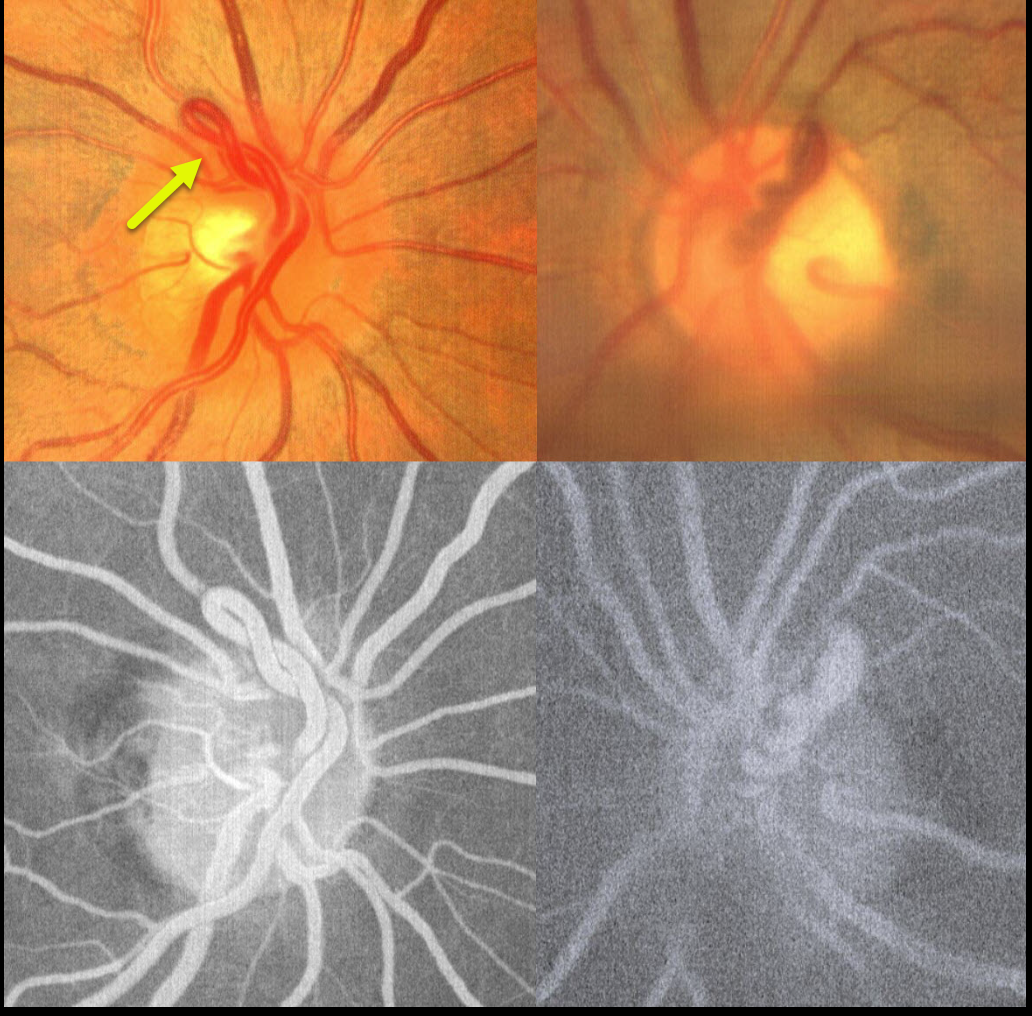
RETINAL VASCULAR LOOP
Kanwaljeet Harjot Madan
Originally posted on @retina.rocks September 16, 2025
This healthy 32YO female was referred for possible asymptomatic disc neovascularization. Vision was 20/20 OU. Fundus photography shows bilateral vascular loops that do not leak angiographically.
Learning Points:
A retinal vascular loop is a benign congenital anomaly that presents as an elevated and twisted bundle of vessels projecting from the disc into the vitreous cavity. Is it usually unilateral and arterial. Although usually just an incidental finding, these can rarely cause a branch arterial or venous occlusion (see Retinal Cases Brief Rep 2014;8;124-126 and Ophthalmology 2020;127;124-126). They should also not be confused with disc neovascularization, which consists of much smaller caliber vessels that leak on fluorescein angiography.
RETINAL VASCULAR LOOP
Originally posted on @retina.rocks August 2, 2022
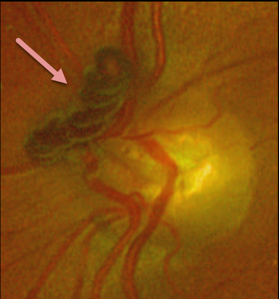
This 77YO female presented with this completely asymptomatic yet prominent retinal vascular loop. Observation was recommended.
Learning Points:
These benign congenital prepapillary vascular loops can be unilateral or bilateral. Although usually just an incidental finding, they can rarely cause a branch arterial or venous occlusion (see Retinal Cases Brief Rep 2014:8;124-126 and Ophthalmology 2020;127:124-126).
They should also not be confused with disc neovascularization, which consists of much smaller caliber vessels that leak on fluorescein angiography.
RETINAL VASCULAR LOOP
Originally posted on @retina.rocks October 13, 2021
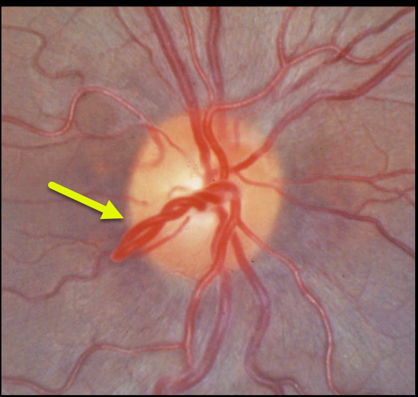
This patient has benign congenital prepapillary vascular loops, which can be unilateral or bilateral.
Learning Points:
Although usually just an incidental finding, they can rarely cause a branch arterial or venous occlusion (see Retinal Cases Brief Rep 2014;8;124-126 and Ophthalmology 2020;127;124-126).
They should also not be confused with disc neovascularization, which consists of much smaller caliber vessels that leak on fluorescein angiography.
RETINAL VASCULAR LOOP
Originally posted on @retina.rocks August 4, 2020
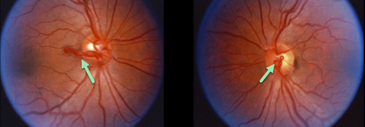
This patient has benign bilateral congenital prepapillary vascular loops. These can be either unilateral or bilateral.
Learning Points:
Although usually just an incidental finding, they can rarely cause a branch retinal arterial or venous occlusion (see Retinal Cases Brief Rep 2014;8;124-126 and Ophthalmology 2020;127;124-126).
They should also not be confused with disc neovascularization, which consists of much smaller caliber vessels that leak on fluorescein angiography.


