

Retinal Venous Malformation (Retinal Macrovessel)
<< Back to Cases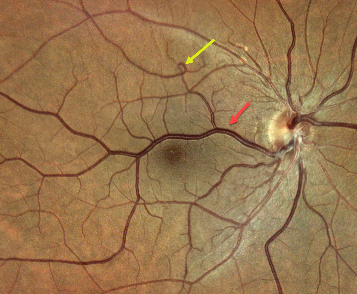
RETINAL VENOUS MALFORMATION
Ananya Kaginalkar, Vaishali Vasavada and Samaresh Srivastava
Originally posted on @retina.rocks July 10, 2026
This 57YO male presented with 3 weeks of blurred vision in his left eye. Vision was 20/20 OD and 20/40 OS. Anterior segments showed a moderate nuclear sclerotic cataract OS, accounting for his acuity. The left fundus was normal.
Color photography shows a large aberrant retinal vein, which crosses the horizontal meridian and drains nearly the entire right macula. There is a small vascular loop superiorly.
Learning Points:
Retinal vessels virtually always respect the horizontal meridian. Retinal venous malformations are congenital anomalous vessels that, by definition, cross the horizontal. Originally described by Gary Brown et al as congenital retinal macrovessels (Arch Ophthalmology 1982;100:1430-1436), these vessels are usually located in the macular region and are virtually always venous. These lesions are now called retinal venous malformations, since about one-quarter of patients have associated venous anomalies in the brain (Pichi et al, JAMA Ophthalmol 2018;136:372-379).
RETINAL VENOUS MALFORMATION
Ayushi Gupta and Vishal Agrawal
Originally posted on @retina.rocks October 7, 2025
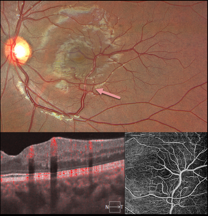
This 17YO female presented for a routine eye examination. Vision was 20/20 in her normal OD and 20/20 OS. Color imaging shows an inferior retinal venous malformation that drains the inferior and superotemporal macula. En face and B-scan OCT angiography show these vessels to lie in the inner retina.
Learning Points:
Retinal vessels virtually always respect the horizontal meridian. Retinal venous malformations are congenital anomalous vessels that, by definition, cross the horizontal. Originally described by Gary Brown et al as congenital retinal macrovessels (Arch Ophthalmology 1982;100:1430-1436), these vessels are usually located in the macular region and are virtually always venous. These lesions are now called retinal venous malformations, since about one-quarter of patients have associated venous anomalies in the brain (Pichi et al, JAMA Ophthalmol 2018;136:372-379).
RETINAL VENOUS MALFORMATION
The European VitreoRetina Society (EVRS) and P.C. Ranjan
Originally posted on @retina.rocks August 29, 2025
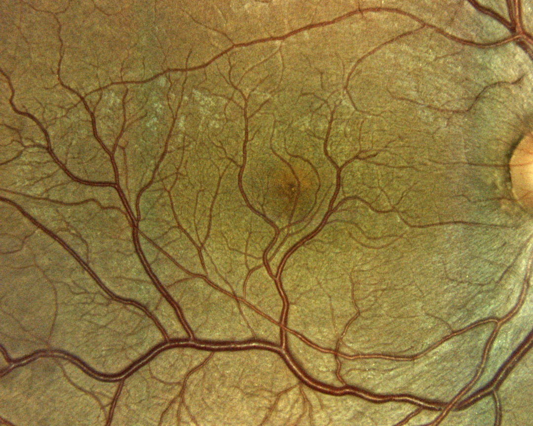
This 50YO male presented for a routine eye examination. Vision was 20/20 OD and 20/20 in his normal OS. Color imaging shows an inferior retinal venous malformation that drains nearly the entire macula.
Learning Points:
Retinal vessels virtually always respect the horizontal meridian. Retinal venous malformations are congenital anomalous vessels that, by definition, cross the horizontal. Originally described by Gary Brown et al as congenital retinal macrovessels (Arch Ophthalmology 1982;100:1430-1436), these vessels are usually located in the macular region and are virtually always venous. These lesions are now called retinal venous malformations, since about one-quarter of patients have associated venous anomalies in the brain (Pichi et al, JAMA Ophthalmol 2018;136:372-379).
RETINAL VENOUS MALFORMATION
Evgenii Chernov
Originally posted on @retina.rocks September 10, 2024
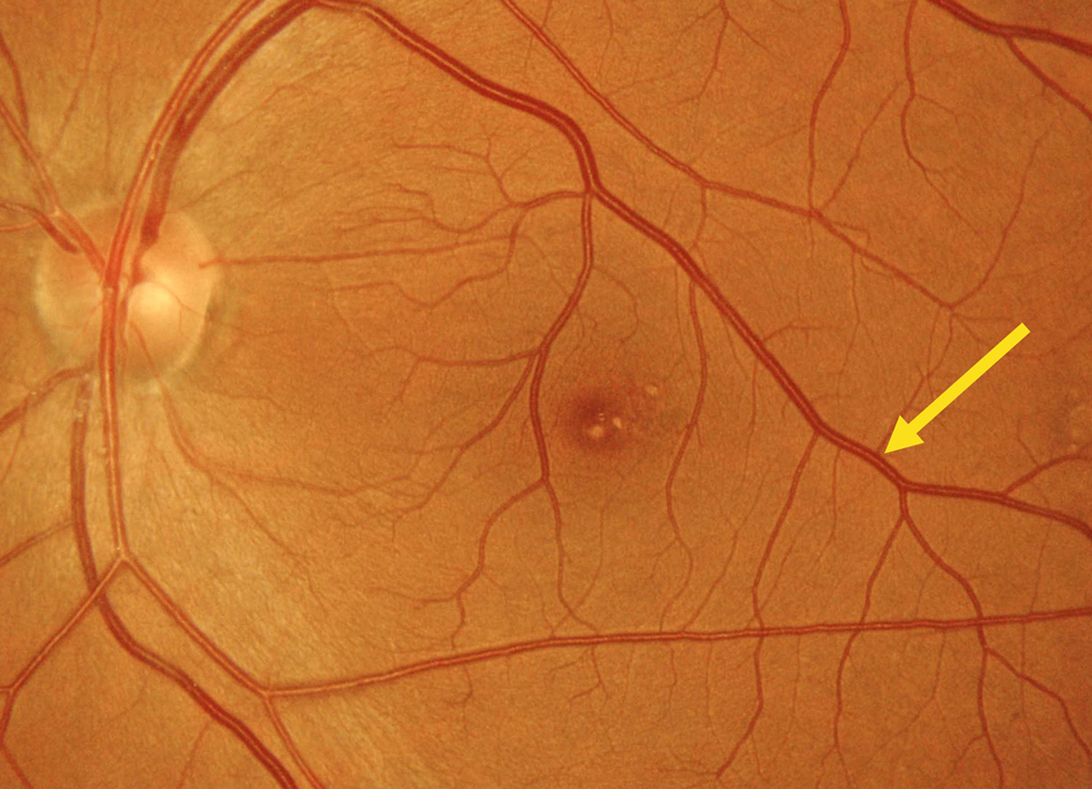
This 28YO female was referred for an asymptomatic funduscopic finding. Vision was 20/20 OU.
Color imaging shows a superior retinal venous malformation. This vessel drains nearly the entire macula as it extends into the inferotemporal midperiphery. Some mixed foveal drusen are also noted.
Learning Points:
Retinal vessels virtually always respect the horizontal meridian. Retinal venous malformations are congenital anomalous vessels that, by definition, cross the horizontal. Originally described by Gary Brown et al as congenital retinal macrovessels (Arch Ophthalmology 1982;100:1430-1436), these vessels are usually located in the macular region and are virtually always venous.
These lesions are now called retinal venous malformations since about one-quarter of patients will have associated venous anomalies in the brain (Pichi et al JAMA Ophthalmol 2018;136:372-379).
RETINAL VENOUS MALFORMATION
Will Gibson
Originally posted on @retina.rocks January 22, 2024
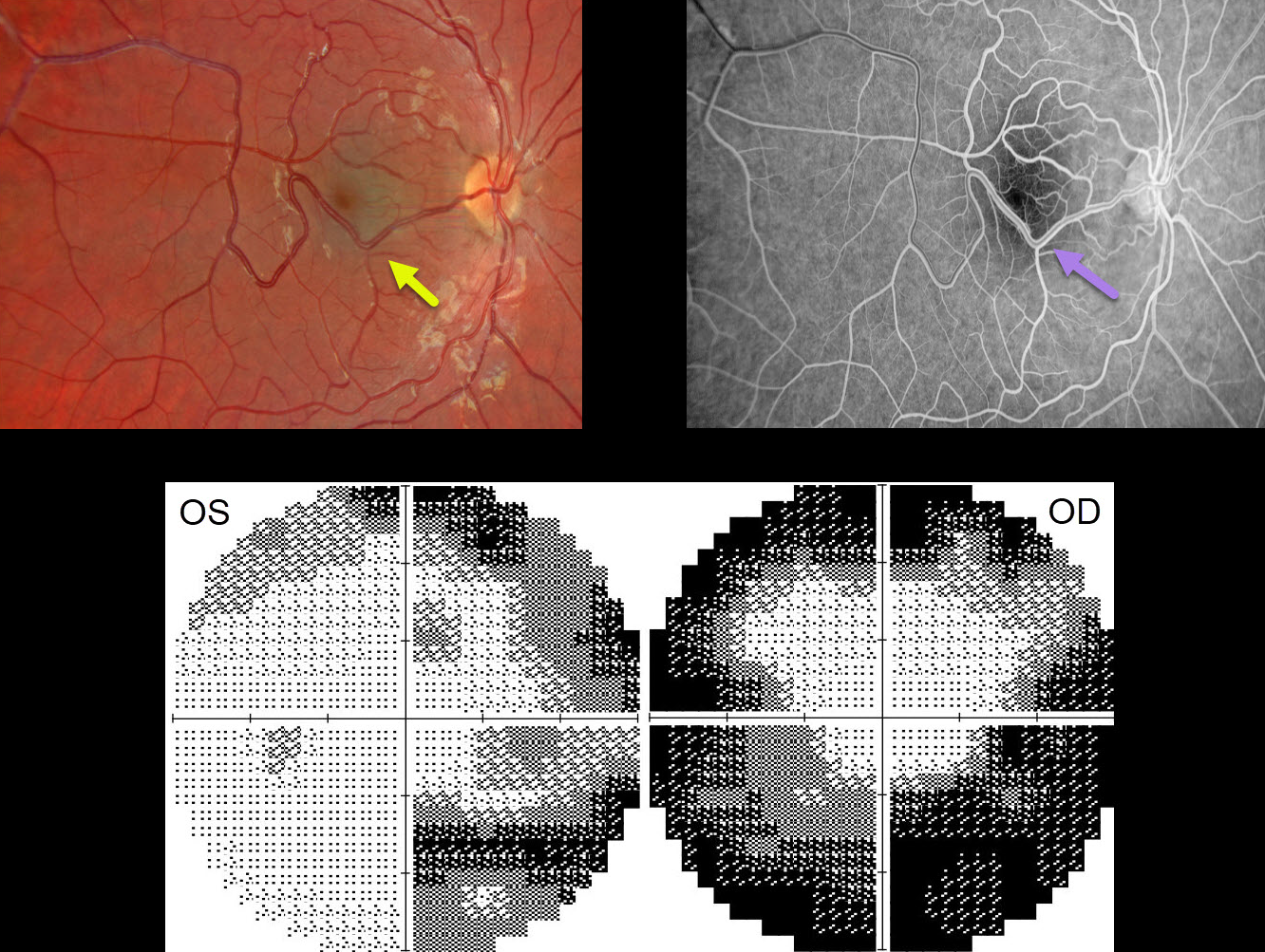
This 17YO male presented with bilateral vision loss and pain on eye movements 1 week following a COVID-19 infection. There was no prior ocular or medical history. Vision was 20/70 OD and 20/50 OS.
Color imaging of the right eye shows a large temporal retinal venous malformation. This vessel shows angiographic laminar filling without leakage. The right and left nerves were clinically normal, and the left fundus was normal (not shown).
OCT retinal nerve fiber layer analysis was normal bilaterally. Visual fields show moderate generalized constriction OD and nasal loss OS with a possible vertical cut inferiorly. Subsequent neurologic imaging was consistent with optic neuritis. Additional workup was unremarkable, and neuro-ophthalmic consultation led to the diagnosis of bilateral retrobulbar optic neuritis secondary to COVID-19.
Observation was recommended, and symptoms began to improve within a week. Two months later, all visual symptoms resolved, vision returned to 20/25 OU, and the visual fields normalized.
Learning Points:
Retinal vessels virtually always respect the horizontal meridian. Retinal venous malformations are congenital anomalous vessels that, by definition, cross the horizontal.
Originally described by Gary Brown et al as congenital retinal macrovessels (Arch Ophthalmology 1982;100:1430-1436), these vessels are usually located in the macular region and are virtually always venous.
These lesions are now called retinal venous malformations, since about one-quarter of patients have associated venous anomalies in the brain (Pichi et al, JAMA Ophthalmol 2018;136:372-379).
RETINAL VENOUS MALFORMATION
Originally posted on @retina.rocks August 14, 2023
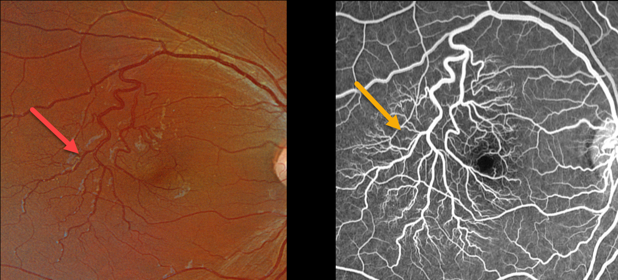
This healthy 13YO female was referred for asymptomatic vascular changes in her right eye. Vision was 20/20 OU.
Optos color RGB imaging shows a prominent superotemporal retinal vein that crosses into the inferotemporal retina. As it descends temporally, it arborizes into numerous smaller branches. There was no leakage angiographically.
Neurologic imaging of our patient was fortunately normal, and observation was recommended.
Learning Points:
Retinal vessels virtually always respect the horizontal meridian. Retinal venous malformations are congenital anomalous vessels that, by definition, cross the horizontal.
Originally described by Gary Brown et al as congenital retinal macrovessels (Arch Ophthalmology 1982;100:1430-1436), these vessels are usually located in the macular region and are virtually always venous.
These lesions are now called retinal venous malformations, since about one-quarter of patients have associated venous anomalies in the brain (Pichi et al, JAMA Ophthalmol, 2018;136:372-379).
RETINAL VENOUS MALFORMATION
Originally posted on @retina.rocks October 12, 2020
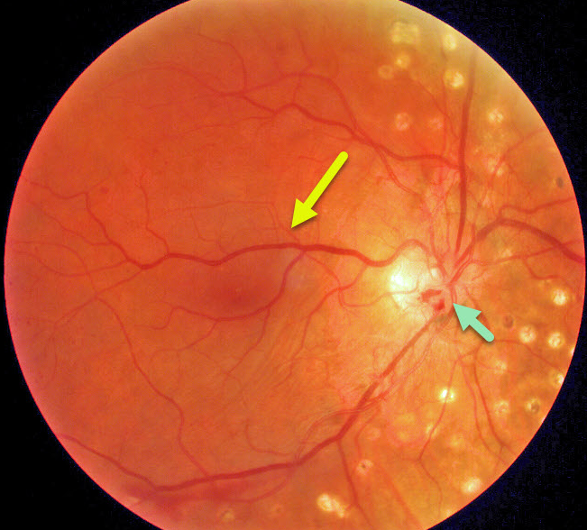
This patient has a retinal venous malformation (formerly known as retinal macrovessel), in addition to full panretinal photocoagulation (PRP) for proliferative diabetic retinopathy. Residual flat disc neovascularization remains despite full PRP.
Learning Points:
A retinal venous malformation is an anomalous retinal vessel that crosses the horizontal raphe (remember that normal retinal vessels respect the horizontal meridian).
These vessels are technically arterio-venous malformations (AVM). Since patients with AVMs can have additional venous anomalies in the brain (24% compared to up to 6% of the population), a brain MRI is recommended to rule out any intracranial abnormalities.


