

Retinitis Pigmentosa (RP) + Misc Inherited Retinal Diseases (IRD)
<< Back to Cases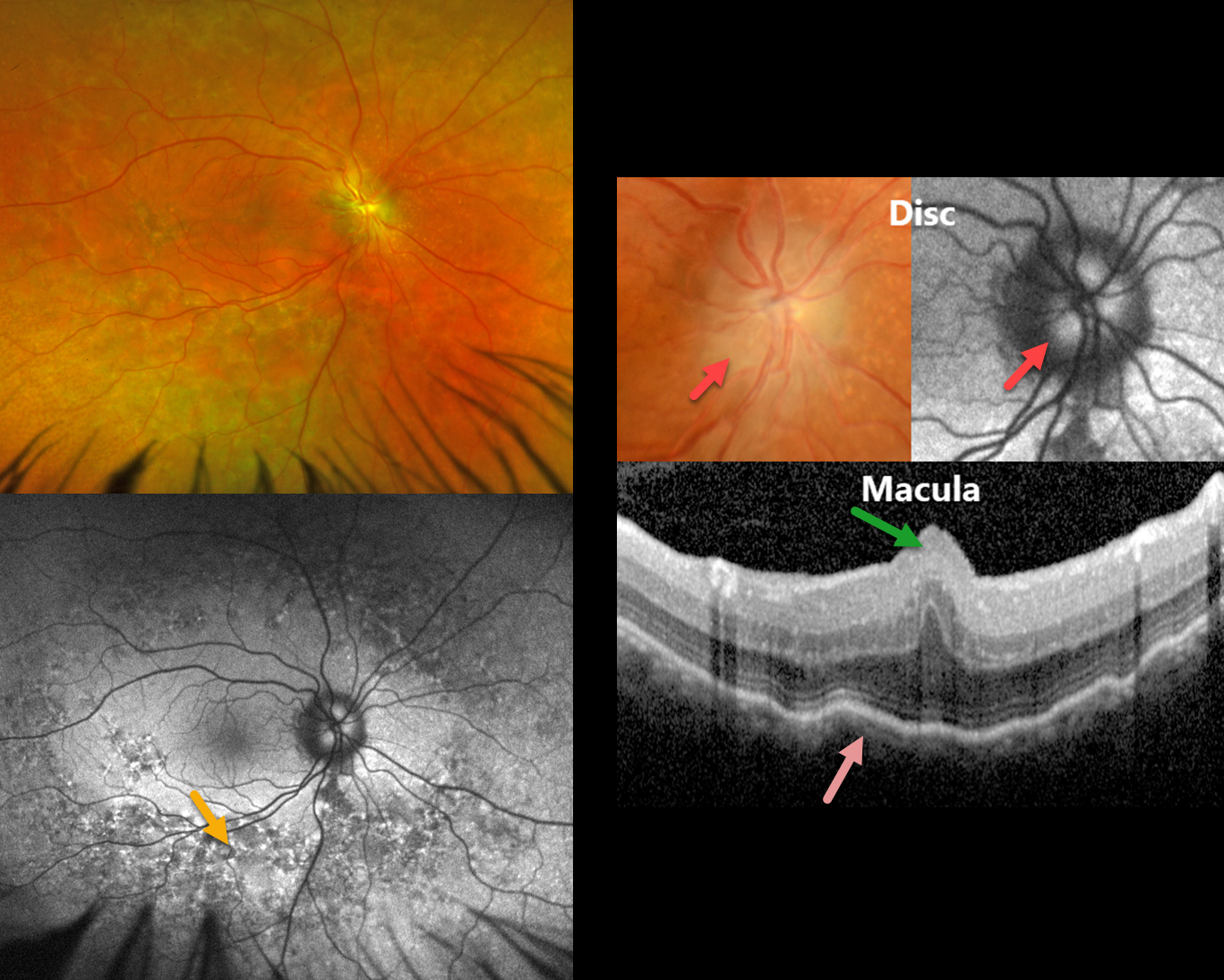
MFRP RETINITIS PIGMENTOSA
João Pedro Marques
Originally posted on @retina.rocks June 9, 2026
This 49YO male has a history of MFRP-related retinitis pigmentosa (RP). We have followed him for 13 years with stable findings of bilateral nanophthalmos (+18D hyperopia, axial lengths 14.82mm OD and 15.14 OS), optic disc drusen (ODD), and choroidal folds. He has compound heterozygosity for 2 pathogenic variants in the MFRP gene.
Optos color RG imaging in his OD shows pigmentary changes throughout the midperiphery along with an ill-defined disc margin. Fundus autofluorescence (FAF) shows variable hyper- and hypo-FAF in the midperiphery. Disc drusen are noted on a more magnified view, which hyper-FAF. Macular OCT shows a central fold, choroidal thickening, and chorioretinal folds. Identical findings were present in his OD (not shown).
Learning Points:
The MFRP (membrane frizzled-related protein) is expressed in the RPE and ciliary epithelium. MFRP functions as a molecular hub on the RPE apical membrane, coordinating protein trafficking and lipid homeostasis. Loss of MFRP leads to DHA accumulation in the RPE, downregulation of visual cycle genes and phototransduction genes, and progressive photoreceptor degeneration.
Biallelic MFRP mutations cause a well-characterized autosomal recessive retinitis pigmentosa (RP) syndrome with nanophthalmos and ODD (Li et al, BJO 2024;108;1679-1687). These eyes are at high risk of angle-closure glaucoma, usually requiring prophylactic laser peripheral iridotomy.
PERICENTRAL RETINAL DEGENERATION
Shraddha Raj Shrivastava, Akansha Sharma and Manish Nagpal
Originally posted on @retina.rocks December 23, 2025
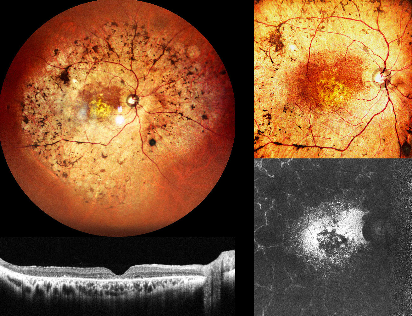
This 47YO male presented with decreased vision in both eyes for 5 years. Ocular family history was negative. Vision was 20/300 OD and 20/660 OS.
Pseudocolor SLO imaging of his right eye shows intraretinal pigment migration (bone-spicules) extending from the macula into the midperiphery. OCT shows outer retinal thinning with disorganization of the outer retinal bands. Fundus autofluorescence (FAF) shows hyper-FAF from the relatively preserved paracentral retina, more central hypo-FAF, and a peculiar peripheral pattern of interlaced hyper-FAF lines around lobular atrophic RPE and retina. Identical findings were noted in his left eye (not shown).
Learning Points:
Pericentral retinitis pigmentosa (RP) is an atypical, mild form of RP characterized by retinal degeneration and intraretinal pigment migration confined to the retinal near midperiphery. Unlike classic RP, pericentral RP tends to progress more slowly, with preserved central visual acuity and less extensive visual field loss over time. Genetically, pericentral RP is heterogeneous (Matsui et al, IOVS 2025;56:6007-6018), with causative variants identified in multiple genes, many of which are also associated with typical RP and other retinal dystrophies, indicating that pericentral RP is a phenotypic variant rather than a distinct clinical entity. Long-term prognosis for central vision is generally favorable in the absence of macular involvement, which unfortunately
UNILATERAL PIGMENTARY RETINOPATHY
Mattie Adams
Originally posted on @retina.rocks November 19, 2025
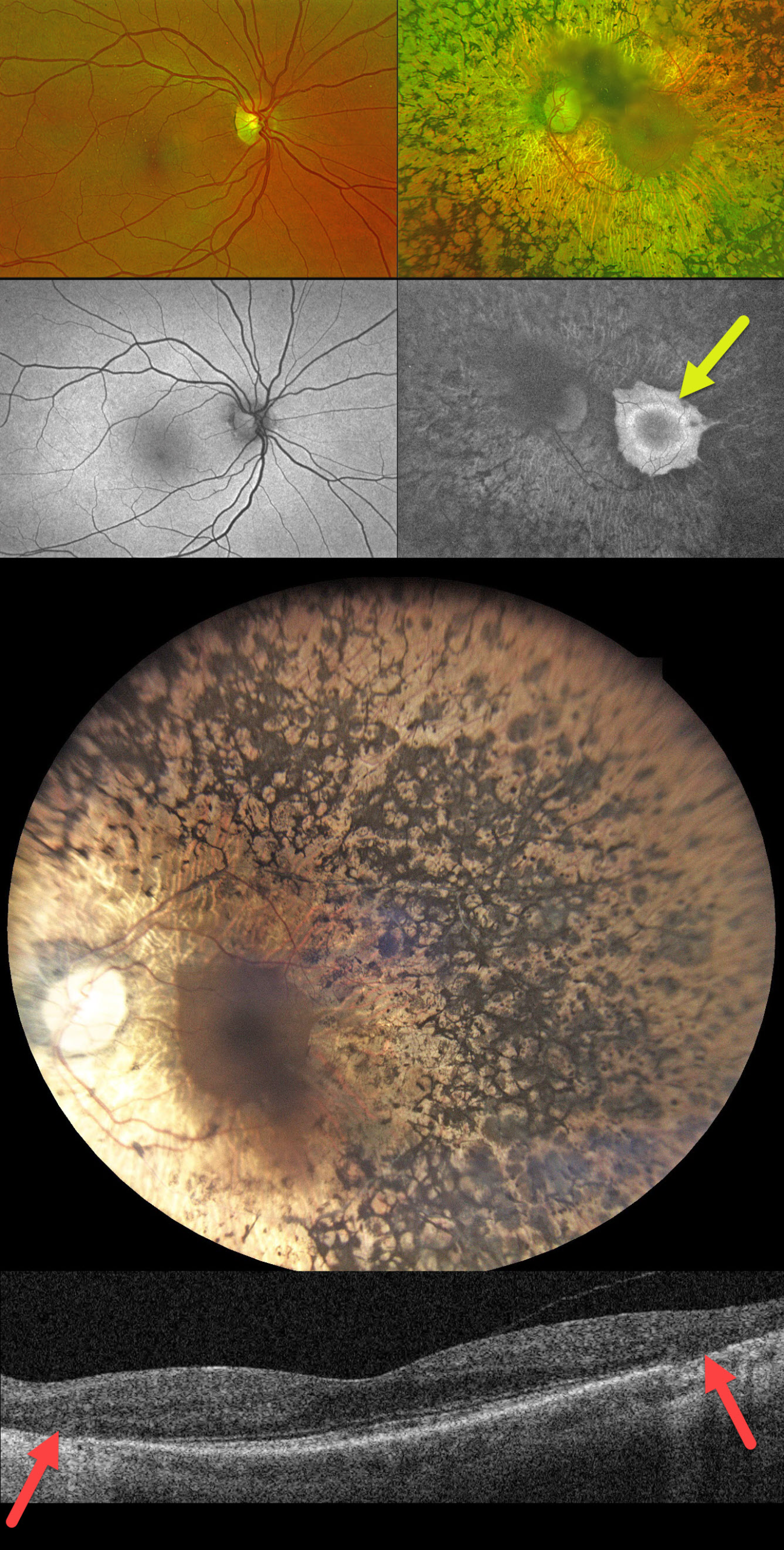
This 55YO female has complained of some blurred vision in her left eye for about 6 years. Family history is completely negative for eye disease. Vision was 20/25 in her normal OD and 20/50 OS.
Optos color RG imaging and fundus autofluorescence (FAF) are completely normal for her right eye. The left eye shows severe apparent retinitis pigmentosa (RP) changes that extend from her peripheral macula into the periphery. FAF shows a somewhat scalloped appearance to the preserved central macula with a hyper-FAF ring around the fovea. OCT scanning shows peripheral macular outer retinal and RPE atrophy. Genetic testing was heterozygous for a pathogenic ABCC6 mutation (c.2787+1G>T, splice donor) and a benign ABCA4 variant (c.5603A>T, p.Asn1868lle).
Learning Points:
Unilateral pigmentary retinopathy, also referred to as unilateral RP, is an extremely rare disorder (Alina-Cristina et al, Romanian J Ophth 2016;60:47-52). Patients require long-term follow-up to rule out a delayed bilateral presentation of an inherited retinal disorder (IRD). Unilateral pseudo-RP, chorioretinal findings with intraretinal pigment migration, is much more common, including trauma, prior retinal detachment, and infectious or inflammatory disorders such as AZOOR. The funduscopic and FAF characteristics of our patient’s left eye strongly suggest a unilateral pigmentary retinopathy rather than one of these other imitators.
With our patient’s genetic testing showing two different heterozygous mutations for recessive disorders, her results were essentially negative. However, this does not definitively rule out an IRD. In a real-world study of the Invitae IRD panel, McClard et al yielded a positive diagnostic result in only 51% of patients (J Vitreoretinal Diseases 2022;6:351-357). Each genetic panel has its own limitations regarding which genes are tested, as well as false positives and false negatives (Pulido et al, Retina 2022;42:1-3).
RETINITIS PIGMENTOSA WITH CME
Rohan Jain and Manish Nagpal
Originally posted on @retina.rocks September 15, 2025
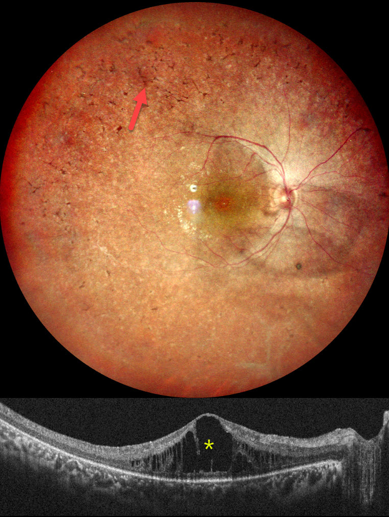
This healthy 18YO female complained of night blindness for a few years. Her mother and sister have a history of retinitis pigmentosa (RP). Vision was 20/90 OU.
Pseudocolor SLO imaging of her right eye shows typical RP scarring with intraretinal pigment migration extending from the macula into the retinal periphery. There is loss of the foveal reflex and cystoid macular edema (CME). Identical findings were noted in the OS (not shown).
Learning Points:
RP is a clinically and genetically heterogeneous group of inherited retinal disorders, which can present sporadically or with any inheritance pattern (autosomal dominant, autosomal recessive, sex-linked, or mitochondrial). It is characterized by diffuse, progressive dysfunction of predominantly rod photoreceptors, followed by RPE degeneration with intraretinal pigment migration. Visual impairment usually presents as night blindness and progressive visual field loss.
CME is observed in up to 20% of RP patients, often without angiographic leakage. Pathological mechanisms include vitreous traction/epiretinal membranes or a breakdown of the blood-retinal barrier due to inflammation or RPE/Muller cell dysfunction (Gaudric et al, Progress in Retinal and Eye Research 2022;91:101092). First-line treatment is topical or oral carbonic anhydrase inhibitors (CAIs). Other options for refractory CME include steroids (topical, periocular, or intravitreal), anti-VEGF injections, or vitrectomy for a tractional component. Our patient was given topical CAIs but was immediately lost to follow-up.
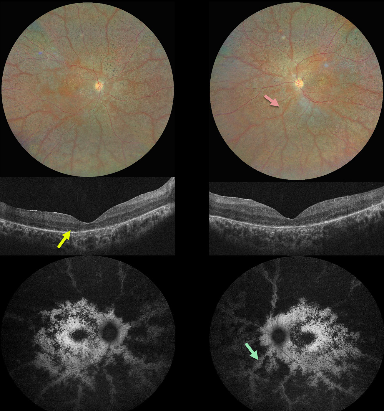
PRESERVED PARA-ARTERIOLE RPE IN RP
Ayushi Gupta and Vishal Agrawal
Originally posted on @retina.rocks June 16, 2025
This 16YO male complained of night blindness for a few years. There was no family history of eye disease. Vision was 20/200 OU.
Color photography shows bilateral findings consistent with retinitis pigmentosa (RP), including diffuse chorioretinal scarring, intraretinal pigment migration, and narrow retinal vessels. The retinal tissue beneath and immediately surrounding the arterioles is relatively spared, and this is most dramatically seen on fundus autofluorescence. OCT scanning shows variable loss of the outer retinal bands and RPE. Our patient could not afford genetic testing and was immediately lost to follow-up.
Learning Points:
Preserved para-arteriole retinal pigment epithelium (PPRPE) in retinitis pigmentosa, originally described by Heckenlively (Br J Ophthalmol 1982;66:26-30), has the unique phenotype, as seen in our patient, of diffuse RP degeneration with relative sparing of the RPE beneath the retinal arterioles. It was later found to be caused by a mutation in the crumbs cell polarity complex component 1 (CRB1) gene (den Hollander et al, Nature Genetics 1999;23:217-221). CRB1 is part of a group of proteins that help determine the structure and orientation of photoreceptors. It may also be involved in forming connections between different types of retinal cells.
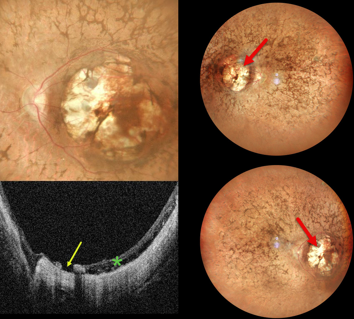
RETINITIS PIGMENTOSA WITH MACULAR COLOBOMA
Ankit Jain and Manish Nagpal
Originally posted on @retina.rocks January 7, 2025
This 32YO female was born with severe vision loss and nystagmus. There was no family history of eye disease. Vision was counting fingers bilaterally.
Pseudocolor SLO imaging shows bilateral symmetric macular colobomas with diffuse retinitis pigmentosa (RP) findings, including vascular narrowing, diffuse pigmentary changes, and intraretinal pigment migration (bone spicules). OCT shows thinned and variably disorganized retinal bands within the colobomas, and a small macular hole is noted nasally OS.
Learning Points:
Macular colobomas are rarely associated with RP (Parmeggiani et al, Eye 2004;18:421-428). North Carolina macular dystrophy, caused by a mutation in the PRDM13 gene (Small et al, Ophthalmology 2016;123:9-18), is characterized by macular coloboma-like lesions. However, these eyes do not have peripheral RP findings. Genetic testing in our patient was not performed, and observation was recommended.
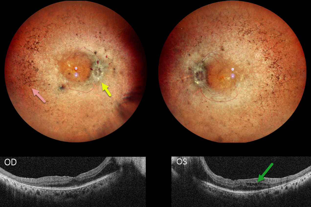
RETINITIS PIGMENTOSA WITH PAPILLEDEMA
Tejaswita Verma and Manish Nagpal
Originally posted on @retina.rocks November 20, 2024
This 30YO male presented with 3 weeks of decreased vision. His father has a history of retinitis pigmentosa (RP). Vision was 20/30 OD and 20/80 OS.
Pseudocolor SLO imaging shows typical bilateral RP scarring with intraretinal pigment migration extending from the peripheral maculas into the retinal peripheries. Bilateral macular puckers and disc swelling with right peripapillary inner retinal fluid are also seen. OCT scanning confirms the epimacular membranes. Bilateral pericentral outer macular atrophy is noted, along with some cystic edema in the left inner and outer nuclear layers.
Learning Points:
RP is a clinically and genetically heterogeneous group of inherited retinal disorders. The disease can present sporadically or with any inheritance pattern (autosomal dominant, autosomal recessive, sex-linked, or mitochondrial). It is characterized by diffuse, progressive dysfunction of predominantly rod photoreceptors, with subsequent RPE degeneration and intraretinal pigment migration. Visual impairment usually presents as night blindness and progressive visual field loss.
Bilateral optic disc edema is not typical for RP. Unfortunately, we were unable to work up our patient for a secondary cause because he was lost to follow-up immediately.
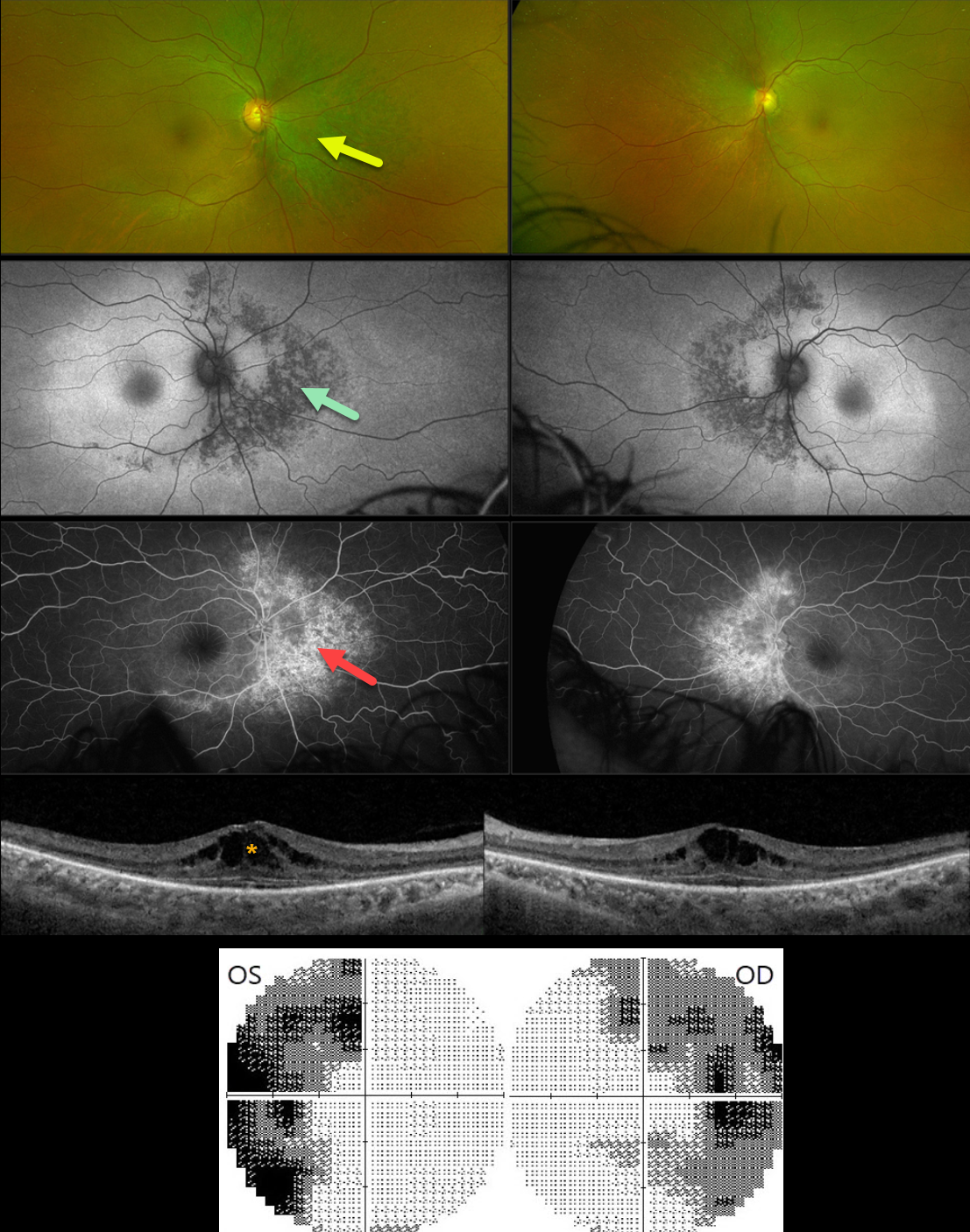
BITEMPORAL HEMIANOPIA WITH RETINITIS PIGMENTOSA
Mattie Adams
Originally posted on @retina.rocks August 12, 2024
This 54YO female was referred for asymptomatic retinal changes. Vision was 20/30 OD and 20/20 OS.
Visual fields showed bitemporal hemianopia. What is your diagnosis?
Optos color RG imaging shows pigmentary changes with intraretinal pigment migration extending superiorly, nasally, and inferiorly from the optic nerves. These changes are hypoautofluorescent and appear as angiographic window defects. Bilateral cystoid edema is seen on Triton swept-source OCT.
Learning Points:
Although bitemporal hemianopia usually signifies retrobulbar pathology at the optic chiasm, retinal disease can sometimes also cause similar field loss. Our patient’s retinitis pigmentosa (RP) changes were centered around each optic nerve, causing the hemianopia. Genetic testing revealed double heterozygous pathogenic ARSG mutations. This genotype has been associated with atypical Usher syndrome (Fowler et al, Ophthalmic Genetics 2021;42:338-343).
INHERITED RETINAL DISEASE
Yuenpang Cheung and Stephen Tsang
Originally posted on @retina.rocks June 3, 2024
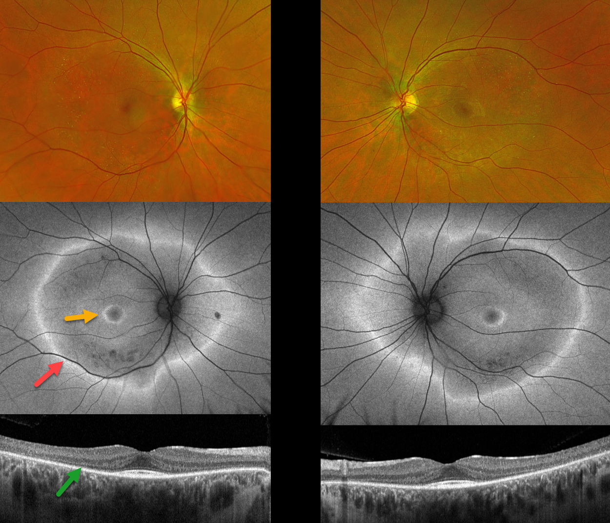
This 32YO female has a known history of an unspecified inherited retinal disease (IRD) diagnosed elsewhere 3-4 years earlier. Genetic testing was negative. She has difficulty seeing in the dark and avoids night driving. More recently, she’s aware of a midperipheral ring of bilateral vision loss. Vision was 20/20 OU.
Optos color RG imaging is fairly normal bilaterally except for drusen-like deposits in the peripheral maculas. Fundus autofluorescence shows a large hyper-FAF ring surrounding each posterior pole, along with a smaller hyper-FAF macular ring. OCT scanning shows outer retinal atrophy sparing the macular center.
Learning Points:
Fundus autofluorescence (FAF) visualizes endogenous fluorophores, compounds that spontaneously fluoresce without an external dye. The main fundus fluorophore is lipofuscin, which resides within the RPE lysosomes. The photoreceptor outer segments absorb some of the autofluorescent excitatory light, and thus normally diminish the FAF signal.
In IRDs, loss of outer segments unmasks this signal, leading to increased FAF. Many patients with an IRD will show a hyper-FAF ring between clinically normal and abnormal retina, which likely represents retina ‘at risk’, showing the junction of a functional and dysfunctional retina (Oishi et al, Int J Retina Vitreous 2019;5(S1);23).
INHERITED RETINAL DISEASE
Seif Anwar
Originally posted on @retina.rocks April 4, 2024
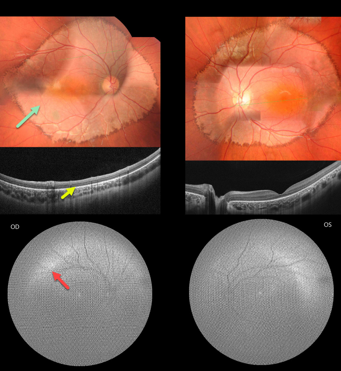
This healthy 6YO boy presented with diminished vision noticed by his parents and teacher. There was no family history of eye disease. Vision was 20/200 OU.
Fundus photography shows fairly symmetrical, creamy, pale subretinal lesions encircling each posterior pole. The margins of these lesions have a somewhat irregular hyperpigmented border.
OCT shows outer retinal thinning and loss of the outer retinal band detail, along with a thinned or absent RPE band.
Fundus autofluorescence (FAF) was of extremely poor quality, and possibly shows superior and temporal hyper-FAF just outside each macula. It was quite difficult to obtain imaging in our patient, and these studies required multiple sessions.
Learning Points:
Unfortunately, we do not have a specific diagnosis for our patient’s presumed sporadic inherited retinal disease (IRD). The location and bilaterality of the multimodal findings suggest a cone-mediated disorder. Our patient’s funduscopic appearance somewhat resembles that found in autosomal recessive CDH3 disease, which includes sparse scalp hair and occasional limb abnormalities (Hull et al, JAMA Ophthalmol 2916;134:992-1000). Genetic testing could not be performed due to cost constraints.
POSTERIOR STAPHYLOMA
Muhammad Arif Ozir
Originally posted on @retina.rocks December 26, 2023
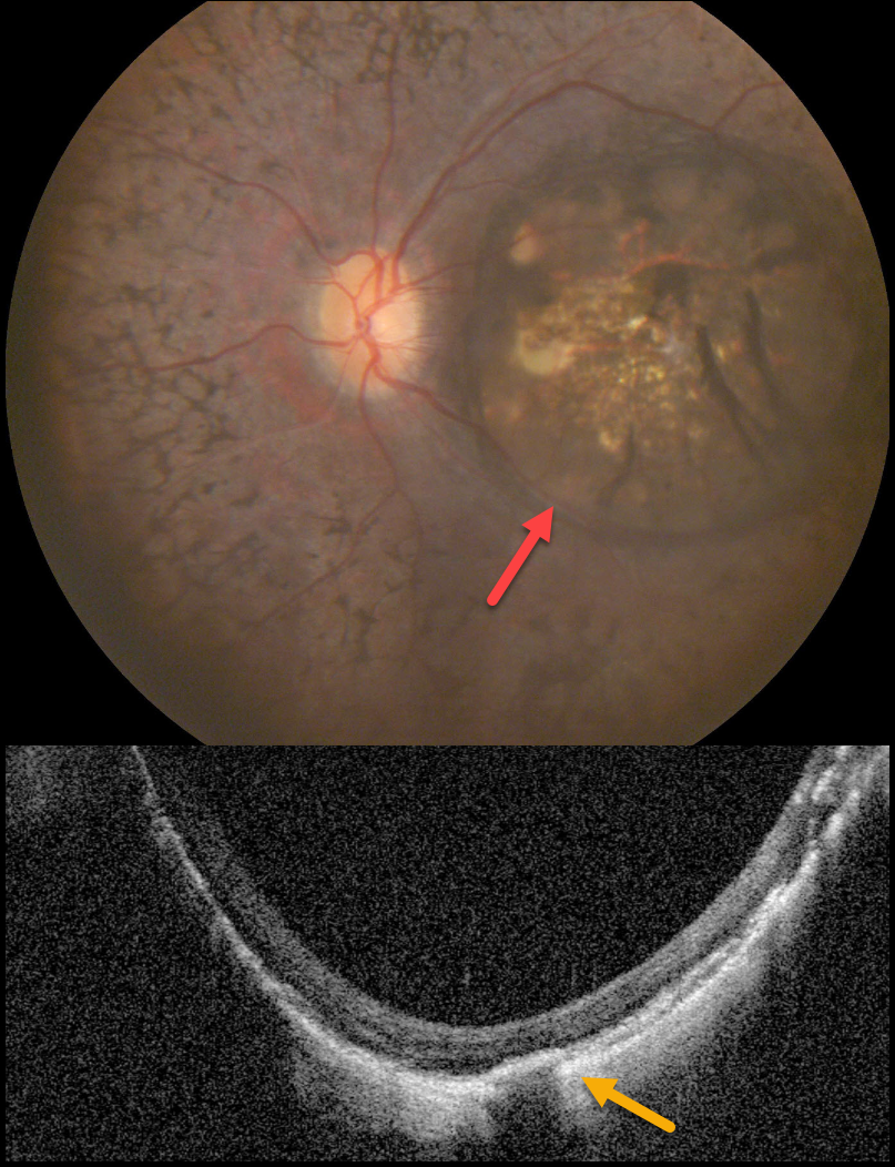
This 20YO male presented with a four-year history of bilateral night blindness and progressive vision loss. Vision was no light perception OU.
Color photography of his left eye shows a variably pigmented colobomatous lesion occupying virtually the entire macula. Macular OCT scanning shows loss of the outer retinal bands, a thinned to non-visualized choroid, and posterior bowing of the sclera. Identical findings were noted in his right eye (not shown).
Learning Points:
Posterior staphylomas are most commonly noted with pathologic myopia, although they have been reported in non-highly myopic eyes with retinitis pigmentosa (Xu et al, Retina 2019;39:1299-1304).
Macular staphylomatous/colobomatous lesions have also been described in other inherited retinal diseases, including North Carolina macular dystrophy, which is caused by an MCDR1 mutation (Small et al, JAMA Ophthalmology 2016;134:355-356).
Unfortunately, our patient was subsequently lost to follow-up, so genetic testing was not possible.
INHERITED RETINAL DISEASE
Yuenpang Cheung, Stephanie Choi and Stephen Tsang
Originally posted on @retina.rocks November 14, 2023
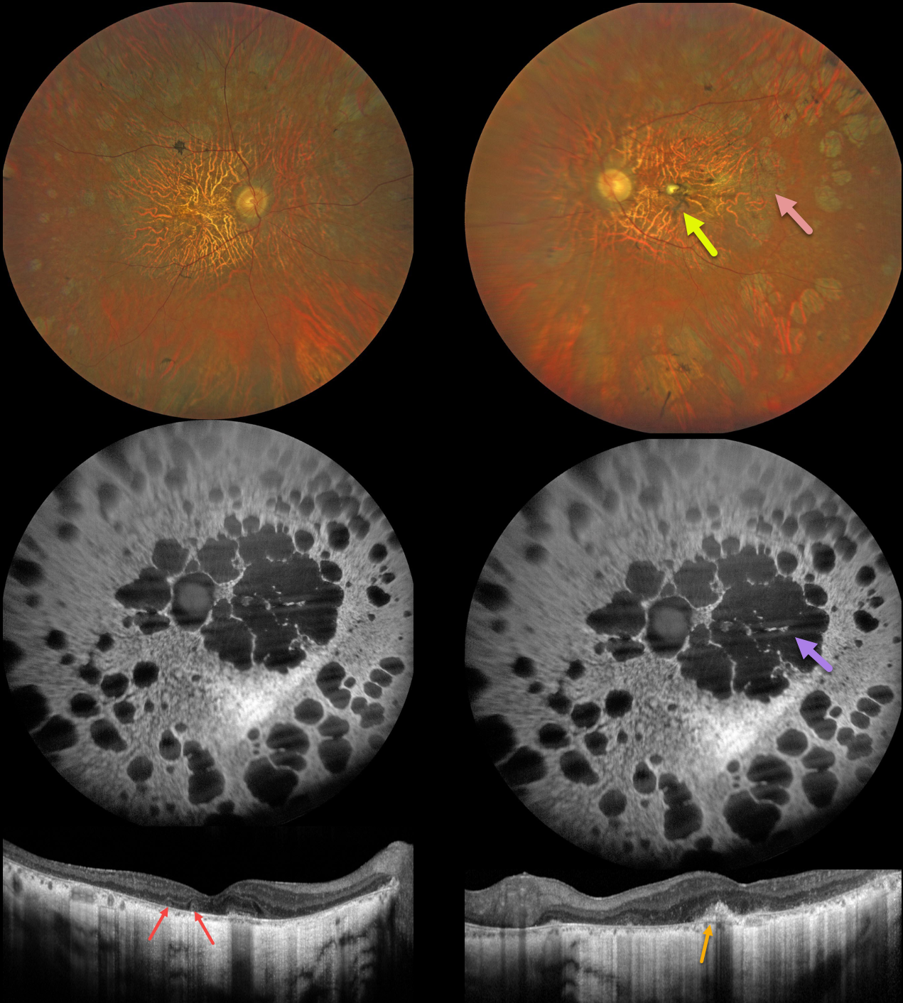
This 56YO male has a history of nyctalopia since childhood with a known mutation in the RDS/peripherin gene. Vision was 20/60 in the right eye and counting fingers in the left eye. The patient has been taking vitamin A supplementation. He has an older brother with a more advanced inherited retinal disorder (IRD).
Fundus imaging shows bilateral diffuse areas of retinal pigment epithelial atrophy. In addition, there is a yellow elevated dome-like lesion with surrounding pigment in the left fovea.
Fundus autofluorescence (FAF) shows hypo-FAF in the areas of atrophy. OCT shows diffuse outer retinal/RPE atrophy with choroidal thinning. In addition, there are a few outer retinal tubulations OD and a subretinal hyperreflective elevated lesion OS. Electrophysiology revealed extinguished rod and diminished cone responses.
Learning Points:
Mutations in the RDS/peripherin gene have been implicated in a number of inherited conditions, including retinitis pigmentosa, pattern dystrophy, retinitis punctata albescens, cone-rod dystrophy, and adult-onset vitelliform macular dystrophy.
RETINITIS PIGMENTOSA + MYOPIC DEGENERATION
Originally posted on @retina.rocks August 30, 2023
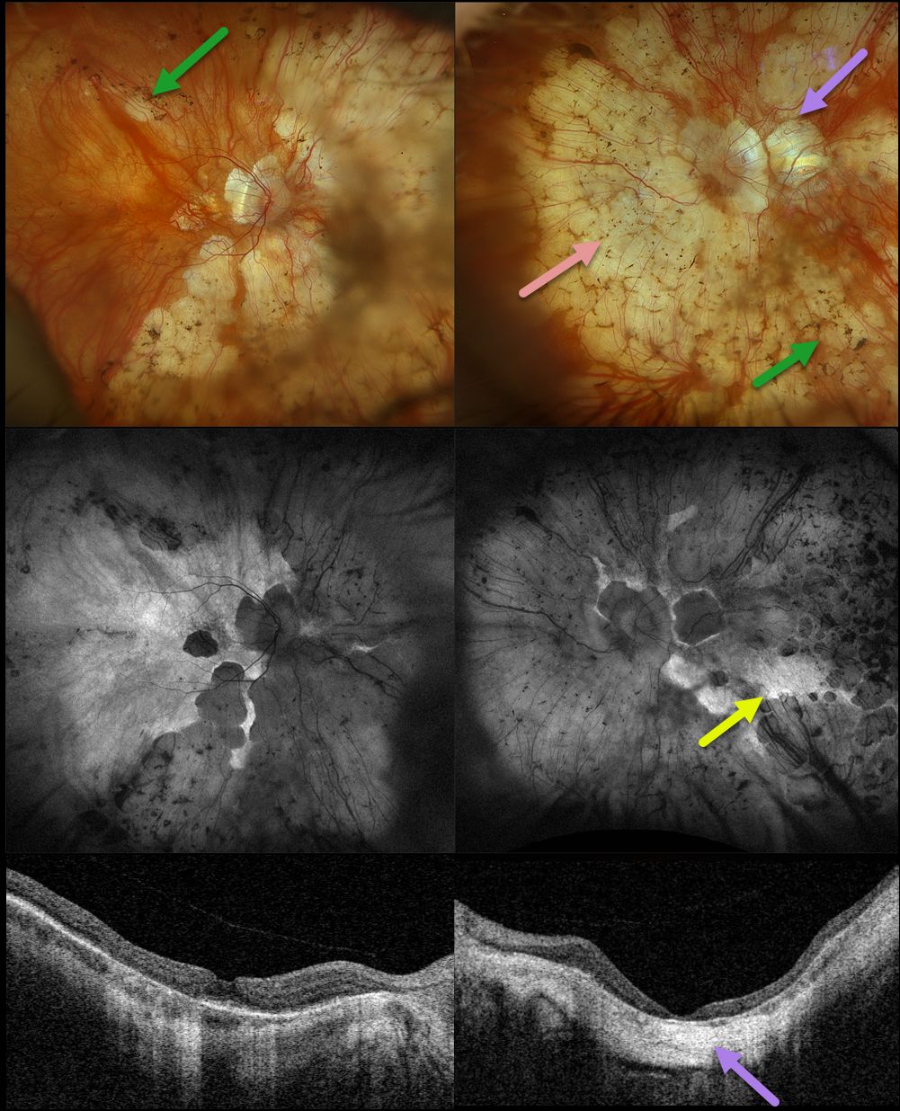
This 78YO female has been followed by us for several years for retinitis pigmentosa (RP) and degenerative myopia. Vision is stable at 20/30 OD and hand motion OS. Multimodal imaging shows a fascinating fusion of findings for these two disorders.
Optos color RGB imaging shows typical RP findings of bone spicules (intraretinal pigment migration) extending from outside the arcades into the peripheries, superimposed on severe degenerative myopic changes.
Fundus autofluorescence (FAF) shows hypo-FAF within the areas of myopic atrophy, with hyper-FAF more peripherally from the RP-induced outer retinal atrophy.
A macular staphyloma is also seen in the left eye and confirmed on swept-source OCT. The degenerative myopic changes just spare the right macular center, which has no staphyloma.
Learning Points:
Genetic testing showed pathogenic heterozygous mutations for OCA2 and RPGR. Fortunately, the RP spares each macula. Her myopic degeneration has been stable, and we are hopeful that her central vision will be preserved in her right eye.
RETINITIS PIGMENTOSA WITH CHOROIDAL CAVITATIONS
Omar Mulki and Faisal Fayyad
Originally posted on @retina.rocks August 16, 2023
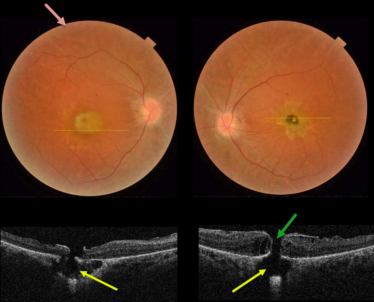
This 32YO female presented with a known history of severe vision loss from retinitis pigmentosa (RP). Her parents are first-degree cousins, and there was no family history of RP. Vision was hand motion OD and counting fingers OS.
Fundus photography shows bilateral irregular yellow central macular pigmentary changes, with a central hyperpigmented spot OS.
OCT scanning through each macula shows a full-thickness macular hole with an underlying defect in the RPE. Each macular hole is suspended over a choroidal cavitation.
Surgical repair was offered with appropriate counseling regarding the poor visual prognosis, and the patient elected to be observed.
Learning Points:
Initially described by Freund et al (Arch Ophthalmol 2003;121:197-204), intrachoroidal cavitations are most commonly found in high myopes as a peripapillary yellow-orange subretinal lesion.
Ornek and Ornek reported a patient with pathologic myopia and a unilateral macular hole communicating with a choroidal cavitation (Retinal Cases 2020;14:328-330).
Although macular holes can rarely be associated with RP (Jin et al, Retina 2008;28:610-614), to the best of our knowledge, ours is the first occurrence of RP with bilateral macular holes and underlying choroidal cavitations.
RETINITIS PIGMENTOSA
Yuenpang Cheung
Originally posted on @retina.rocks August 9, 2023
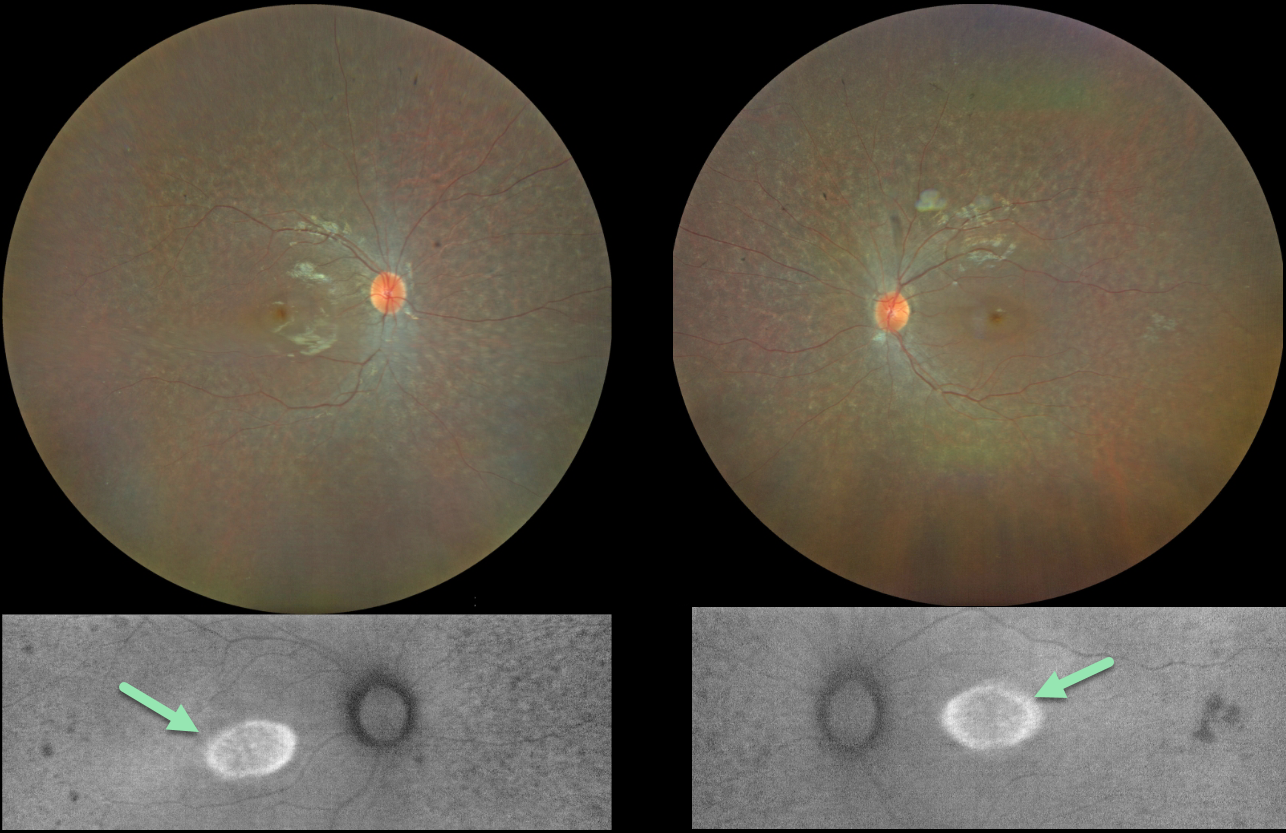
This 13YO girl has a history of congenital deafness, autism, and ADHD. Vision was 20/30 OU.
Color imaging shows bilateral and symmetrical moth-eaten pigmentary changes with an oval area of foveal hyper-autofluorescence.
Fundus autofluorescence (FAF) visualizes endogenous fluorophores, compounds that spontaneously fluoresce without an external dye.
The main fundus fluorophore is lipofuscin, which resides within the RPE lysosomes. The photoreceptor outer segments absorb some of the autofluorescent excitatory light, and thus normally diminish the FAF signal.
In inherited retinal diseases (IRDs), loss of outer segments unmasks this signal, leading to increased FAF. Many patients with an IRD will show a hyper-FAF ring between clinically normal and abnormal retina, which likely represents retina ‘at risk’, showing the junction of a functional and dysfunctional retina (Oishi et al, Int J Retina Vitreous 2019;5(S1);23).
USHER SYNDROME
Emma Oreskovic and Natasa Draca
Originally posted on @retina.rocks July 19, 2023
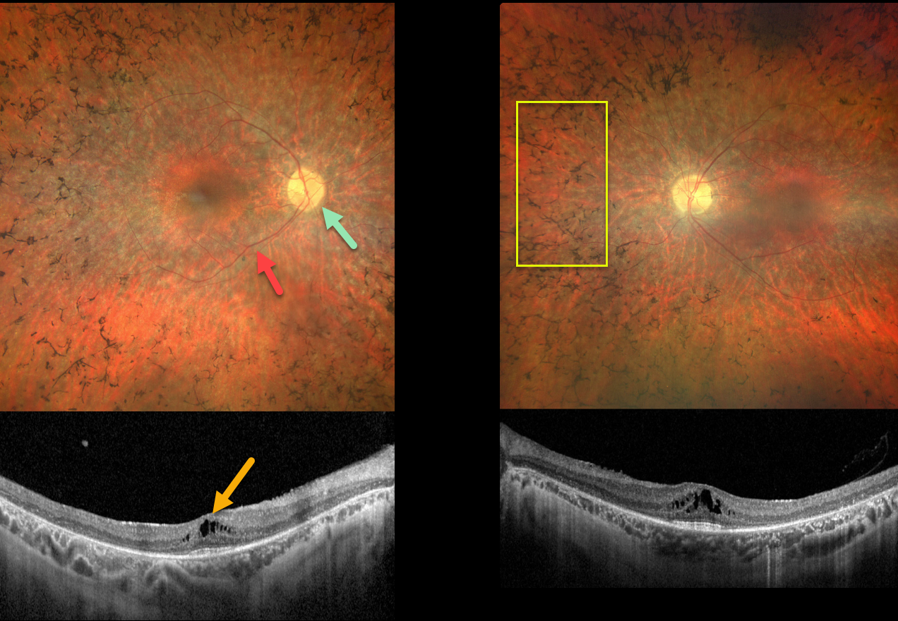
This 33YO female has a known history of retinitis pigmentosa (RP) and congenital neurosensory hearing loss. Her nyctalopia and peripheral vision loss are stable, and vision was 8/20 OD and 15/20 OS.
Color photography of both eyes shows symmetrical pigmentary changes and intraretinal pigment migration (bone spicules) extending from the peripheral maculas into the retinal peripheries. The central maculas look fairly normal, but show schisis in the central inner nuclear layer. The nerves are slightly pale, and the retinal vessels are attenuated.
Genetic testing was heterozygous for a PCHD15 pathogenic variant.
Learning Points:
Usher syndrome (USH) affects over 400 000 persons worldwide and is a major cause of deafness with blindness. Ten USH genes have been identified to date, and three clinical subtypes (USH1-3) have been described. Mutations in the PCDH15 gene that encode for the protocadherin-15 protein cause autosomal recessive Usher syndrome type 1.
Although our patient clinically has USH, genetic testing failed to reveal a causative gene (she only had a single pathogenic gene for a recessive disorder). The negative genetic test result does not mean this patient doesn’t have USH; rather, it reminds us that this test must be interpreted with caution and always with the clinical findings in mind (see Mustafi et al, Ophthalmology Retina 2022;6:702-710).
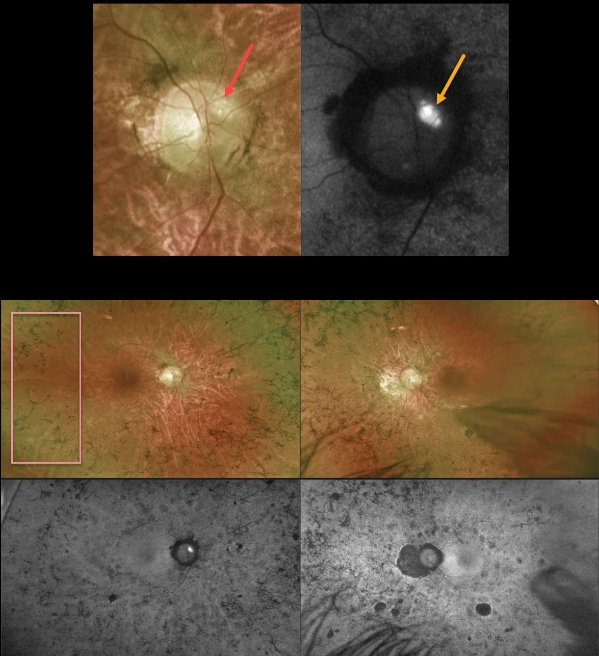
OPTIC NERVE DRUSEN + RETINITIS PIGMENTOSA
Originally posted on @retina.rocks April 17, 2023
This 43YO male with a known history of retinitis pigmentosa (RP) was referred for further evaluation. He noted a gradual worsening of vision bilaterally. Vision was 20/80 OD and 20/200 OS.
Optos color imaging of the right optic nerve shows several large optic disc drusen (ODD) that markedly hyperautofluoresce. Ultrawide-field imaging shows diffuse, classic bilateral RP scarring.
Learning Points:
ODD are refractile calcific concretions that form anterior to the lamina cribrosa. They are thought to be extruded, extracellular, calcified mitochondria and may be inherited as an autosomal dominant trait. They are present in about 3% of RP patients (Russell et al, Eye 2022;36:2213).
Most patients are asymptomatic, but some can develop progressive visual field defects or central vision loss. There is no treatment for the drusen themselves, although symptomatic macular neovascularization can be treated with thermal laser, photodynamic therapy, or anti-VEGF injections.
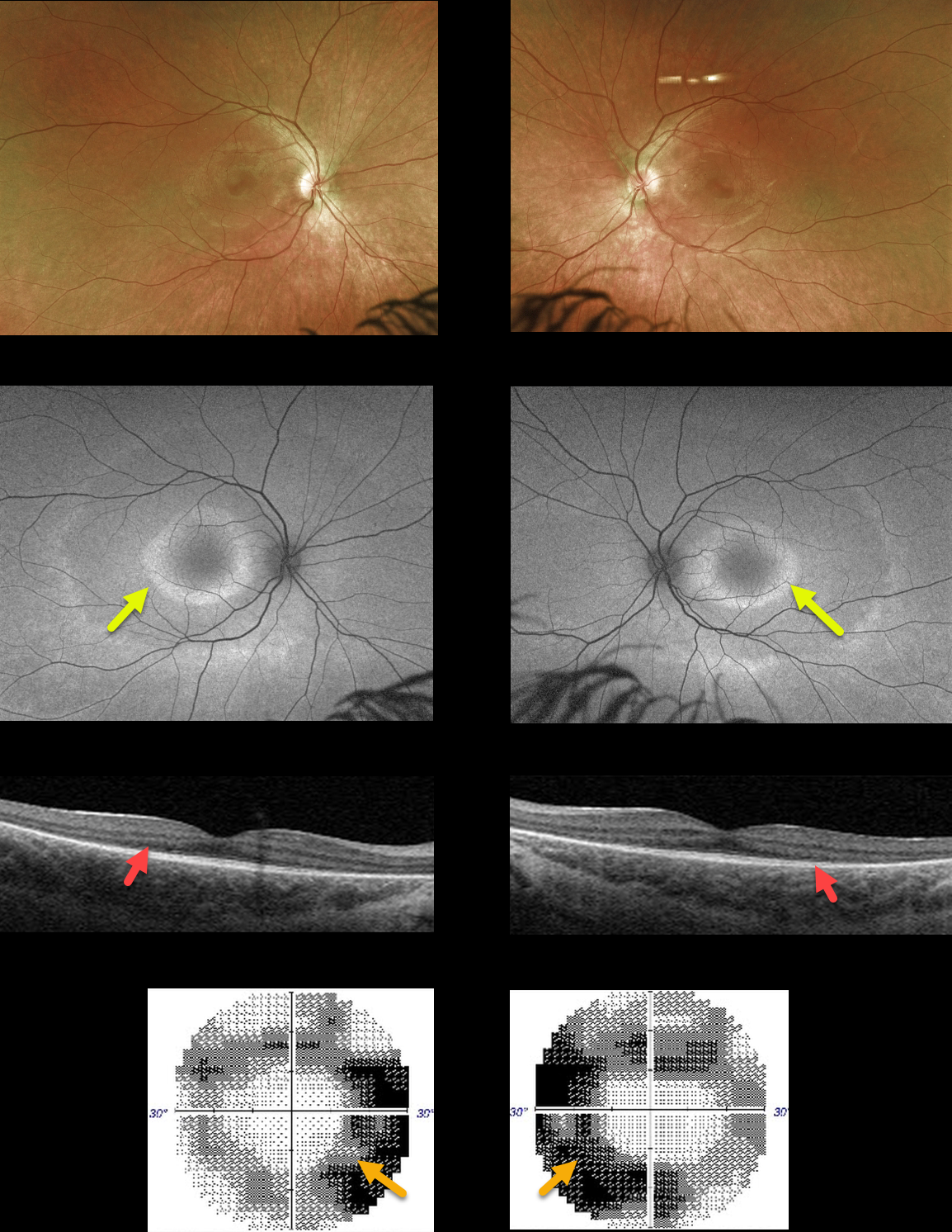
RETINITIS PIGMENTOSA
Originally posted on @retina.rocks March 14, 2023
This 22YO healthy female presented with a year or two of mildly symptomatic problems with distance vision. Vision was 20/25 OD and 20/30 OS. There was no family history of eye disease, and her hearing was normal.
Optos color imaging shows bilaterally symmetrical doughnut-shaped rings of subtle pigmentary changes around each macular center. Fundus autofluorescence (FAF) shows these rings to be hyper-FAF. Swept-source OCT shows outer retinal atrophy. 30-2 visual fields are constricted.
Learning Points:
FAF visualizes fluorophores, endogenous compounds that spontaneously fluoresce without the need for an external dye. The main fundus fluorophore is lipofuscin, which resides within the RPE lysosomes. The photoreceptor outer segments absorb some of the autofluorescent excitatory light, and thus normally diminish the FAF signal.
In RP, loss of the outer segments unmasks this signal, leading to increased FAF. Many patients with RP will show a hyper-FAF ring between clinically normal and abnormal retina, which likely represents retina ‘at risk’, showing the junction of a functional and dysfunctional retina.
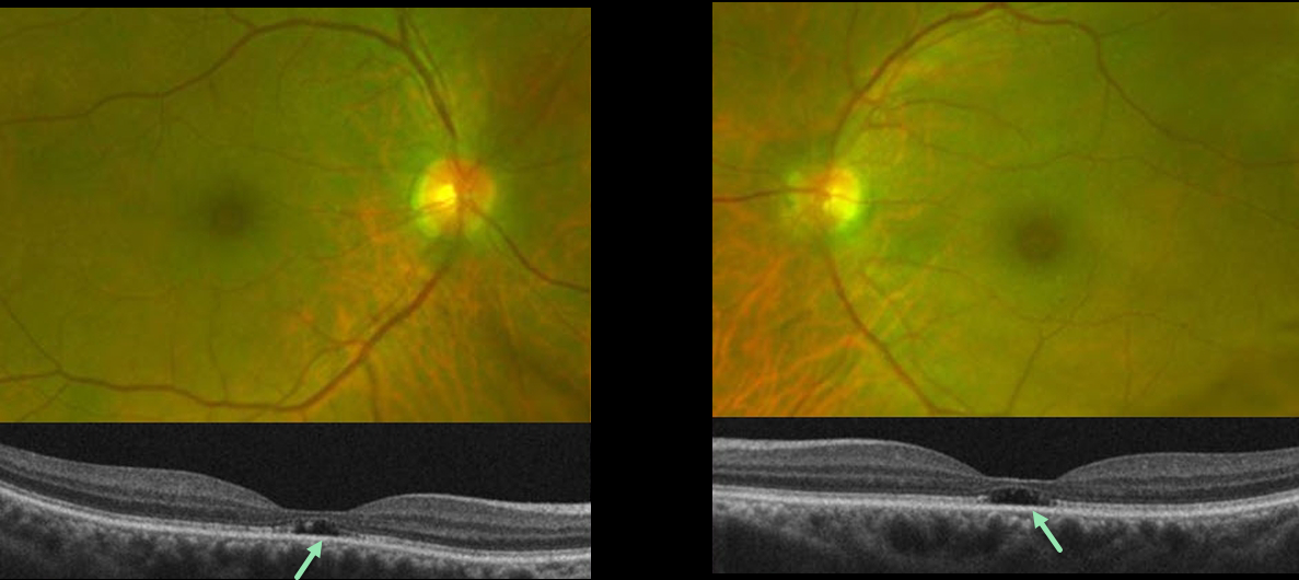
OUTER FOVEAL MICRODEFECTS FROM POSSIBLE IRD
Emily Shepherd
Originally posted on @retina.rocks March 10, 2023
This 65YO presented complaining of difficulty seeing at a distance for some time. She gave a history of radial keratotomy in both eyes about 20 years prior. There was no significant past medical history or family history of eye disease. Vision was 20/60 OD and 20/70 OS.
Optos color imaging shows mild symmetrical pericentral pigmentary change. Fundus autofluorescence and fluorescein angiography were unremarkable (not shown). However, OCT B-scans show bilateral subfoveal loss of the outer retinal tissue external to the external limiting membrane.
An ERG showed rod receptor dysfunction and abnormal dark adaptation with delayed adaptation, while a multifocal ERG was consistent with normal cone function.
Our patient underwent genetic testing and was found to have a heterozygous mutation for FLVCR1. This gene is associated with autosomal recessive posterior column ataxia with retinitis pigmentosa.
A recent report by Kuehlewein et al (Graefe’s Arch Clin Exp Ophthalmol 2019;257:629-638) showed a variety of phenotypes, none of which matched our patient. It is therefore difficult to say whether our patient’s findings are isolated (given the lack of funduscopic, autofluorescence, and angiographic findings beyond the OCT outer defects) or represent an atypical manifestation (given the abnormal electrophysiology) of FLVCR1 disease.
She is scheduled to return for follow-up imaging and visual field testing.
Learning Points:
Outer foveal microdefects (Cohen et al, Ophthalmology Retina 2021;5:553-561) have been described in numerous conditions, including macular telangiectasia, tamoxifen use, ABCA4 disorders (cone-rod dystrophies, Stargardt disease, and fundus flavimaculatus), phototoxicity, trauma, and vitreomacular traction disorders.
In our experience, in patients with inherited retinal diseases (IRD), these findings are most common with a pathogenic ABCA4 mutation (Leng et al, Retina 2012;32:1411-1419 and Wang et al Invest Ophthalmol Vis Sci 2022;53(5):28). These defects, caused by focal loss of the outer retinal layers, leave an optically empty space which can be confused with subretinal fluid but are more of an optical gap within the outer retina.
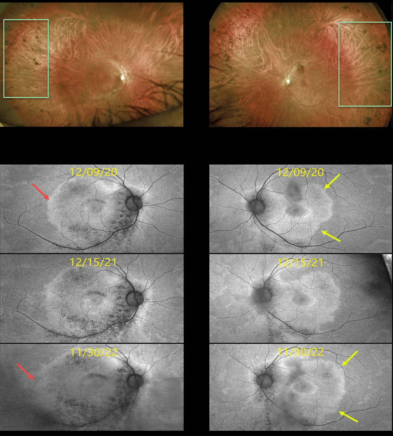
RETINITIS PIGMENTOSA
Originally posted on @retina.rocks January 4, 2023
This 53YO male presented on 12/9/20 with a history of known retinitis pigmentosa (RP). He noticed bilateral blurred vision that had been worsening for about 6 months. Vision was 20/50 OD and 20/30 OS.
Optos color imaging shows fairly normal appearing maculas with a fairly-well circumscribed border of pigmentary loss and migration extending anteriorly from the mid-peripheries.
However, fundus autofluorescence (FAF) reveals subclinical findings of increased macular hyper-FAF sparing each fovea, with hypo-FAF more inferiorly. Subsequent genetic testing revealed a pathogenic RP1 mutation.
FAF shows gradual progressive changes over the 2 years we have been following him. The temporal margins of the hyper-FAF in his right and left eyes have significantly enlarged. This is due to the unmasking of the underlying RPE with progressive photoreceptor atrophy.
Learning Points:
FAF visualizes fluorophores, endogenous compounds that spontaneously fluoresce without the need for an external dye. The main fundus fluorophore is lipofuscin, which resides within the RPE lysosomes.
The photoreceptor outer segments absorb some of the autofluorescent excitatory light, and thus normally diminish the FAF signal.
In RP, loss of the outer segments unmasks this signal, leading to increased FAF. Many patients with RP will show a hyper-FAF ring between clinically normal and abnormal retina, which likely represents retina ‘at risk’, showing the junction of functional and dysfunctional retina.
See Schmitz-Valckenberg et al (Progress Retinal Eye Research 2021;81:100893) for an excellent general review of FAF.
For further reading on monitoring RP with FAF, see Robson et al (Retina 2011;31:1670-1679) and Lee et al (Ophthalmology Retina 2018;2;1062-1070).
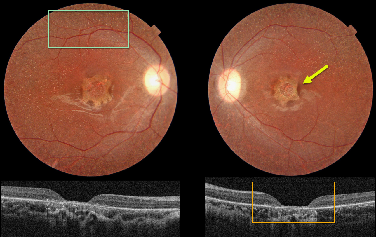
UNKNOWN INHERITED RETINAL DISEASE
Nilesh Kumar and Ashish Sharma
Originally posted on @retina.rocks September 8, 2022
This 19YO female has stable 20/200 vision bilaterally with some form of inherited retinal disease (IRD).
Color photography shows a central one-disc-diameter area of scalloped atrophy with tiny white dots extending from the peripheral macula to beyond the arcades.
OCT shows central neurosensory retinal atrophy with a thin remaining rim of featureless tissue and underlying RPE loss.
Learning Points:
These findings do not fit neatly into any specific diagnosis. Stargardt/ABCA4 disease usually shows more pericentral than central atrophy, and usually involves just the outer retina. The subretinal flecks are much larger and yellow in appearance than the tiny whitish dots seen in our patient.
Fundus albipunctatus is a form of congenital stationary night blindness with somewhat similar white dots, but foveal atrophy and severe central vision loss are usually absent (though it may be associated with an RDH5 mutation).
Finally, retinitis punctata albescens presents with white dots similar to those in fundus albipunctatus, but it is a progressive disorder that can later develop more typical retinitis pigmentosa findings. Foveal atrophy is not a feature of this disease.
Fundus autofluorescence (FAF) is often helpful in differentiating these disorders. The lesions in ABCA4 disease are hyper-FAF. The global FAF signal is markedly diminished in fundus albipunctatus.
Unfortunately, our patient did not get FAF imaging. And of course, genetic testing would hopefully be diagnostic, but this was also not available.
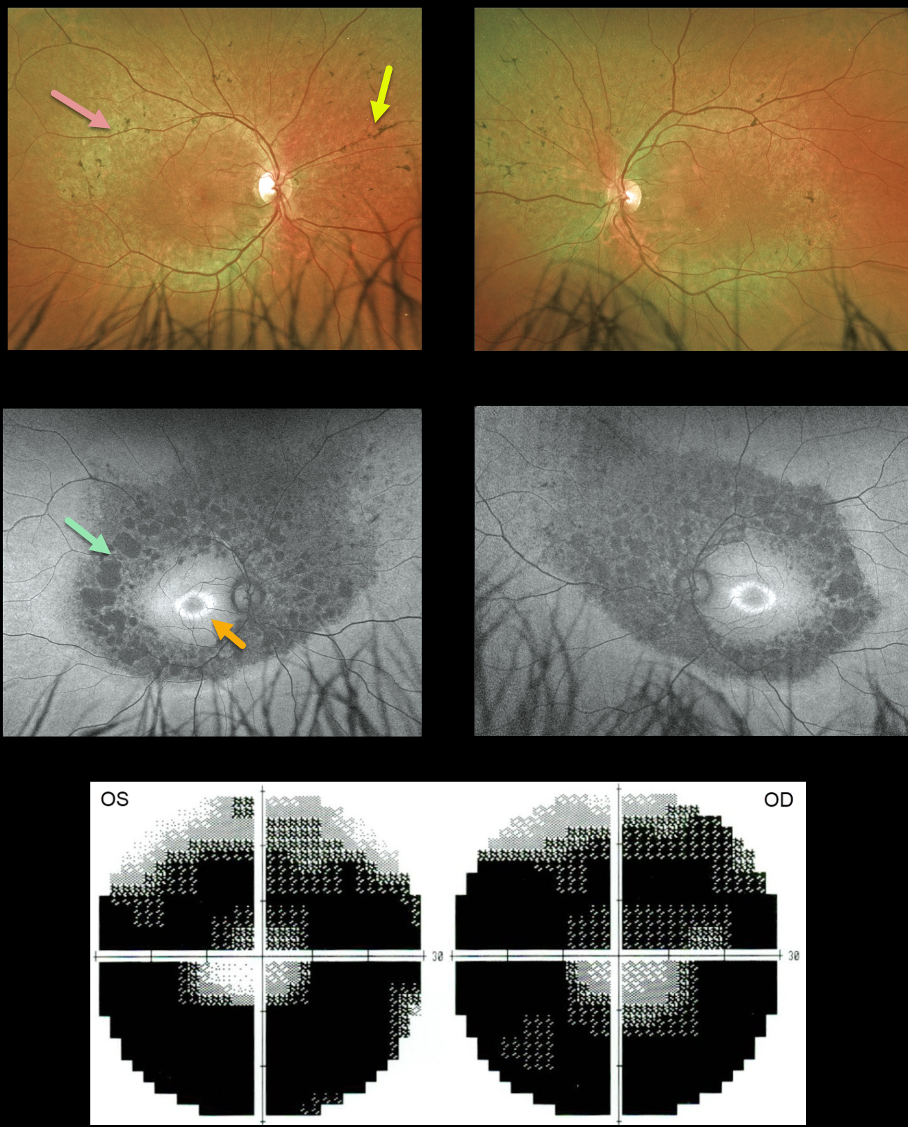
RETINITIS PIGMENTOSA
Originally posted on @retina.rocks July 4, 2022
This 34YO male was referred for relatively asymptomatic retinal findings. He had complained of some blurred distance vision for several years. Vision was 20/25 OU.
Optos color imaging shows fairly normal maculas with midperipheral encircling pigment loss and intraretinal pigment migration. Fundus autofluorescence (FAF) shows a bull’s-eye-shaped area of macular hyper-FAF, and areas of midperipheral and nasal hypo-FAF corresponding to the pigmentary changes clinically. Visual field testing shows marked bilateral constriction. Invitae genetic testing was negative.
Learning Points:
Patients with retinitis pigmentosa (RP) classically have pale optic nerves, narrowed retinal blood vessels, variable outer retinal and RPE loss, and intraretinal pigment migration (bone spicules) due to loss of the inhibitory effect of the degenerated outer segments, allowing the RPE to migrate along the retinal capillaries.
A negative genetic test does not mean this patient doesn’t have RP; rather, it reminds us that such testing must be interpreted with caution and always with the clinical findings in mind.
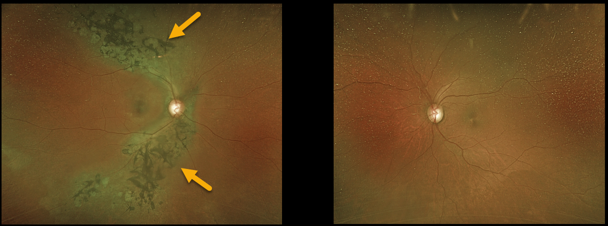
TRAUMATIC PSEUDO-RP
Originally posted on @retina.rocks May 31, 2022
A 67YO female was referred for asymptomatic retinal findings in her right eye. She gave a history of unspecified head trauma as a child.
Optos imaging shows chorioretinal scarring with intraretinal pigment migration, extending superotemporally and inferotemporally from the optic nerve. The left fundus was completely normal.
Learning Points:
Photoreceptor loss from numerous causes, including blunt trauma, retinal detachment, macular telangiectasia, and retinitis pigmentosa (RP), can allow for intraretinal RPE migration with this shared phenotype.
Unilateral RP is exceedingly rare and requires normal electrophysiologic findings and long-term observation to ensure delayed findings in the fellow eye.
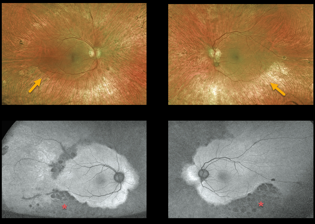
ALAGILLE SYNDROME
Originally posted on @retina.rocks January 26, 2022
This 24YO man with Alagille syndrome presented with vision of 20/200 OD and 20/50 OS. There was a long history of bilateral poor vision. He was adopted and is a ward of the state.
Optos color imaging shows fairly normal maculas with midperipheral encircling pigment loss with some pigment migration.
Fundus autofluorescence (FAF) shows extensive areas of midperipheral and nasal hypo-FAF.
Learning Points:
Alagille syndrome, also known as arteriohepatic dysplasia, is an autosomal dominant, genetically and phenotypically heterogeneous disorder characterized by distinct facial features, including a prominent forehead, deep orbital sulcus, and pointed chin.
It is caused by loss of function of the JAG1 gene, which is located on chromosome 20p12. The JAG1 gene encodes transmembrane proteins responsible for cell signaling during embryonic development, notably in the Notch signaling pathway.
Patients develop intrahepatic cholestatic liver disease, heart abnormalities, hypogonadism, and intellectual disability. Virtually all patients have peripheral retinal pigmentary changes, and three-quarters have anterior chamber angle abnormalities, including Axenfeld and Rieger anomalies (our patient’s slit lamp examinations were normal).
For a recent, comprehensive review of the spectrum of ocular findings in Alagille syndrome, see da Palma et al., Invest Ophthalmol Vis Sci 2021; 62 (7):27.
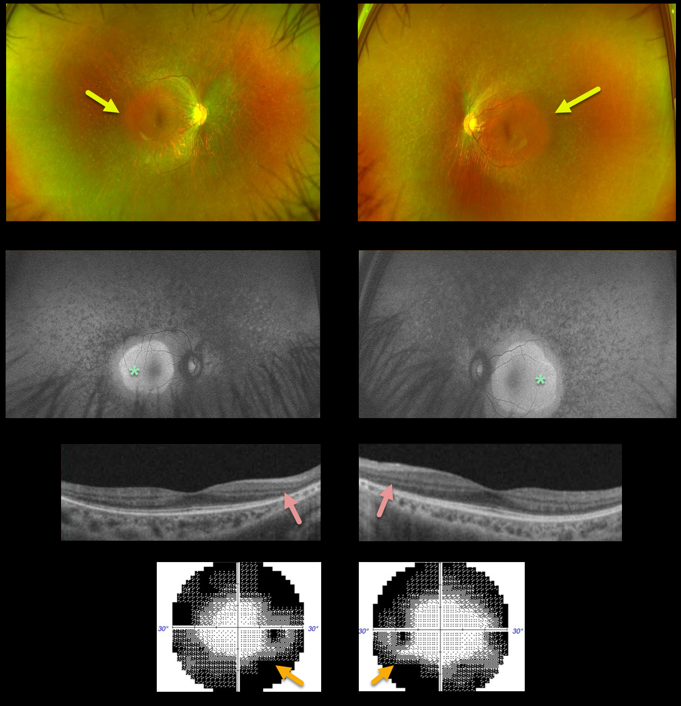
RETINITIS PIGMENTOSA
Alex Hynes
Originally posted on @retina.rocks December 14, 2021
This 25YO male presented with complaints of nyctalopia and peripheral vision loss. Vision was 20/25 OU.
Optos color imaging shows a diffuse moth-eaten appearance to the RPE, sparing each macula. Minimal bone spicules are noted. Fundus autofluorescence (FAF) shows macular hyperautofluorescence.
OCT scanning shows peripheral outer retinal thinning. Visual field testing shows classing bilateral constriction. Genetic testing showed no pathogenic mutations.
Learning Points:
Patients with retinitis pigmentosa (RP) classically have pale optic nerves, narrowed retinal blood vessels, variable outer retinal and RPE loss, and intraretinal pigment migration (bone spicules) due to loss of the inhibitory effect of the degenerated outer segment,s allowing the RPE to migrate along the retinal capillaries. In real life, patients often lack all these features.
As in this case, intraretinal pigment migration may be minimal or absent but often appears over time (see Takahashi et al, AJO 2018;195:176-180).
A truly normal fundus in a patient with RP is extremely unusual, as there are usually very subtle clinical and FAF findings.
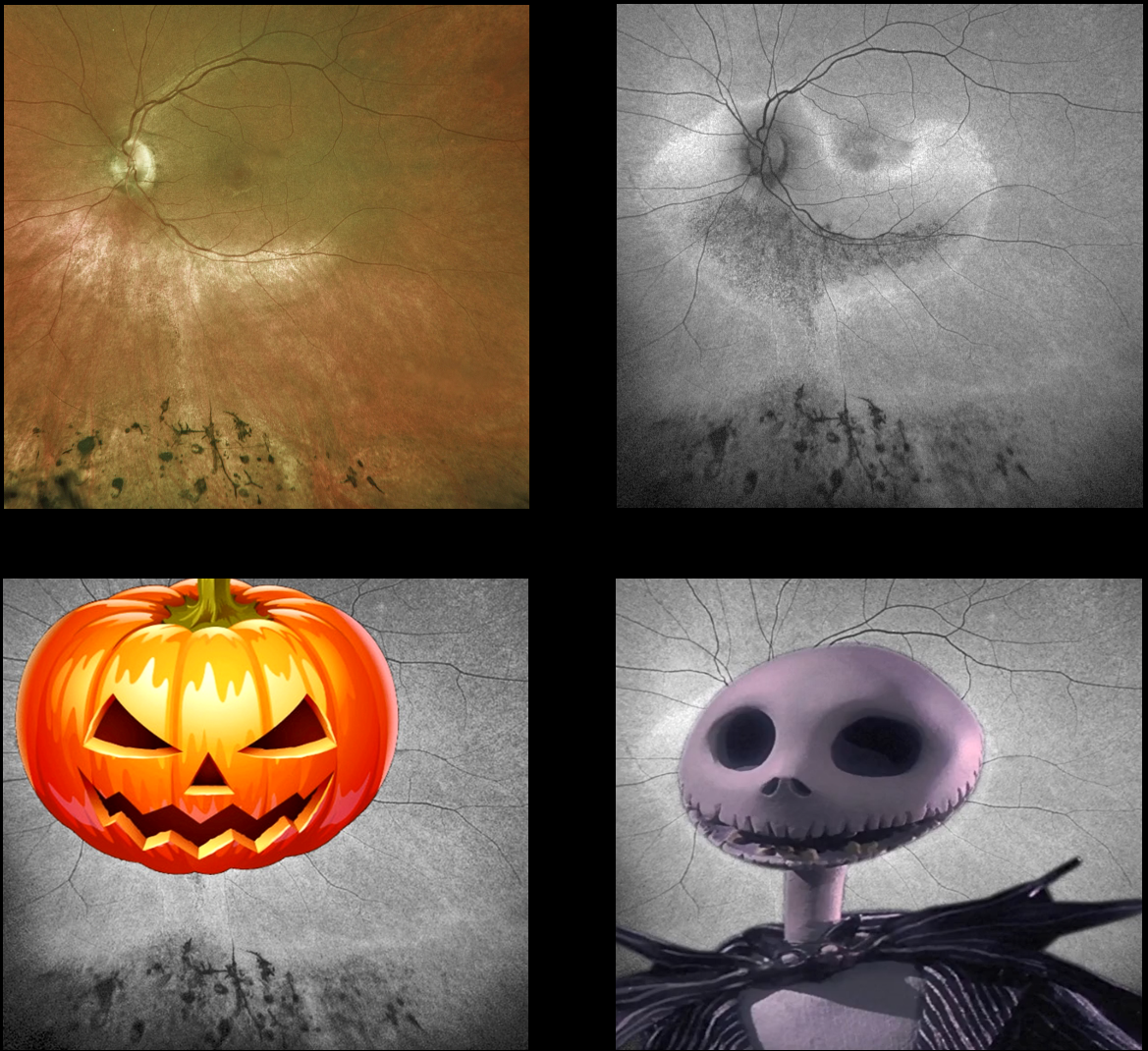
HALLOWEEN RETINOPATHY
Originally posted on @retina.rocks October 29, 2021
This is a patient with retinitis pigmentosa, but do you see Jack O’Lantern or Jack Skellington? Maybe something else spooky?
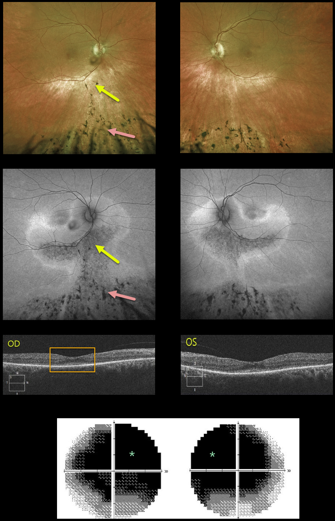
SENIOR-LOKEN SYNDROME
Originally posted on @retina.rocks August 13, 2021
This 51YO female presented with 2 years of progressive night vision loss. There was a history of a prior traumatic brain injury, along with a renal transplant in 2011. VA was 20/60 OD and 20/50 OS.
Optos color and fundus autofluorescence (FAF) imaging show extremely symmetrical findings. A gutter of pigment loss extends along the inferotemporal arcades and inferior to the optic nerve into the inferior peripheries, where there is more extensive chorioretinal scarring with intraretinal pigment migration.
The gutters are classic findings in subretinal exudation, most commonly central serous chorioretinopathy (Yannuzzi et al, Ophthalmology 1984;91:1554-1572). The intraretinal pigment migration can also be seen following blunt trauma (pseudo-RP).
OCT B-scanning shows outer retinal thinning and a probable thickened choroid bilaterally, possibly supporting the central serous diagnosis. However, the marked symmetry of all these findings made us feel that trauma or central serous was unlikely. VF testing shows marked diffuse depression.
Subsequent workup included full field electrophysiology and genetic testing. Dark-adapted ERG showed normal amplitudes, but the light-adapted and cone flicker responses were severely reduced. Her genetic testing revealed pathologic mutations in the NPHP1, ABHD12, LRP5, and PDE6A genes.
The NPHP1 mutation, coupled with her retinal findings and kidney history, confirmed a rare genetic autosomal recessive renal-retinal syndrome known as Senior-Løken Syndrome.
Learning Points:
Senior-Løken Syndrome was first described independently in 1961 by Senior et al as juvenile familial nephropathy and tapetoretinal degeneration (AJO 1961;52:625-633) and Løken et al as hereditary renal dysplasia and blindness (Acta Paediatrica 1961;50:177-184).
Cystic kidney disorders known as nephronophthisis are associated with retinal findings that fall within the broader category of retinal ciliopathies (Adams et al, Ophthalmic Genetics 2007;28:113-125), including Leber congenital amaurosis and some variants of retinitis pigmentosa. Less than 200 cases of Senior-Løken have been reported.
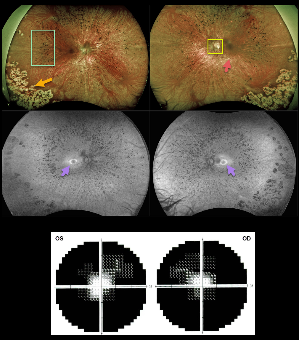
RETINITIS PIGMENTOSA
Originally posted on @retina.rocks June 2, 2021
This 35YO female has classic findings for retinitis pigmentosa. Optos imaging shows somewhat pale nerves, narrowed retinal vessels, and peripheral intraretinal pigment migration (bone spicules). There are also confluent cobblestone-like changes in the inferotemporal peripheries.
Fundus autofluorescence (FAF) shows a hyper-FAF ring around the central macula, indicating preserved photoreceptors that may be at-risk for future degeneration.
Visual fields are markedly constricted bilaterally.
Learning Points:
Photoreceptors normally inhibit RPE cells, so any process involving photoreceptor loss can cause intraretinal RPE migration that mimics retinitis pigmentosa, including chronic retinal detachment and blunt trauma.
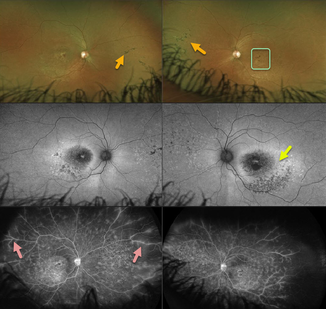
USHER SYNDROME
Originally posted on @retina.rocks April 23, 2021
This 26YO female presented with progressive bilateral vision loss and photophobia. Vision was 20/60 OD and 20/200 OS. She had moderate hearing loss since birth, and denied any family history of eye disease.
Ultra-wide-field imaging reveals areas of nasal mid-peripheral bone spicules (intraretinal pigment migration) and ‘beaten-bronze’ macular atrophy.
There is a ring of hyper-autofluorescence surrounding each macula, which identifies at-risk retina for future degeneration. Fluorescence angiography (FA) shows variable window defects due to pigmentary changes, with diffuse peripheral vascular staining and leakage.
Learning Points:
The association with neurosensory deafness/hearing loss and retinitis pigmentosa (RP) is called Usher Syndrome. About 10% of children who are deaf/hearing impaired have RP.
Vascular leakage is not uncommon with inherited retinal diseases. Photoreceptors normally inhibit RPE cells, and photoreceptor damage allows for RPE intraretinal migration along retinal capillaries. Vascular endothelial cells adjacent to these translocated RPE cells are thin and fenestrated, similar to the choriocapillaris, and may be the cause of this vascular leakage (see Li et al, Ophthalmology 1995;102;8805-816).
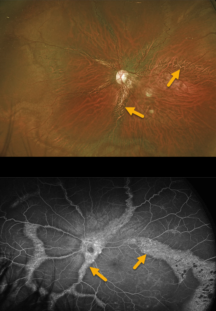
UNKNOWN DIAGNOSIS
Originally posted on @retina.rocks March 3, 2021
This unknown shows extensive and unusual, radial and circumferential scarring of the posterior pole in this 37 YO male’s left eye. The right eye (not pictured) had a failed retinal detachment repair at an outside practice many years earlier, resulting in no light perception vision and an opaque, white cornea.
Since the radial changes seem to mostly follow the arcades, pigmented paravenous retinochoroidal atrophy is considered in the differential, but there are no areas of intraretinal pigment migration (bone spicules), and the more peripheral connecting circumferential scarring would be unusual. No electrophysiology has been performed.
This patient presented like this years ago without symptoms, all findings have been stable, and we have been following him since with 20/30 vision.
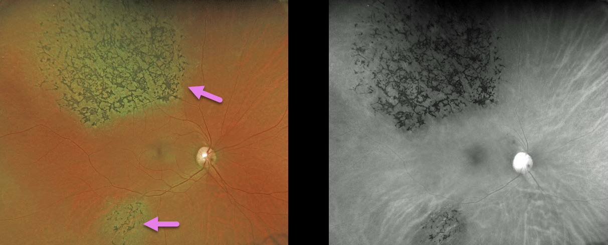
TRAUMA
Originally posted on @retina.rocks September 29, 2020
A 49YO female was involved in a motor vehicle accident 30 years ago.
Her retina showed signs of pigmentary retinopathy from the ocular injury, which could be mistaken for retinitis pigmentosa (RP). Unlike typical RP, our patient had multifocal, sectoral, unilateral findings.
Learning Point:
Intact photoreceptors inhibit RPE migration. Photoreceptor loss from numerous causes, including blunt trauma, retinal detachment, macular telangiectasia, and retinitis pigmentosa, can allow intraretinal RPE migration with this shared phenotype.
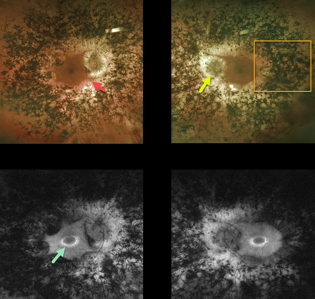
RETINITIS PIGMENTOSA
Originally posted on @retina.rocks June 11, 2020
This patient with retinitis pigmentosa (RP) surprisingly has good central vision 20/30 OU.
The fundus photos show all the classic findings for RP, including attenuated retinal vessels, extensive bone spicules (intraretinal migration of the RPE along retinal capillaries), and optic disc pallor.
Fundus autofluorescence for this patient shows a ring of hyperautofluorescence around each central macula. This ring identifies at-risk retina for future degeneration and can help monitor disease progression.
Learning Points:
RP is a retinal degenerative disorder that causes nyctalopia and progressive vision loss.
Photoreceptors normally inhibit RPE cells, so any process with photoreceptor loss can cause similar localized RPE migration.
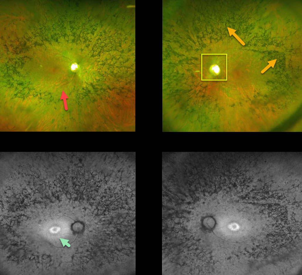
RETINITIS PIGMENTOSA
Originally posted on @retina.rocks January 24, 2020
This patient with retinitis pigmentosa (RP)surprisingly has good central vision 20/30 OU.
The fundus photos show all the classic findings for RP, including attenuated retinal vessels, bone spicules (intraretinal migration of the RPE along retinal capillaries), and optic disc pallor.
Fundus autofluorescence for this patient shows a ring of hyperautofluoresence around each central macula. This ring identifies at-risk retina for future degeneration and can help monitor the progression of this disease.
Learning Points:
Retinitis pigmentosa is a retinal degenerative disorder that causes nyctalopia and progressive vision loss.
Photoreceptors normally inhibit RPE cells, so any process with photoreceptor loss can cause similar localized RPE migration.


