

Retinocytoma and Retinoblastoma
<< Back to Cases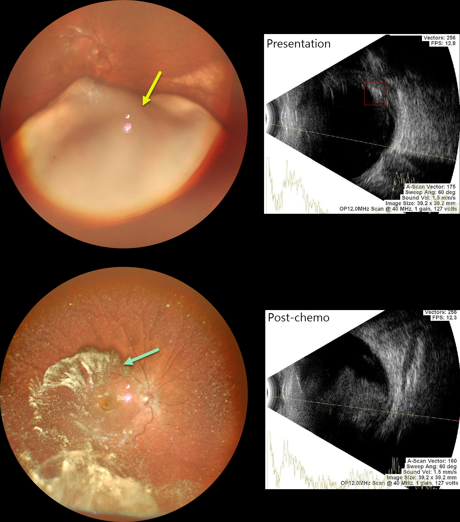
RETINOBLASTOMA
Malvika Singh, Akansha Sharma, Shraddha Raj Shrivastava, Anand Gandhi and Manish Nagpal
Originally posted on @retina.rocks August 17, 2026
This previously healthy 2YO girl presented with leukocoria in her right eye. Family history was negative.
Examination under anesthesia revealed a large smooth endophytic lesion filling more than half of the vitreous cavity, extending into the inferior macula. B-scan ultrasonography revealed intralesional calcifications. The left eye was normal.
MRI showed no extra-ocular or CNS lesions. We diagnosed her with ICRB Group D retinoblastoma in the right eye. She underwent 3 cycles of chemotherapy.
Repeat examination under anesthesia, 9 weeks following initial presentation, showed marked tumor regression, although there was widespread significant vitreous seeding. MRI did not show any optic nerve infiltration or CNS spread. Enucleation of this eye was scheduled
Learning Points:
Retinoblastoma is the most common primary intraocular malignancy of childhood, with a peak incidence before age five. Presenting signs frequently include leukocoria and strabismus. The disease may be heritable or nonheritable, and delayed diagnosis can result in orbital extension, metastatic spread, and decreased survival. Early identification and intervention with chemotherapy, focal therapies such as laser photocoagulation or cryotherapy, and, in select cases, intra-arterial or intravitreal chemotherapy maximize the chances of ocular salvage and life preservation.
RETINOBLASTOMA
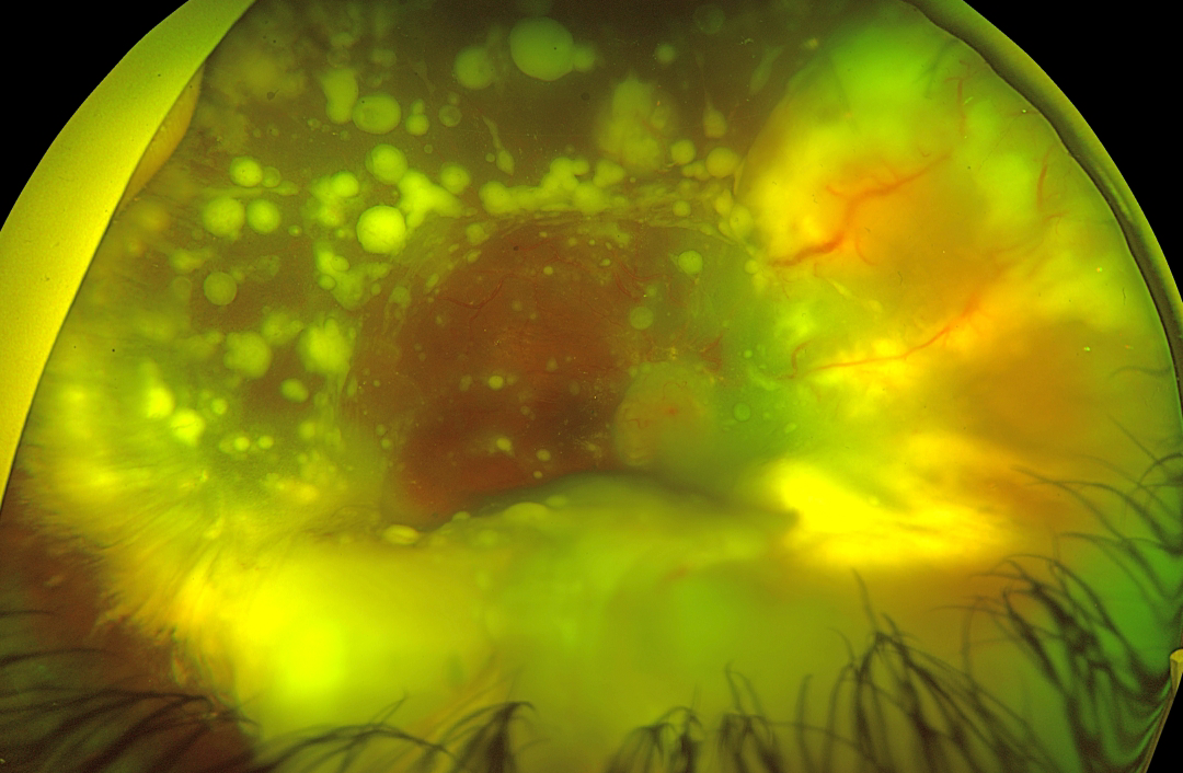
Victor Ribeiro de Sant'Ana, Thailor Dartora, Laísa Ferraz de Arruda, Gabriela Mousse de Carvalho and Rodrigo Jorge
Originally posted on @retina.rocks December 8, 2025
RETINOCYTOMA
The European VitreoRetinal Society and Dibya Prabha
Originally posted on @retina.rocks November 7, 2025
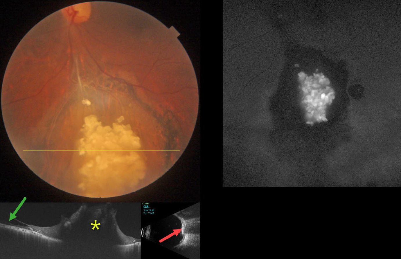
This healthy 43YO male presented with gradual bilateral blurring. Vision was 20/20 in his normal OD and 20/30 OS.
Color photography shows an elevated, glistening, inner retinal yellow-white mulberry-like lesion in the inferior midperiphery. Surrounding subretinal pigmentary changes are noted. OCT through this lesion shows a markedly thickened, disorganized, hyperreflective retina. Retinoschisis is noted nasally. The lesion is markedly hyperreflective on B-scan ultrasonography and hyper-autofluorescent on fundus autofluorescence.
Learning Points:
Retinocytomas are fleshy, opalescent, benign tumors that are felt to represent spontaneously regressed retinoblastoma (RB). They appear identical to previously radiated RB tumors. The risk of a germline RB1 mutation in sporadic unilateral retinoblastoma or retinocytoma is about 10-20%.
Clinically, retinocytomas can resemble astrocytic hamartomas. However, retinocytomas often have underlying RPE changes and chorioretinal scarring (as in our patient), which would not be present with astrocytic hamartomas.
The brain MRI was normal, and observation was recommended.
RETINOBLASTOMA
Ankit Jain and Manish Nagpal
Originally posted on @retina.rocks March 24, 2025
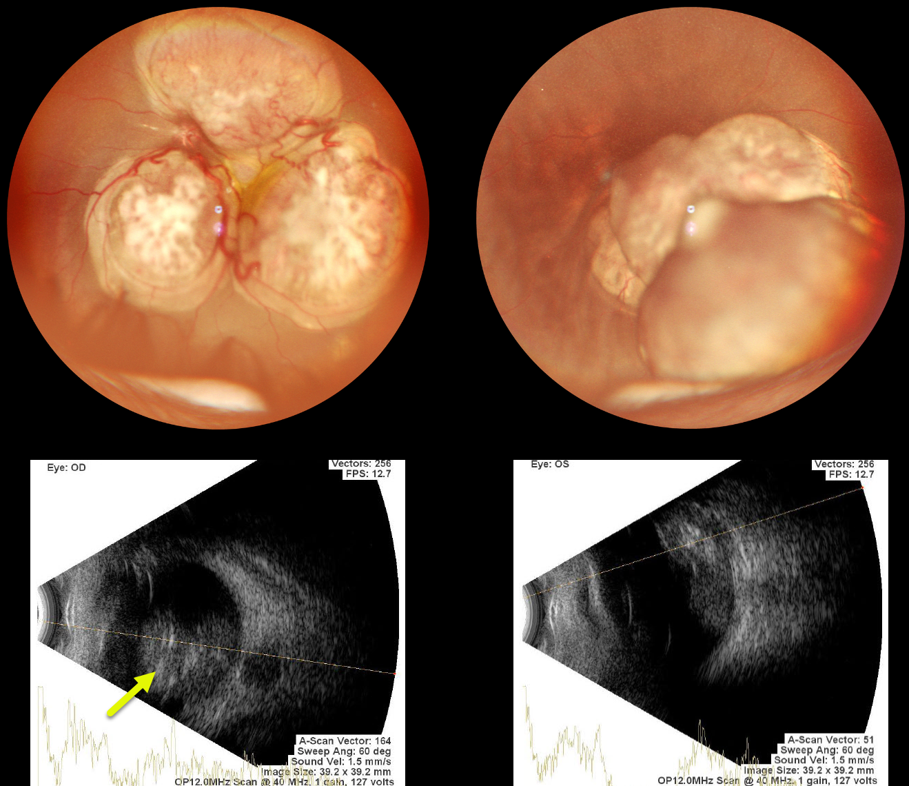
This 8-month-old girl presented with bilateral nystagmus, leukocoria, and inability to focus on objects. Family history was completely negative.
Examination under anesthesia revealed bilateral lobular white retinal masses projecting into each vitreous. B-scan ultrasonography shows intralesional hyperreflectivity suggestive of calcification. She was immediately referred to ocular oncology.
Learning Points:
Retinoblastoma (RB) is the most common primary intraocular tumor of childhood. The RB1 gene, located on chromosome 13 (13q14.2), encodes a tumor suppressor protein. The vast majority of cases have no family history. Bilateral and multifocal RB occurs in 20-35% of patients and is virtually always due to a germinal mutation. These patients are also at risk for secondary tumors, including pinealoblastoma.
Treatment options include systemic, intra-arterial, and intravitreal chemotherapy; focal therapy (laser photocoagulation, cryotherapy, plaque, or external beam radiotherapy); and enucleation. We assume our patient has a new germline mutation given the negative family history.
RETINOCYTOMA
Originally posted on @retina.rocks December 9, 2024
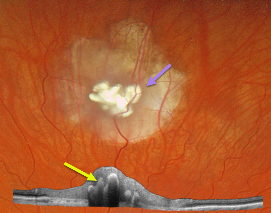
This healthy 30YO male was referred for an asymptomatic retinal lesion in his left eye. There was no family history of eye disease. Vision was 20/25 OU.
Optos color RGB imaging shows a nodular, calcified retinal lesion with surrounding atrophic chorioretinal scarring. Topcon swept-source OCT scanning shows a disorganized retina overlying the hyperreflective calcifications, which shadow posteriorly.
Learning Points:
Retinocytomas are fleshy, opalescent, benign tumors that are felt to represent spontaneously regressed retinoblastoma (RB). They appear identical to previously radiated RB tumors. As with typical RB, these patients’ offspring are at 50% risk of developing RB and thus require genetic counseling.
Clinically, retinocytomas can resemble astrocytic hamartomas. However, retinocytomas often have underlying RPE changes and chorioretinal scarring (as in our patient), which are not present in astrocytic hamartomas.
Brain MRI scanning was normal. Genetic testing for the retinoblastoma (RB1) and tuberous sclerosis (TSC1/TSC2) genes was suggested, but the patient denied this for now due to cost concerns.
RETINOBLASTOMA
Originally posted on @retina.rocks May 2, 2022
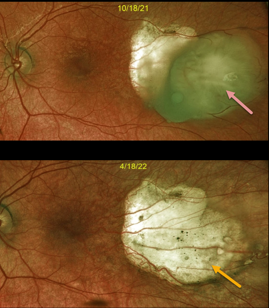
This is a 3YO female with a history of bilateral retinoblastoma and prior enucleation of the right eye. It is a follow-up of a previously posted case (11/7/21).
Optos imaging of the left eye shows an active retinoblastoma with complete regression following 6 months of treatment, which included intra-arterial chemotherapy and cryotherapy.
Learning Points:
Small tumors can be treated with focal therapies such as laser therapy, cryotherapy, or radioactive plaque therapy.
Larger tumors are treated with chemotherapy (most commonly intra-arterial through the ophthalmic artery and occasionally systemic). Chemotherapy has dramatically decreased the need for enucleation.
RETINOBLASTOMA
Originally posted on @retina.rocks November 17, 2021
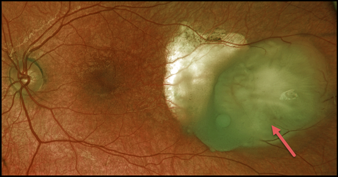
This 3YO female has a history of bilateral retinoblastoma and prior enucleation of the right eye. Optos color RG imaging of her left eye shows a still active retinoblastoma, despite prior treatment, which included intra-arterial chemotherapy and cryotherapy.
Learning Points:
Small tumors can be treated with focal therapies such as laser therapy, cryotherapy, or radioactive plaque therapy.
Larger tumors are treated with chemotherapy (most commonly intra-arterial through the ophthalmic artery and occasionally systemic). Chemotherapy has dramatically decreased the need for enucleation.
RETINOCYTOMA
Originally posted on @retina.rocks April 16, 2021
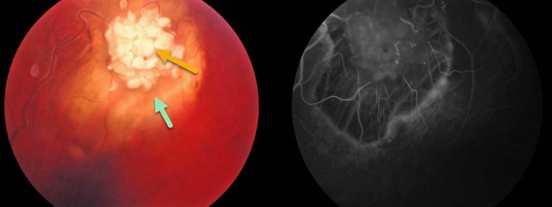
This 57YO male presented with a retinocytoma without any history of retinoblastoma (RB) or treatment for RB.
Clinically, retinocytomas can resemble astrocytic hamartomas. However, retinocytomas often have underlying RPE changes and chorioretinal scarring, which are not present in astrocytic hamartomas.
Learning Points:
Retinocytomas are fleshy, opalescent, benign tumors that are thought to spontaneously regress in Rb. They appear identical to previously radiated Rb tumors.
Similar to typical RB, these patients’ offspring are at a 50% risk for developing Rb and thus require genetic counseling.
RETINOCYTOMA
Originally posted on @retina.rocks July 20, 2020
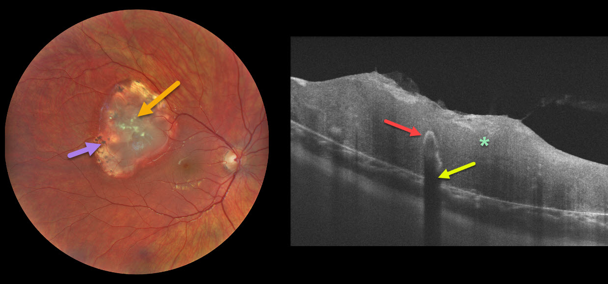
This patient presented with a retinocytoma of the right eye. The OCT line scan shows a disorganized, thickened retina. Typical intralesional calcification is noted. This calcium is highly reflective, causing shadowing of the underlying retina.
Clinically, retinocytomas can resemble astrocytic hamartomas. However, retinocytomas often have underlying RPE changes and chorioretinal scarring, which are not present in astrocytic hamartomas.
Learning Points:
Retinocytomas are fleshy, opalescent, benign tumors that are felt to be spontaneously regressed retinoblastoma (RB). They appear identical to previously radiated RB tumors.
Similar to typical RB, these patients’ offspring are at a 50% risk for developing RB and thus require genetic counseling.
RETINOBLASTOMA
Originally posted on @retina.rocks May 7, 2020
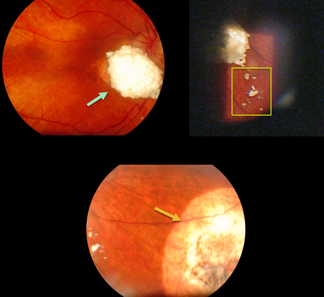
This patient was treated as a child for retinoblastoma, although unfortunately we don’t know the details of her prior treatment.
She has a calcified, elevated peripapillary lesion with overlying vitreous seeds. There is also a scar in the inferonasal midperiphery from another destroyed tumor. The multifocal nature of her tumors indicates that this was a genetic mutation, and that she likely has the retinoblastoma gene.
Learning Points:
Small tumors can be treated with focal treatments such as thermal laser, cryotherapy or radioactive plaque therapy. Larger tumors are treated with chemotherapy (most commonly intra-arterial through the ophthalmic artery and occasionally systemic). Chemotherapy has dramatically decreased the need for enucleation.


