

Retinopathy of Prematurity (ROP)
<< Back to Cases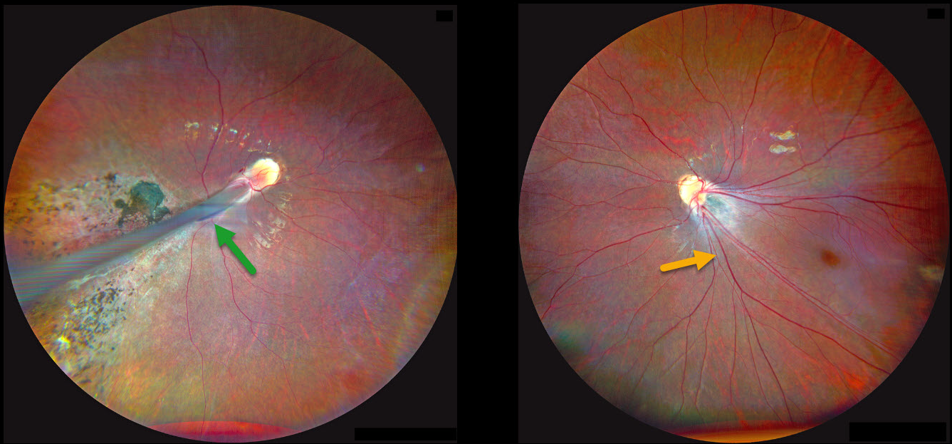
RETINOPATHY OF PREMATURITY
Kanwaljeet Harjot Madan
Originally posted on @retina.rocks June 23, 2025
This 4YO girl’s parents noticed that her right eye was deviating outwards for several months, along with decreased vision bilaterally. Her mother gave a history of prematurity at 28 weeks of gestational age with a low birth weight of 1175 g and oxygen exposure. Vision was counting fingers OD and 20/80 OS. There was a large right exotropia.
Fundus photography OD shows a prominent retinal fold extending from the optic disc into the inferotemporal periphery. Diffuse chorioretinal scarring is noted surrounding the retinal fold. The retinal vessels OS are dragged inferotemporally with marked foveal ectopia.
Learning Points:
Retinopathy of prematurity (ROP) is a potentially blinding vasoproliferative disease occurring in premature infants. Although multifactorial, current screening guidelines are based primarily on low birth weight and gestational age (Kim et al, Surv Ophthalmology 2018;63:618-637). Screening and treatment for these eyes is extremely subspecialized, with treatment options primarily including timely scatter laser photocoagulation and intravitreal anti-VEGF injections (Yonekawa et al, Retina 2017:37:2208-2225).
Patients with ROP have an increased risk for vision-threatening complications throughout their life, including vitreous hemorrhage and retinal detachment (Hsu et al, Ophthalmology Retina 2023;7:72-80). We will therefore monitor our patient yearly.
RETINOPATHY OF PREMATURITY
Originally posted on @retina.rocks April 23, 2025
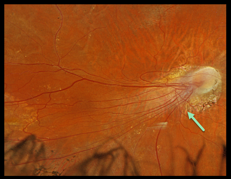
This 70YO female presented with stable 20/100 vision in her right eye. Her left eye was enucleated almost 60 years previously due to complications of retinopathy of prematurity (ROP).
Optos color RGB imaging shows marked temporal macular dragging with peripapillary and distal temporal chorioretinal scarring.
Learning Points:
Patients with ROP have an increased risk for vision-threatening complications throughout their life, including vitreous hemorrhage and retinal detachment (Hsu et al, Ophthalmology Retina 2023;7:72-80). We will therefore continue to monitor our patient yearly.
RETINOPATHY OF PREMATURITY
Şengül Özdek
Originally posted on @retina.rocks July 1, 2024
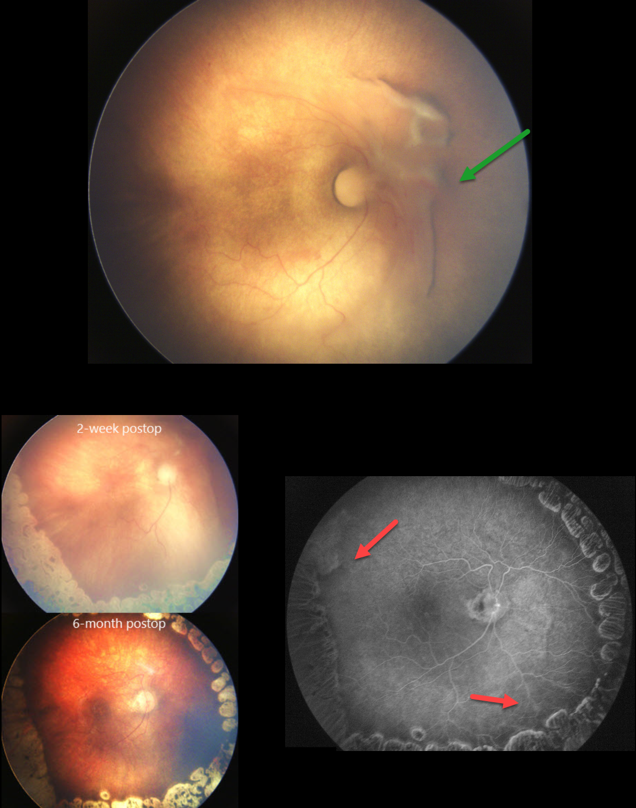
A prematurely born baby at a gestational age of 30 weeks and birth weight of 1120 grams was referred to our clinic at postmenstrual age of 37 weeks for the treatment of stage 4A ROP in her right eye. It was a zone 1 disease, and even the macula was not yet vascularized in either eye. There was a tractional retinal detachment at the nasal side of the right optic disc.
A lens-sparing vitrectomy was done with laser ablation of zone 2. Zone 1 (macula) was not lasered, and an anti-VEGF injection was given at the end of the surgery. The retina was completely attached at both 2-weeks and 6-months postoperatively. The retina was finally completely vascularized in zone 1 six months postoperatively.
Learning Points:
The current ROP classification system is based on the International Classification of Retinopathy of Prematurity, Third Edition (ICROP 3; Ophthalmology 2021;128:e51-e68), and there is no zone half in the zone descriptions. However, especially in developing countries with limited resources in neonatal intensive care units, ROP with avascular macula (even less than zone I, which could be called zone half!), as in the present case, can be seen. Such cases are usually called Aggressive ROP, which can progress to stage 4-5 very quickly.
It is generally recommended to laser the avascular retina during vitrectomy to control active disease. However, in zone 1 cases where the macular vascularization is not yet completed, as in our patient, we leave the zone 1 non-lasered to give a chance for macular vascularization after surgery. An anti-VEGF injection at the end of the surgery was therefore given to control possible disease activation due to the lack of laser treatment to the posterior nonperfused retina. To the best of our knowledge, this is the first presentation of such a treatment approach in the literature.
RETINOPATHY OF PREMATURITY
Originally posted on @retina.rocks May 13, 2021
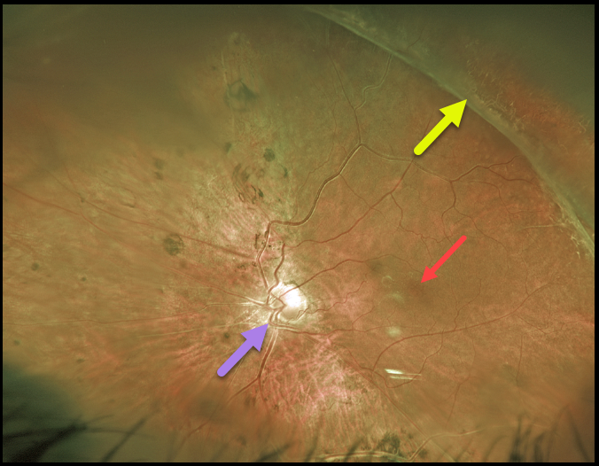
For over 20 years, we’ve been following this 39YO male with stable regressed retinopathy of prematurity in his left eye. Vision is NLP OD and 20/80 OS.
Optos imaging shows that the macular center is dragged temporally due to vitreoretinal traction from regressed neovascularization.
The macular center is usually about 2 disc diameters temporal to the edge of the optic nerve, and our patient’s is about 3 disc diameters. This dragging is usually accompanied by a straightening of the temporal arcades.
However, in our patient, this is masked by situs inversus, a congenital abnormality in which the retinal vessels exit the optic nerve with a nasal bend before coursing temporally.
Learning Points:
These outcomes can be minimized through aggressive neonatal screening and treatment with laser photocoagulation and anti-VEGF agents.


