

Retinoschisis Degenerative
<< Back to Cases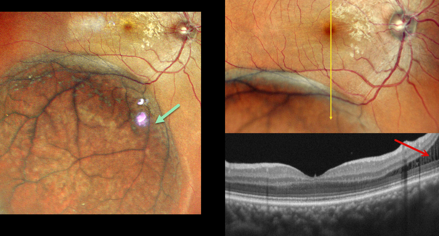
DEGENERATIVE RETINOSCHISIS
Malvika Singh and Manish Nagpal
Originally posted on @retina.rocks August 25, 2026
This asymptomatic 40YO female was referred for a retinal detachment in her right eye. Vision was 20/20 OD and 20/20 OS in her normal OS.
Pseudocolor SLO imaging shows a large area of inferotemporal degenerative retinoschisis (DRS) extending just into the distal macula. OCT scanning confirms schisis without retinal detachment. Observation was recommended.
Learning Points:
Uncomplicated DRS typically follows a stable or slowly evolving course that should not be treated. The development of inner-layer and outer-layer holes essentially creates a full-thickness retinal break. Progression of subretinal fluid beyond the DRS margins (combined schisis-rhegmatogenous retinal detachment, RRD), in the presence of both inner- and outer-layer holes, is the only DRS scenario requiring intervention.
It is often clinically difficult to differentiate pure DRS from a combined schisis-RRD or from RRD. OCT scanning through the area of interest, as in our patient, often provides definitive evidence for the proper diagnosis and management (Paris et al, Survey Ophthalmology 2026;71:71-80).
COMBINED RETINOSCHISIS-RHEGMATOGENOUS RETINAL DETACHMENT
Originally posted on @retina.rocks August 12, 2026
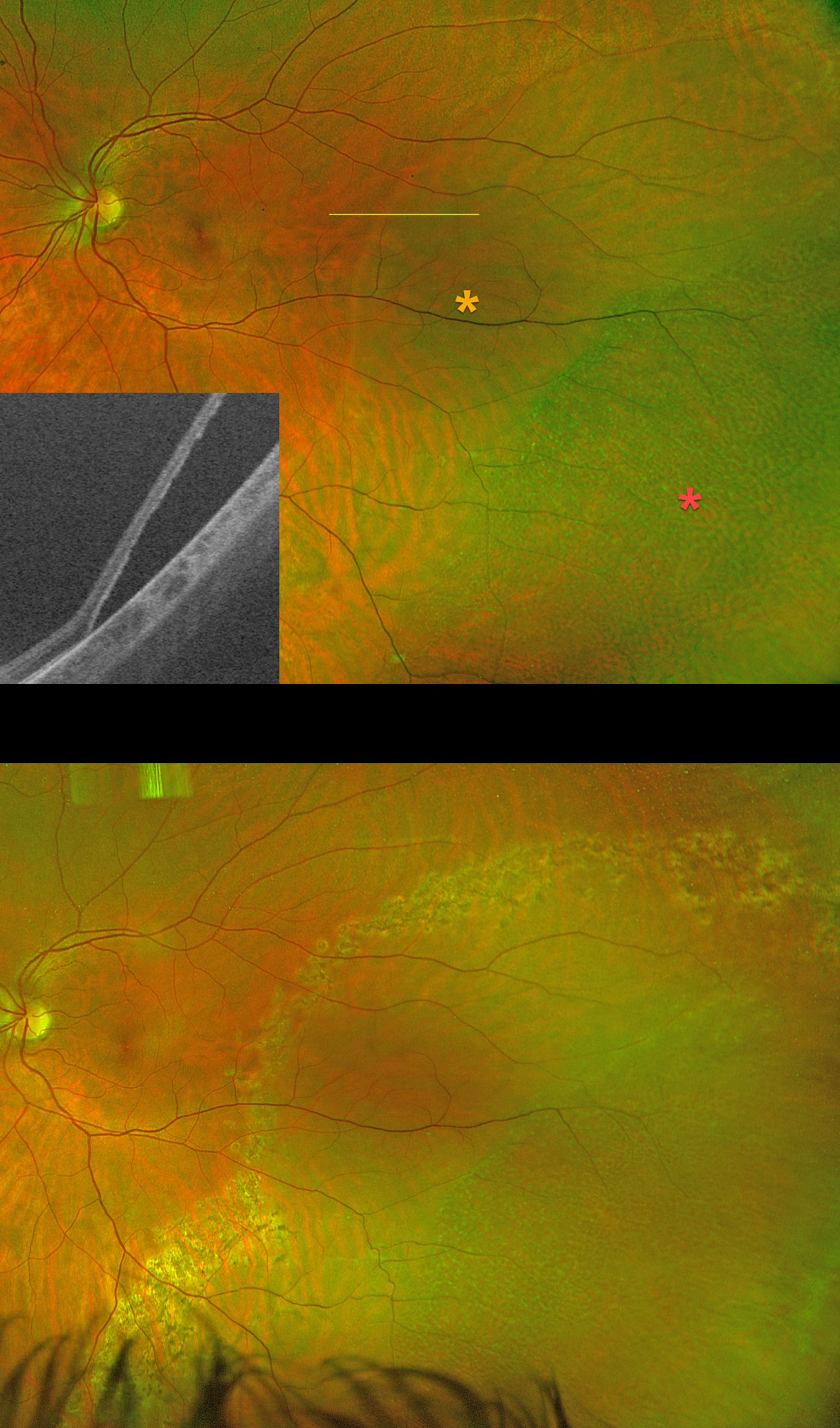
This 35YO pregnant female presented with 3 days of flashes in her left eye. She had a family history of retinal tears in her father and rhegmatogenous retinal detachment (RRD) in her maternal uncle. Vision was 20/20 OS.
Silverstone color RG imaging shows a macula-on combined retinoschisis-RRD. There was no posterior vitreous detachment. Schisis is noted more peripherally, with the rhegmatogenous fluid noted more posteriorly. Swept-source OCT confirms the presence of a full-thickness retinal detachment. Treatment options were discussed, including laser demarcation, primary scleral buckle with cryotherapy, and pars plana vitrectomy. She elected for laser retinopexy, which was performed that day.
She delivered a healthy baby, and her retina has remained stable over the past 9 months as of her last visit.
Learning Points:
Isolated degenerative retinoschisis should not be treated. However, it can progress to a combined schisis-RRD in the presence of breaks in both the inner and outer layers. Asymptomatic combined detachments can be demarcated, as in our patient. Symptomatic detachments are usually treated with pars plana vitrectomy.
SPONTANEOUSLY FLATTENED DEGENERATIVE RETINOSCHISIS
Kris Garbig and Doug Garbig
Originally posted on @retina.rocks June 10, 2026
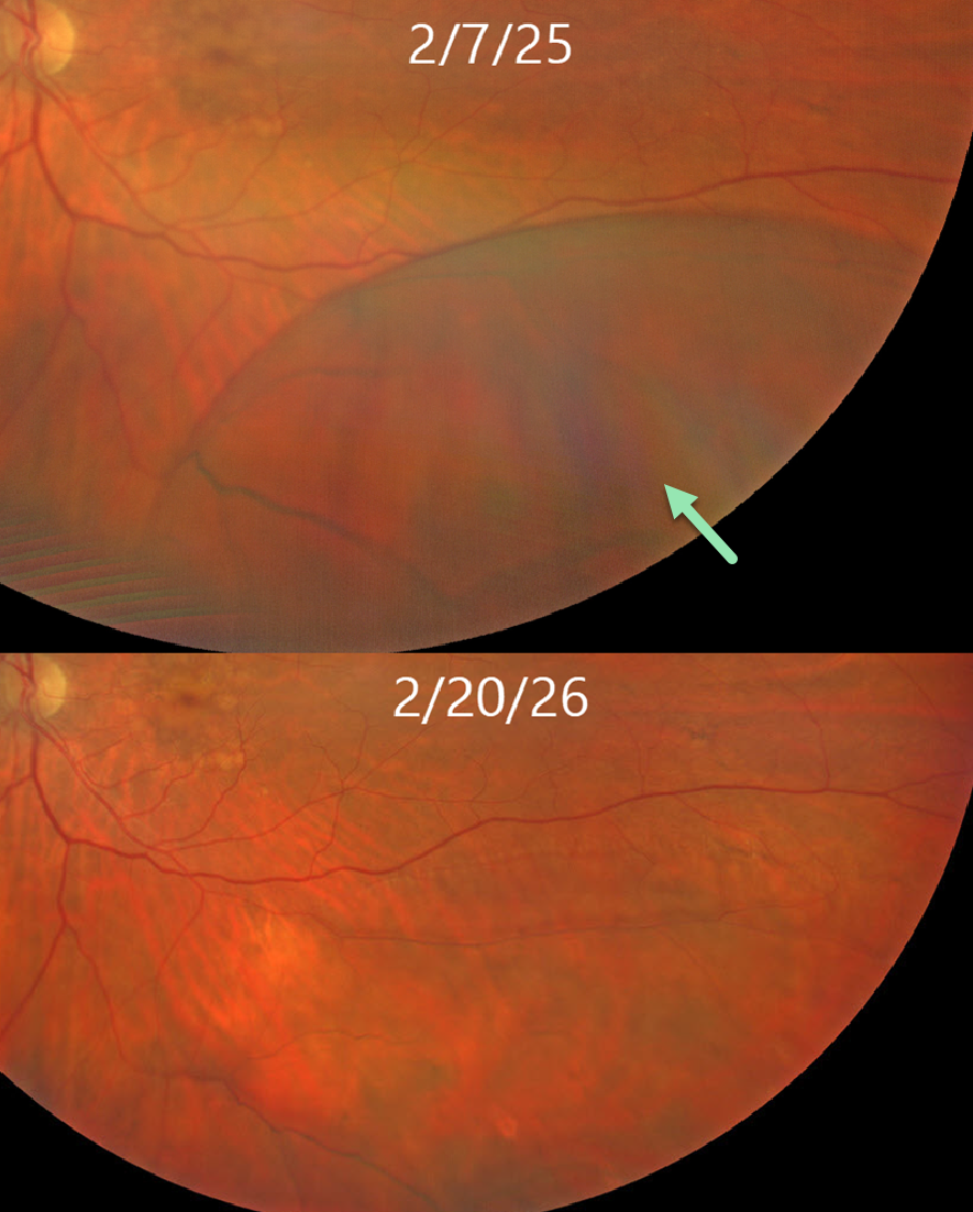
This 77YO female presented for routine examination on 2/7/25 with asymptomatic degenerative retinoschisis (DRS) inferotemporally in her left eye. When she returned one year later, the schisis had completely spontaneously flattened.
Learning Points:
The dynamics of DRS cavities were noted by Byer in 1972 when he reported the first case of spontaneous regression (Arch Ophthalmology 1972;88:207-209). Since adopting UWF imaging in our practice, we have found that schisis is often not static, with DRS enlarging or flattening. Inner- and outer-layer breaks also appear over time, often independent of changes in the schisis cavity.
LATTICE DEGENERATION WITH SUBCLINICAL DEGENERATIVE RETINOSCHISIS
Originally posted on @retina.rocks March 5, 2026
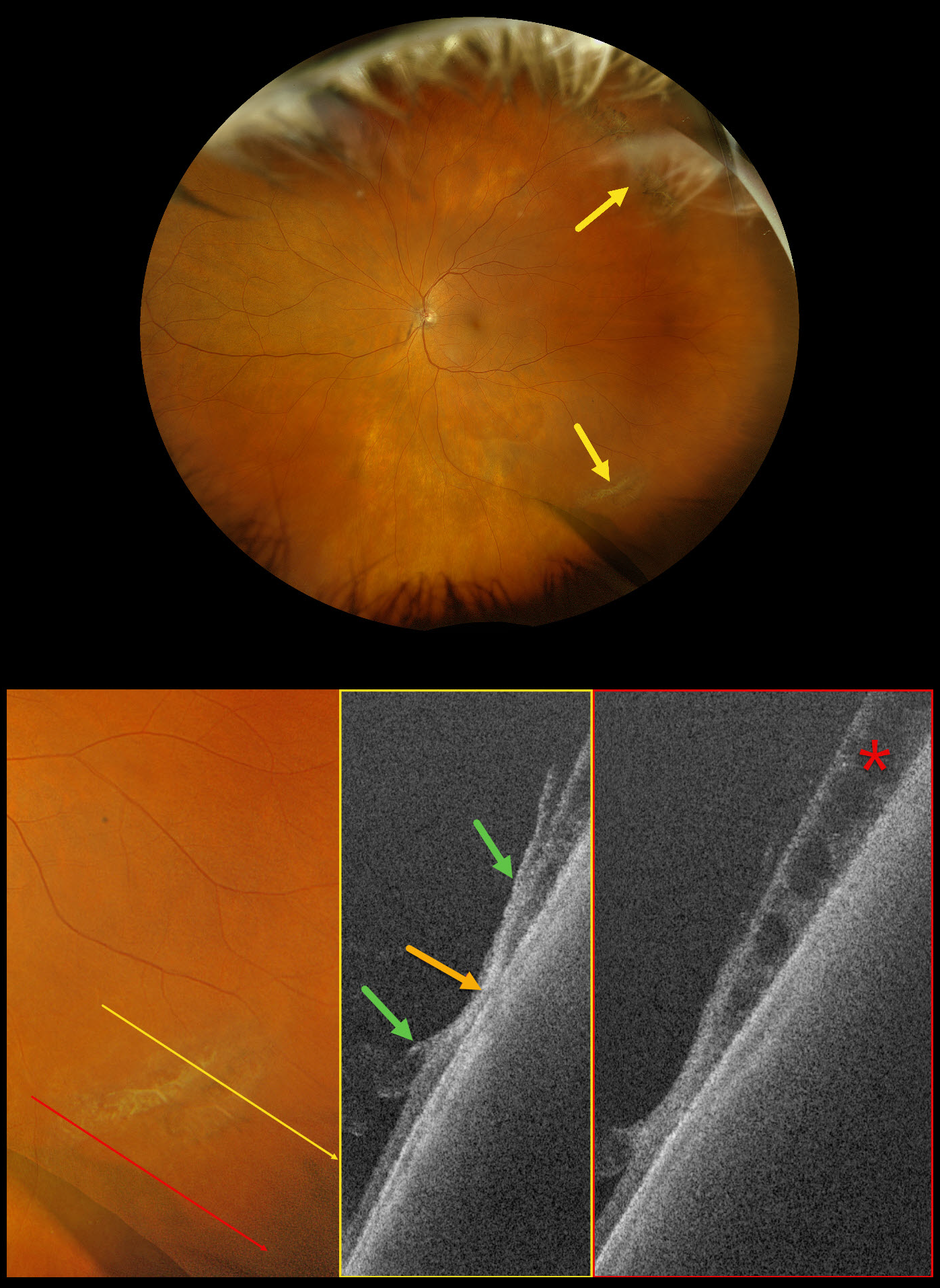
This 73-year-old female was referred for a symptomatic posterior vitreous detachment (PVD) with a retinal break in her left eye. Vision was 20/20.
Optos Silverstone RGB imaging shows supero- and inferotemporal lattice degeneration with some atrophic retinal holes. Swept-source OCT through the lattice shows a thinned, disorganized retina with areas of vitreoretinal adhesion along its margins. A more inferior line scan shows degenerative retinoschisis, which could not be visualized clinically or on the Optos image.
Our patient showed no retinal breaks from her symptomatic PVD, and observation was recommended.
Learning Points:
Ultrawidefield imaging has revolutionized our specialty, making many of us realize how limited our clinical examinations are compared with our imaging devices. In our patient’s case, the RGB image captured the fundus nearly from ora to ora. Swept-source OCT provided a near histologic view of the lattice pathology, including atrophic retina centrally, with a firm area of vitreoretinal adhesion along its margins. And finally, it revealed subclinical degenerative retinoschisis.
DEGENERATIVE RETINOSCHISIS + SITUS INVERSUS
Sayena Jabbehdari and Ahmed Sallam
Originally posted on @retina.rocks March 5, 2025
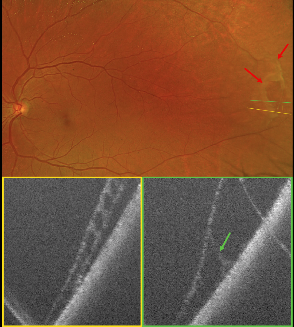
This 64YO female was referred for a possible asymptomatic rhegmatogenous retinal detachment (RRD) in her left eye. Vision was 20/20 in her normal OD and 20/20 OS.
Optos color RG imaging shows superotemporal degenerative retinoschisis with a few outer-layer holes (OLH). OCT confirms the schisis as well as the outer layer hole.
Learning Points:
Clinically, degenerative retinoschisis usually appears as a smooth, dome-shaped peripheral retinal elevation without the outer retinal corrugations classically seen in RRD (Oquendo et al, AJO 2024;268:212-221). Degenerative retinoschisis is virtually never prophylactically treated (Byer 1986;93:1127-1137). Although the schisis cavities can enlarge, prophylactic treatment, including thermal laser, does not prevent further spread and should not be performed (Ness et al, Surv Ophthalmology 2022;67:892-907).
Situs inversus of the optic disc was identified as a benign incidental finding in our patient. This occurs in 5% of normal eyes and 70% of eyes with tilted discs (Witmer et al, Surv Ophthalmology 2010;55:403-428). It can be mistaken for other congenital disc anomalies.
DEGENERATIVE RETINOSCHISIS
Vaibhav Sethi
Originally posted on @retina.rocks January 21, 2025
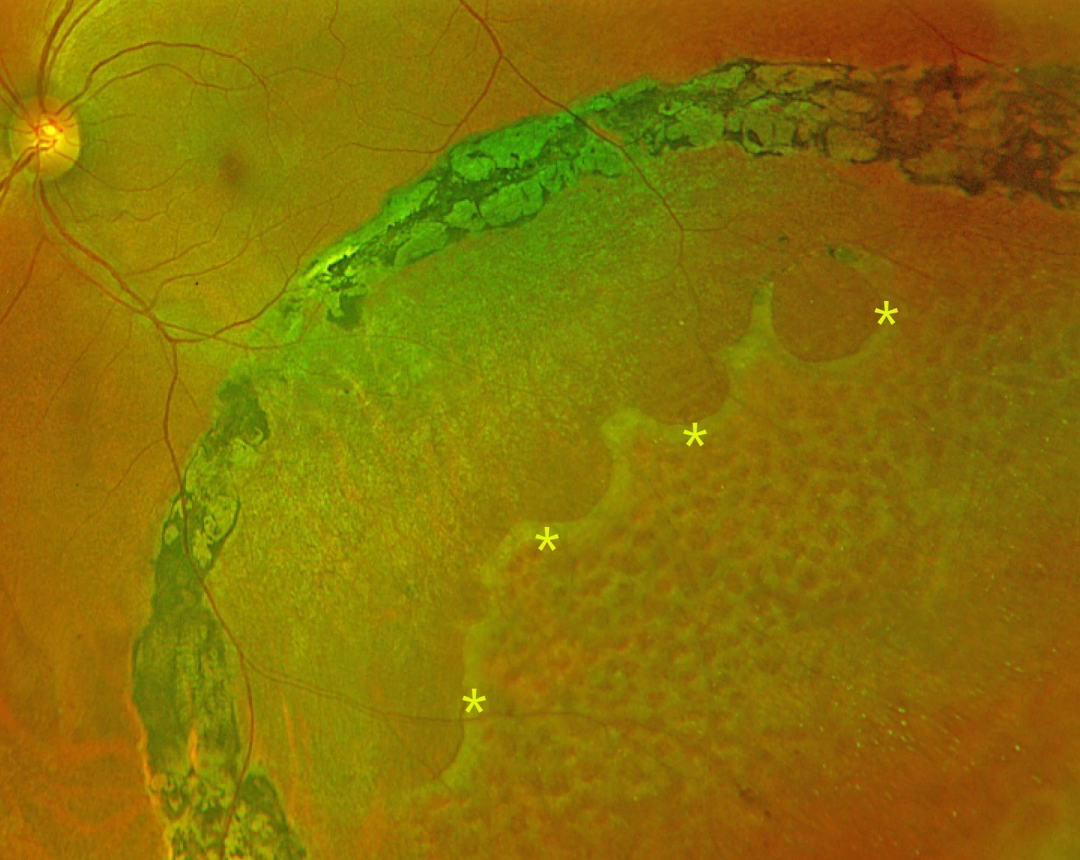
This 47YO female presented with 4 years of flashes in her left eye that began immediately following laser photocoagulation elsewhere. Vision was 20/20 bilaterally.
Optos color RG imaging shows inferotemporal degenerative retinoschisis, demarcated by heavy, confluent laser scarring along its posterior extent. A row of outer-layer holes is noted within the schisis cavity, with innumerable inner-layer holes more anteriorly.
Learning Points:
Degenerative retinoschisis is virtually never prophylactically treated (Byer 1986;93:1127-1137). Although the schisis cavities can enlarge, prophylactic treatment including thermal laser does not prevent further spread, should not be performed (Ness et al, Surv Ophthalmology 2022;67:892-907), and in our patient’s case, caused persistent symptomatic flashes. Vitrectomy surgery can successfully repair combined schisis rhegmatogenous retinal detachment (RRD) when liquid vitreous enters the subretinal space through holes in both the inner and outer layers.
DEGENERATIVE RETINOSCHISIS
Janelle Adeniran
Originally posted on @retina.rocks May 16, 2024
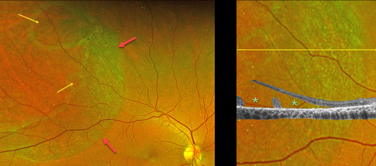
This 55YO male was referred for an asymptomatic possible retinal detachment in his right eye. Vision was 20/30.
Optos color RG imaging shows superotemporal degenerative retinoschisis with two outer-layer holes (OLHs). A broad band of pigmented chorioretinal scarring is noted along the posterior edge of the schisis.
OCT scanning through the smaller outer layer hole inferiorly shows the elevated edges of the OLH with the overlying inner schisis cavity. Despite the posterior pigment suggesting a demarcation line, no detachment of the outer layer was noted, and observation was recommended.
Learning Points:
A combined schisis-retinal detachment occurs when degenerative schisis contains both inner- and outer-layer breaks, essentially forming a full-thickness break that allows liquid vitreous to reach the subretinal space.
DEGENERATIVE RETINOSCHISIS
Originally posted on @retina.rocks April 4, 2024
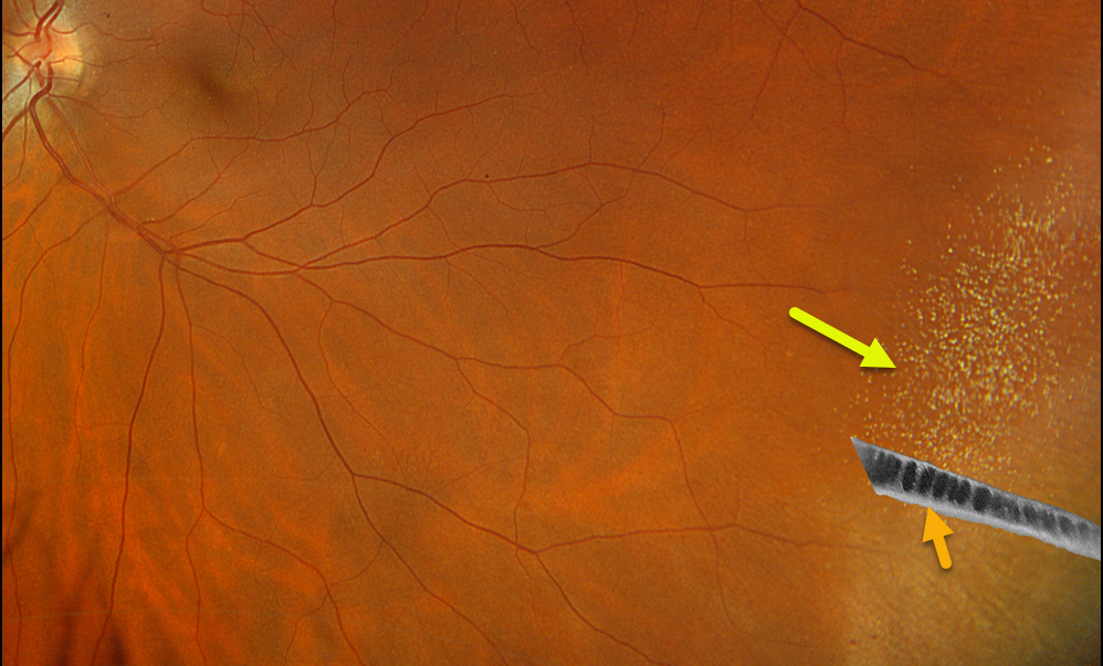
This 52YO female was referred for a possible retinal detachment in her left eye. There were no visual symptoms.
Optos color RGB imaging shows inferotemporal retinoschisis with numerous inner retinal refractile dots. Optos California swept-source OCT confirms schisis with splitting of the outer plexiform layer.
Learning Points:
Degenerative retinoschisis is an age-related splitting of the outer plexiform layer, most commonly located inferotemporally.
It is sometimes difficult to differentiate a schisis from a rhegmatogenous detachment, and ultrawidefield OCT imaging, as in this case, is an amazing diagnostic tool.
DEGENERATIVE RETINOSCHISIS
Originally posted on @retina.rocks February 14, 2024
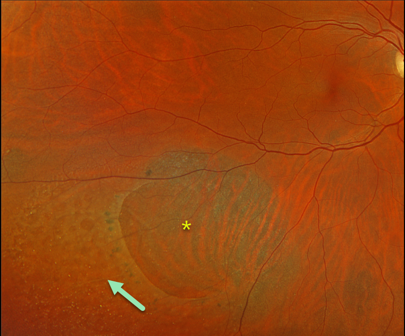
This 61YO female has been followed for several years for asymptomatic unilateral degenerative retinoschisis in her right eye. Vision is 20/30.
Optos color RGB imaging shows a large posterior outer layer hole with surrounding pigment. Innumerable small inner-layer holes are noted more peripherally within the inferotemporal schisis cavity.
Learning Points:
Degenerative retinoschisis is virtually never prophylactically treated (see Byer 1986;93:1127-1137). Patients can rarely develop a combined schisis rhegmatogenous retinal detachment (RRD) when liquid vitreous accesses the subretinal space through both an inner-layer and an outer-layer hole.
We will therefore continue to follow her twice yearly.
DEGENERATIVE RETINOSCHISIS
Originally posted on @retina.rocks January 24, 2024
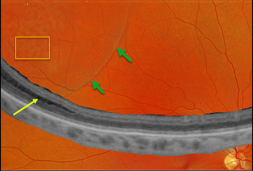
This 46YO female was referred for a possible retinal detachment. She had no visual complaints. Vision was 20/20 OD and 20/20 in her normal OS.
Optos color RGB imaging shows a bullous superotemporal retinoschisis. Numerous inner-layer holes are seen. Optos Silverstone ultrawidefield OCT shows splitting of the outer plexiform layer, confirming a schisis.
Learning Points:
Degenerative retinoschisis is an age-related splitting of the outer plexiform layer, most commonly located inferotemporally. It is sometimes difficult to differentiate a schisis from a rhegmatogenous detachment, and ultrawidefield OCT imaging, as in this case, is an amazing diagnostic tool.
Patients can rarely develop a combined schisis rhegmatogenous retinal detachment (RRD) when liquid vitreous accesses the subretinal space through both an inner-layer and an outer-layer hole. We will follow up with our patient yearly.
DEGENERATIVE RETINOSCHSIS
Originally posted on @retina.rocks June 27, 2023
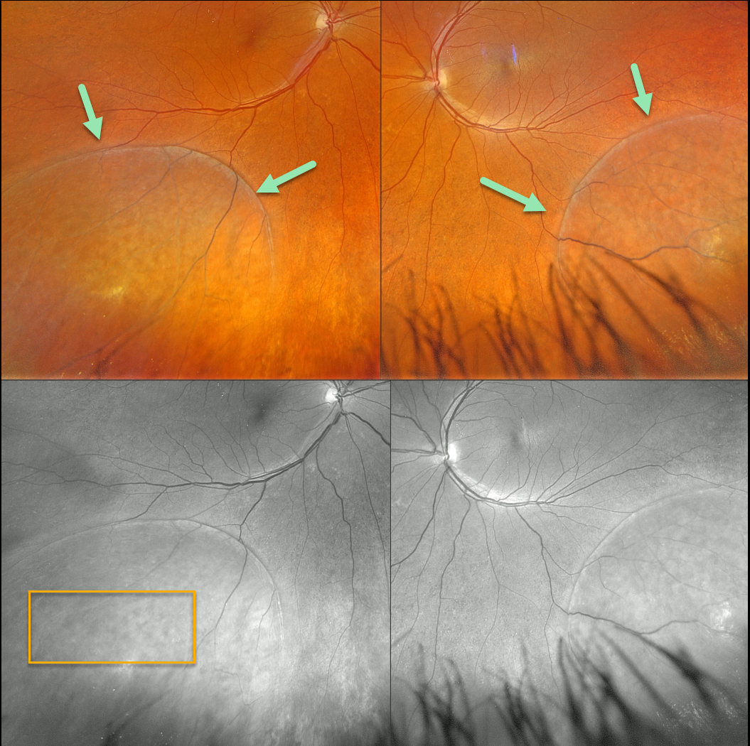
This 50YO female was referred for possible bilateral retinal detachments. Vision was 20/30 OD and 20/40 OS.
Optos color RGB imaging shows bullous, symmetrical inferotemporal retinoschisis. The innumerable inner-layer holes are best seen in the green channel.
Learning Points:
Degenerative retinoschisis is virtually never prophylactically treated (see Byer 1986;93:1127-1137). Patients can rarely develop a combined schisis rhegmatogenous retinal detachment (RRD) when liquid vitreous accesses the subretinal space through both inner and outer retinal holes.
Our patient is at low risk for developing a combined schisis RRD due to the lack of outer-layer breaks. We will follow her yearly.
DEGENERATIVE RETINOSCHISIS
Originally posted on @retina.rocks October 19, 2022
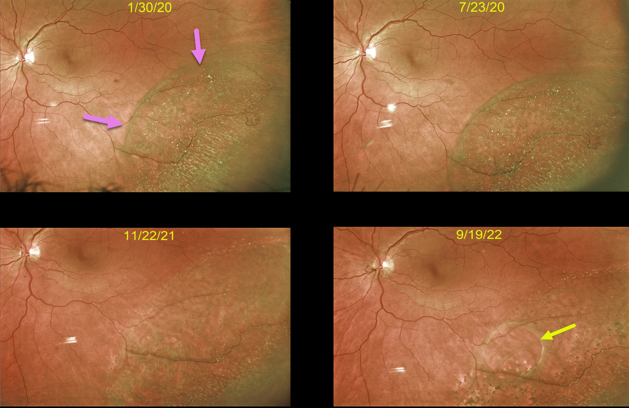
This 59YO female was initially seen with asymptomatic retinoschisis in her left eye. When examined six months later, the schisis appeared stable.
16 months later, the schisis cavity was slightly enlarged in all dimensions with the formation of an early outer layer hole. Most recently, the outer layer hole enlarged, and the schisis spontaneously mostly collapsed, essentially resolving itself. Continued observation was recommended.
Learning Points:
Degenerative schisis cavities can enlarge, and in these cases, prophylactic treatment including thermal laser does not prevent further spread and is therefore not recommended (Ness et al, Surv Ophthalmology 2022;67;892-907).
Schisis very rarely progresses into the fovea (Byer Ophthalmology 1986;93:1127-1137) and even then is compatible with preserved central vision as long as the outer layer remains attached (Watzke et al 2013;33:606-612).
Our patient shows how the life cycle of a schisis cavity is often dynamic, including enlargement, development of inner or outer layer breaks, and resolution.
DEGENERATIVE RETINOSCHISIS
Originally posted on @retina.rocks August 9, 2022
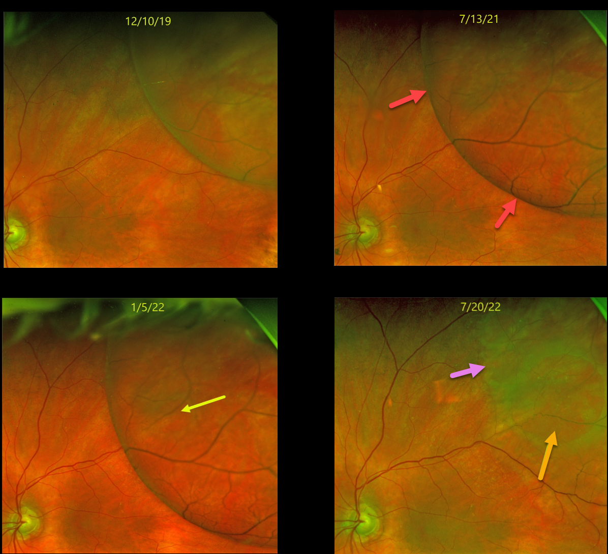
This 80YOF was initially seen with asymptomatic retinoschisis in her left eye. When examined 1.5 years later on 7/13/21, she remained asymptomatic, but the schisis cavity had enlarged in all dimensions.
Another six months later (1/5/22), the schisis cavity continued to enlarge in all dimensions with the formation of an early outer layer hole. Most recently (7/20/22), the outer-layer hole enlarged, and the schisis spontaneously collapsed, essentially resolving itself. Continued observation was recommended.
Learning Points:
Schisis cavities can enlarge, and in these cases, prophylactic treatment, including thermal laser, does not prevent further spread. Schisis very rarely progresses into the fovea (Byer Ophthalmology 1986;93:1127-1137) and even then is compatible with preserved central vision as long as the outer layer remains attached (Watzke et al 2013;33:606-612).
BRVO WITH DEGENERATIVE RETINOSCHISIS
Barbara Parolini and Veronika Matello
Originally posted on @retina.rocks June 13, 2022
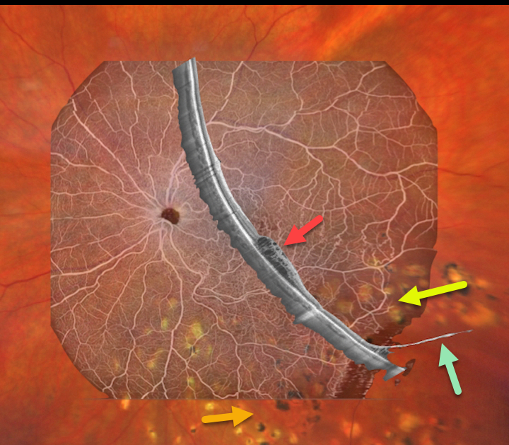
This 67YO female has a history of branch retinal vein occlusion (BRVO) in her left eye. Asymptomatic degenerative retinoschisis was also present bilaterally. Vision was 20/33. She had received prior scatter laser and intravitreal injections about 3 years earlier.
Fortunately, all findings remained stable despite loss to follow-up due to the COVID pandemic.
Clarus 500 wide field imaging shows mild inferotemporal tortuosity with collateral vessels within the involved quadrant. Prior scatter laser scarring for secondary retinal neovascularization, along with prior barrier laser around the posterior edge of the schisis cavity, is noted.
Canon Xephilio S1 OCT angiography highlights the collateral vessels and more peripheral retinal nonperfusion. Xephilio B-scan shows some inferotemporal macular edema, and just catches the more distal inferotemporal schisis.
DEGENERATIVE RETINOSCHISIS
Originally posted on @retina.rocks October 27, 2021
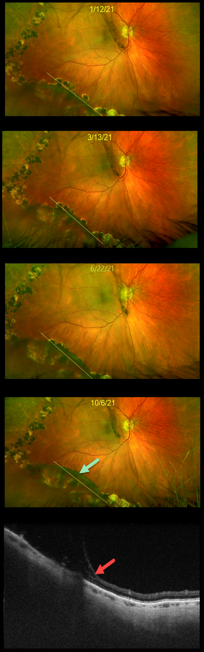
This 71YO female was treated elsewhere with barrier laser around degenerative schisis in her right eye. Over the past 9 months, the inferotemporal schisis cavity has gradually enlarged. The yellow line indicates the original location of schisis. At the most recent visit on 10/6/21, the schisis cavity extended through the barrier laser.
Triton swept-source OCT scanning through this area confirmed pure schisis with no retinal detachment. She remains completely asymptomatic, and we continue to recommend observation.
Learning Points:
This patient is at virtually no risk of a combined schisis-rhegmatogenous detachment due to the absence of inner and outer layer breaks.
Schisis cavities can enlarge, and in these cases, prophylactic treatment, including thermal laser, does not prevent further spread.
Schisis very rarely progresses into the fovea (Byer Ophthalmology 1986;93:1127-1137) and even then is compatible with preserved central vision as long as the outer layer remains attached (Watzke et al 2013;33:606-612).
DEGENERATIVE RETINOSCHISIS
Originally posted on @retina.rocks July 27, 2021
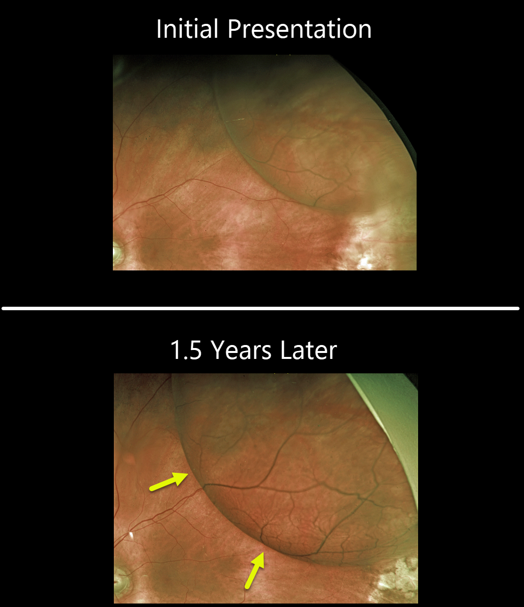
This 80YOF was initially seen with asymptomatic retinoschisis in her left eye. When examined approximately a year and a half later, she remained asymptomatic, but the schisis cavity had enlarged in all dimensions.
Observation was recommended. This patient is at virtually no risk of a combined schisis-rhegmatogenous detachment due to the absence of breaks in the inner and outer layers.
Learning Points:
Retinochisis cavities can enlarge. In these cases, prophylactic treatment, including thermal laser, does not prevent the schisis from enlarging. Schisis very rarely progresses into the fovea (Byer Ophthalmology 1986;93:1127-1137) and even then is compatible with preserved central vision as long as the outer layer remains attached (Watzke et al 2013;33:606-612).
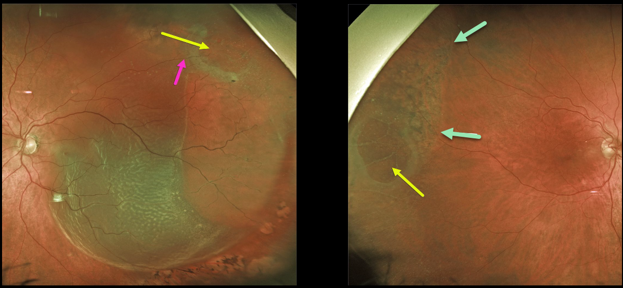
DEGENERATIVE RETINOSCHISIS
Originally posted on @retina.rocks May 27, 2021
This 72YO male presented with an acute macula-off retinal detachment (RD) in his left eye.
Optos imaging shows that this is not a typical rhegmatogenous RD since an outer layer hole (OLH) is noted superotemporally. A retinal vessel can be traced over the OLH, indicating that this is not a full-thickness defect.
The patient also has degenerative retinoschisis with an OLH in his right eye. A shallow subclinical schisis detachment is likely present due to the pigmentary changes throughout the schisis cavity.
Prophylactic treatment is not recommended for this eye, but we are carefully following him, as he does have a small risk of developing an RD here as well.
Learning Points:
This detachment in the left eye does not have a typical full-thickness break, but is caused by degenerative retinoschisis.
Numerous tiny inner-layer holes, along with the visible large OLH, essentially created a full-thickness retinal defect, giving the liquid vitreous access to the subretinal space. The detachment was successfully repaired with vitrectomy surgery.
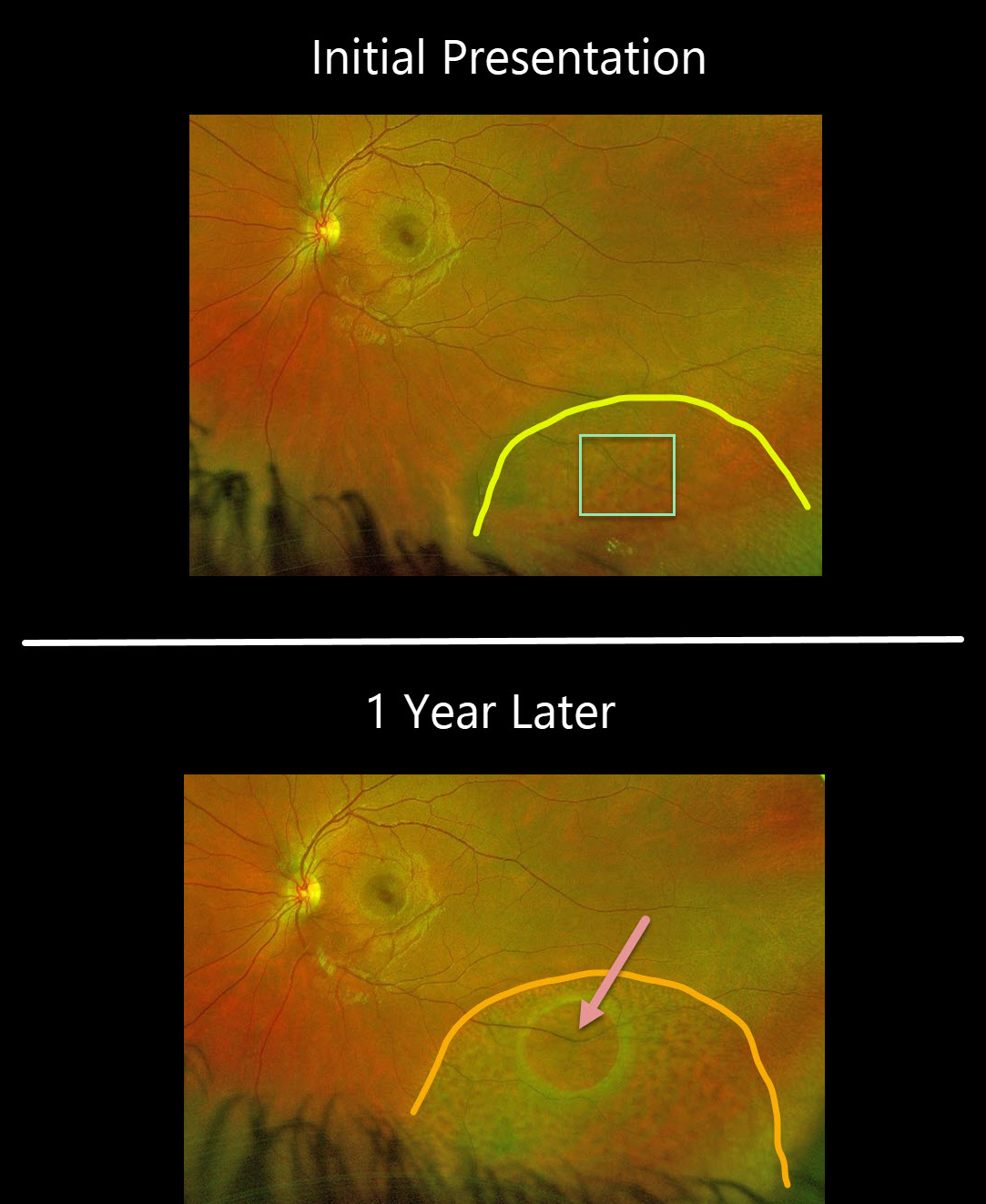
DEGENERATIVE RETINOSCHISIS
Originally posted on @retina.rocks December 25, 2020
This 22YO patient, courtesy of Dr. Brooke Spivey, presented with degenerative retinoschisis with numerous inner-layer holes.
One year later, the schisis cavity had enlarged, with a new posterior outer layer hole. There were no visual symptoms.
Learning Points:
This patient is at a low risk of developing a combined rhegmatogenous schisis detachment due to the presence of both inner and outer layer breaks.
The outer layer remains attached, and no prophylactic treatment was recommended since thermal laser does not prevent schisis from enlarging, schisis very rarely progresses into the fovea (Byer Ophthalmology 1986;93:1127-1137), and even then is compatible with central vision as long as the outer layer remains attached (Watzke et al 2013;33:606-612), and asymptomatic holes are generally not treated.
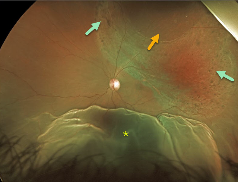
COMBINED RETINOSCHISIS-RHEGMATOGENOUS RETINAL DETACHMENT
Originally posted on @retina.rocks December 7, 2020
This 33YO patient presented with a prominent inferior retinal detachment associated with superotemporal retinoschisis with a large outer layer hole (OLH). The OLH was surrounded by laser-like chorioretinal scarring, although the patient denied prior laser surgery.
At vitrectomy, a superonasal gutter of peripheral detachment extended to an equatorial tear at 10:30 (not visualized in these images). The OLH and schisis were not detached.
Learning Points:
Patients with degenerative retinoschisis and associated inner- and outer-layer holes are at risk of rhegmatogenous retinal detachment.
Despite our patient probably undergoing prior prophylactic laser photocoagulation around the outer layer hole, he still developed a retinal detachment from a presumed new retinal tear outside the area of schisis.
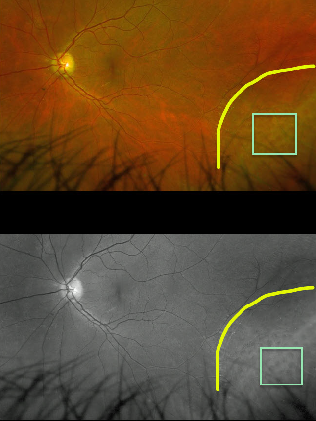
DEGENERATIVE RETINOSCHISIS
Originally posted on @retina.rocks September 4, 2020
This patient has degenerative retinoschisis with numerous inner-layer holes, best seen on the Optos green channel image.
Learning Points:
This patient just needs observation due to the small risk of developing a rhegmatogenous detachment.
This risk would increase if an outer-layer hole were present, since the inner- and outer-layer holes would essentially represent a full-thickness break, thereby giving the liquid vitreous access to the subretinal space.
DEGENERATIVE RETINOSCHISIS
Originally posted on @retina.rocks March 11, 2020
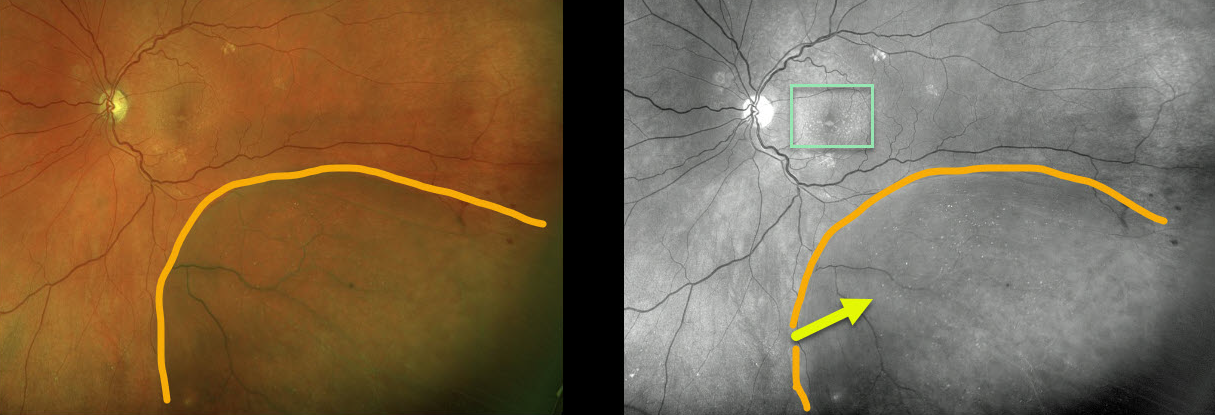
This patient with degenerative retinoschisis has extensive refractile dots, thought to be Muller cell footplates, which are well-visualized in both the Optos pseudocolor and green channel images.
This diabetic patient also has subretinal drusenoid deposits that are best seen with the green channel.
Learning Points:
Degenerative retinoschisis is virtually never prophylactically treated (see Byer 1986;93;1127-1137).
Patients can rarely develop a rhegmatogenous retinal detachment when liquid vitreous accesses the subretinal space thru both an inner and outer layer hole.
DEGENERATIVE RETINOSCHISIS
Originally posted on @retina.rocks January 23, 2020
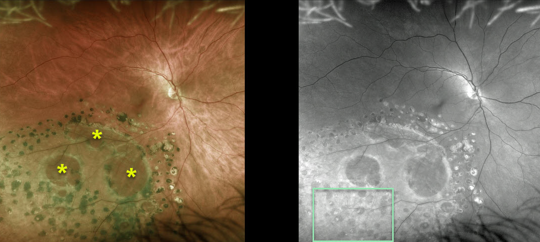
This patient has degenerative retinoschisis. A doctor at an outside practice placed prophylactic barrier laser around the outer layer holes to prevent retinal detachment.
There are also extensive smaller inner layer holes that are best seen in the Optos green-channel image.
Learning Points:
Degenerative retinoschisis is virtually never prophylactically treated (see Byer 1986;93;1127-1137). Patients can rarely develop a rhegmatogenous retinal detachment when liquid vitreous accesses the subretinal space thru both an inner and outer layer hole.


