

Rhegmatogenous Retinal Detachment (RRD) Giant Retinal Tear
<< Back to Cases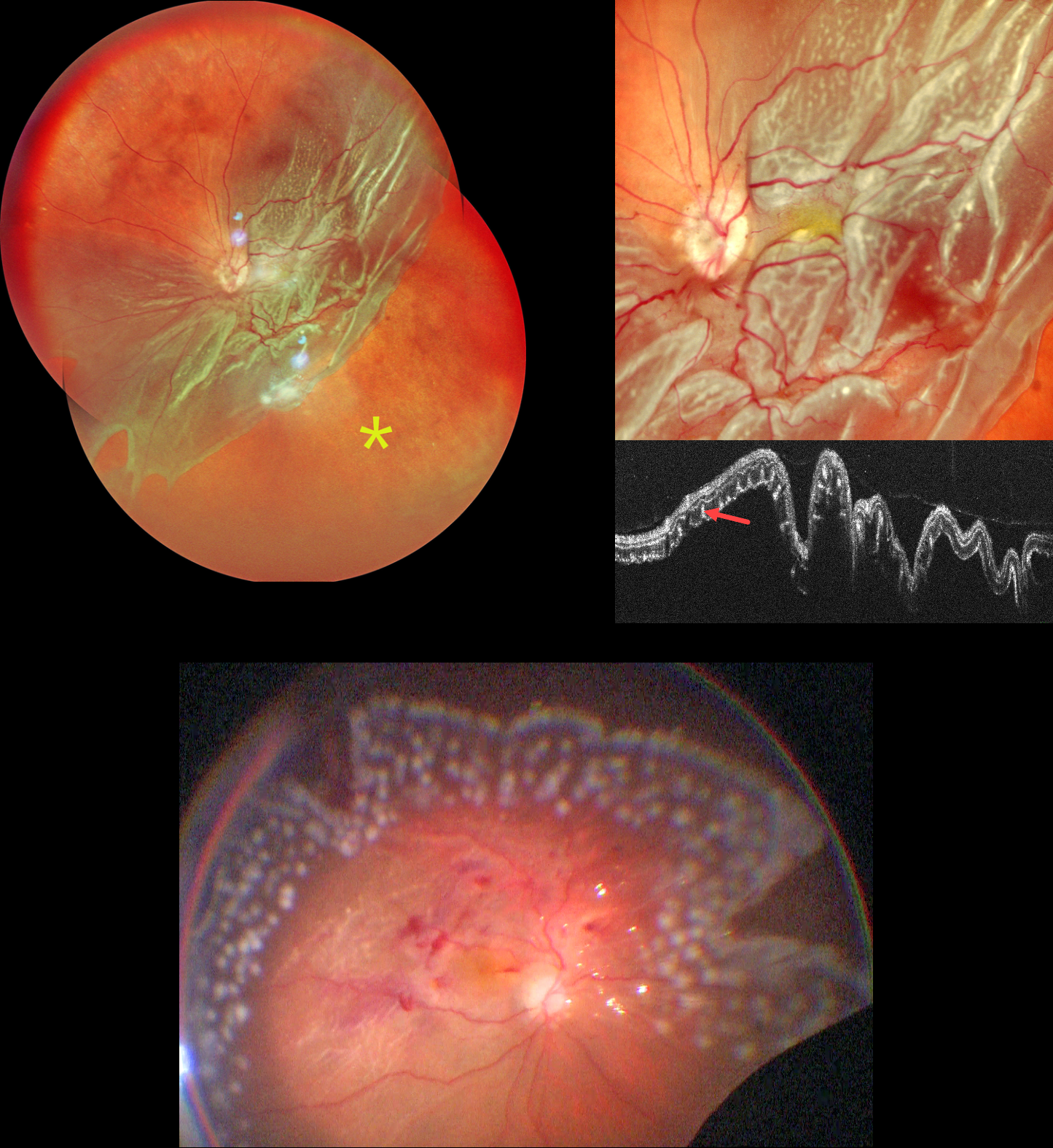
GIANT RETINAL TEAR
Shraddha Raj Shrivastava and Manish Nagpal
Originally posted on @retina.rocks September 1, 2026
This 62YO male presented with 3 months of vision loss in his left eye. Vision was counting fingers.
Pseudocolor SLO imaging shows a giant retinal tear extending from the 2 to 8 o’clock meridians with bare RPE distal to the posterior retina. There are extensive outer retinal corrugations within the posteriorly detached retina. OCT dramatically shows the detached retina, which is redundantly folded onto itself. The outer retinal corrugations appear as numerous hyperreflective outer retinal pillars.
The detachment was emergently repaired with vitrectomy, membrane peeling, endolaser photocoagulation, and silicone oil. The retina was completely attached at the end of the procedure.
Learning Points:
Outer retinal corrugations, also known as hydration outer retinal folds, are one of the key findings that help differentiate a rhegmatogenous retinal detachment from exudative and traction retinal detachments. The cause of these folds remains uncertain but may result from the hydration of glycosaminoglycans in the interphotoreceptor matrix (Dalvin et al, Retinal Cases & Brief Reports 2020;14:355-359).
GIANT RETINAL TEAR
Originally posted on @retina.rocks May 27, 2026
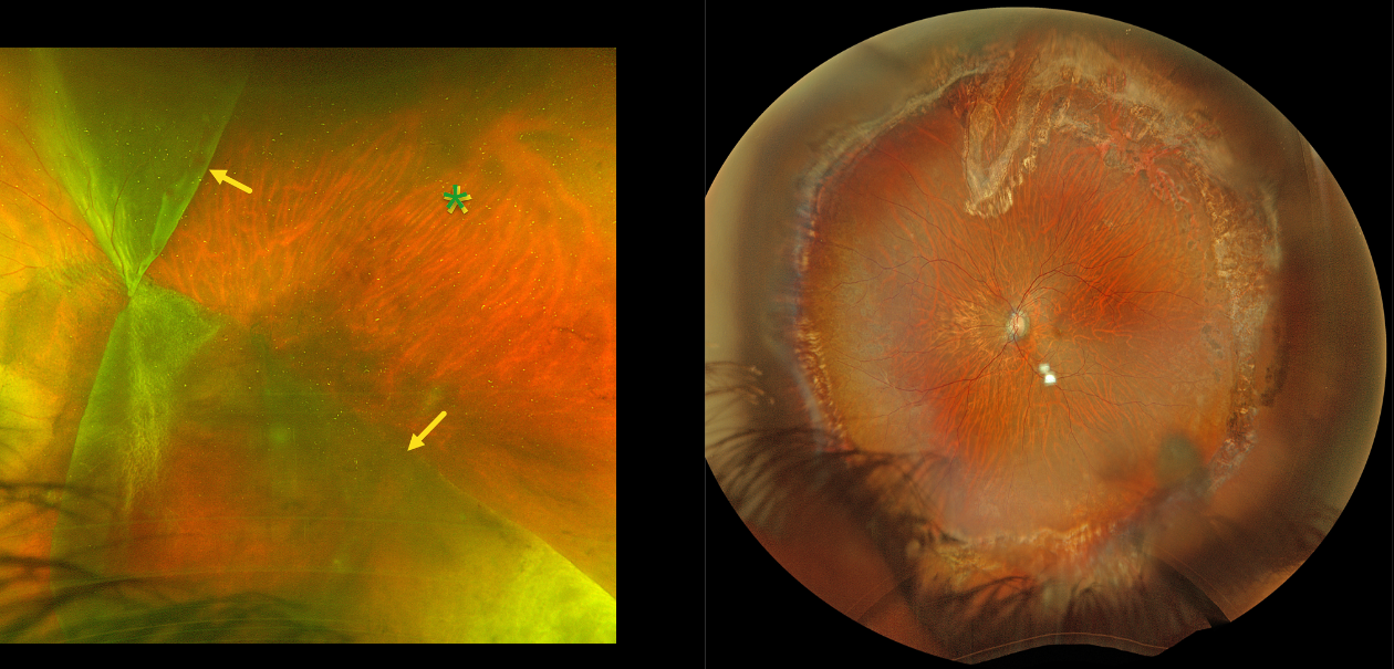
This 44YO male presented with hand motion vision OS from an acute giant tear (GRT) retinal detachment. Optos color RG imaging shows a complex GRT with one flap folded superonasally and the other inferotemporally, creating a sharply defined square region of temporal bare RPE. The optic disc is covered by the giant tear. Emergent vitrectomy was scheduled.
Six months postoperatively, the retina remained completely attached. Vision was 20/80.
GIANT RETINAL TEAR
The European VitreoRetinal Society (EVRS), Shilpi Narnaware and Prashant Bawankule
Originally posted on @retina.rocks April 10, 2026
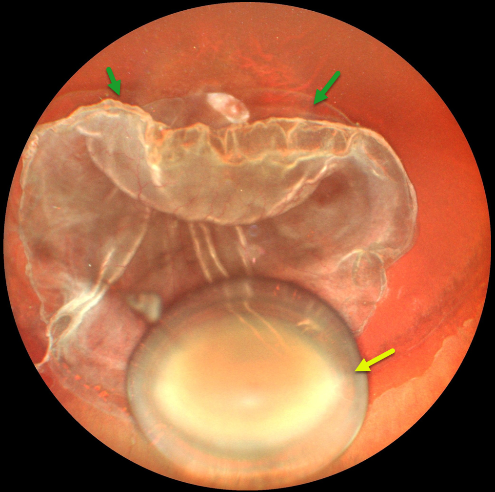
This 32YO male with known Marfan’s syndrome presented with one month of vision loss in his left eye. Examination revealed pathological myopia with bilateral spontaneously dislocated cataracts and a total rhegmatogenous retinal detachment (RRD) OS. Best-corrected vision was 20/80 OD and light perception OS.
Ultrawidefield photography shows a superior >270-degree giant retinal tear, total RRD, rolled edges, and a dislocated cataractous crystalline lens in the inferior vitreous. Emergent pars plana vitrectomy/lensectomy was planned, but unfortunately, he did not show for his surgery and was subsequently lost to follow-up.
GIANT RETINAL TEAR
The European VitreoRetinal Society (EVRS), Akansha Sharma, Manish Nagpal, Vaidehi Sathaye, and Navneet Mehrotra
Originally posted on @retina.rocks March 27, 2026
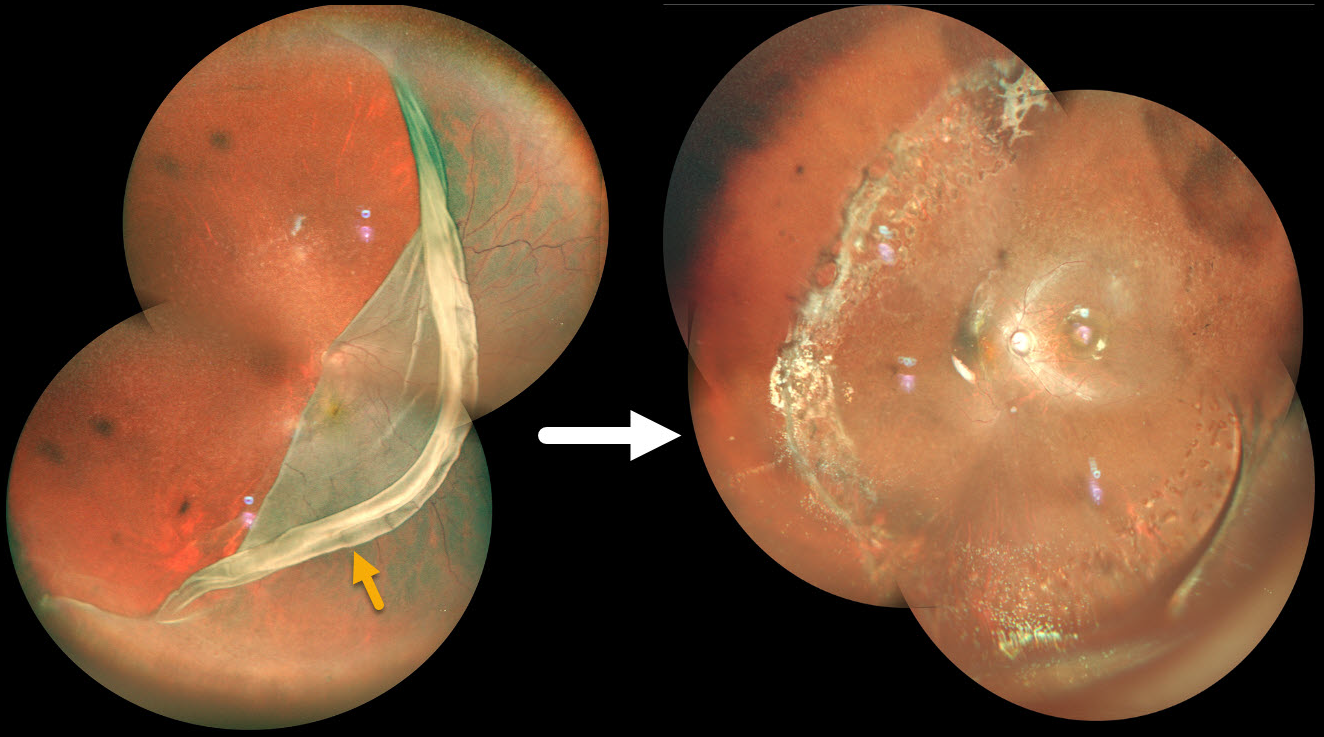
This 15YO male presented with sudden vision loss in his right eye. Vision was hand motion. Pseudocolor SLO imaging shows a giant retinal tear extending from 7:30 to 12 o’clock, with the macular retina folded onto itself nasal to the nerve. Emergent pars plana vitrectomy with silicone oil, followed by removal of the oil 7 months later, successfully repaired the detachment with vision improving to 20/200.
GIANT RETINAL TEAR
Malvika Singh and Manish Nagpal
Originally posted on @retina.rocks August 27, 2025
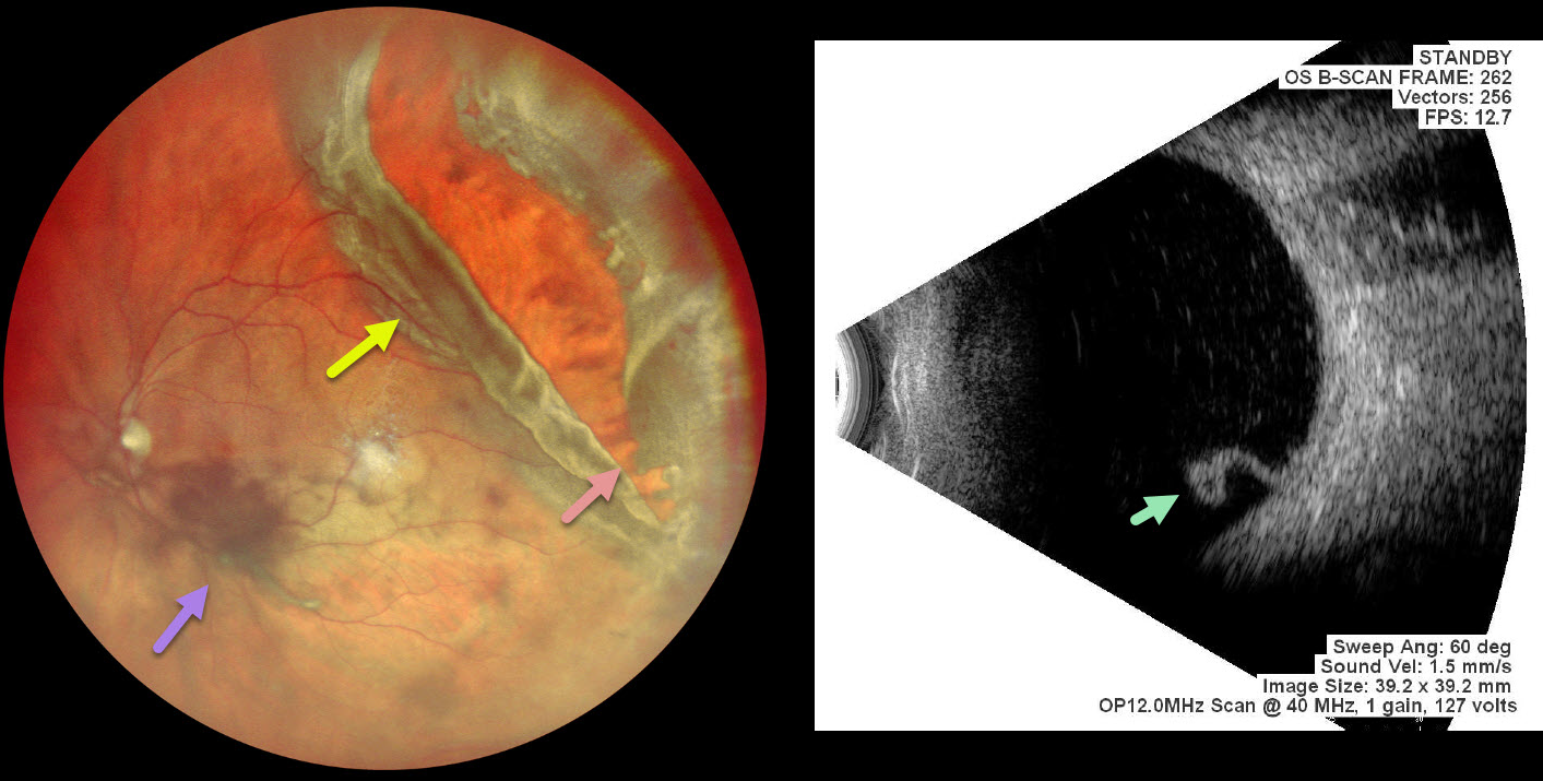
This 56 YO male presented with 4 days of vision loss in his left eye. He gave a history of multiple prior retinal reattachment surgeries in his right eye. Vision was 20/400 in his OD and 20/20 in OS.
Pseudocolor SLO imaging shows a superotemporal rhegmatogenous retinal detachment (RRD) from a giant retinal tear (extends over 3 clock hours) with mild vitreous hemorrhage. The anterior portion of the tear has multiple jagged edges, and the posterior flap has folded over onto itself. The macula is attached clinically and on B-scan ultrasonography, which shows the mobile posterior flap folded over onto itself. Emergency vitrectomy was recommended, but unfortunately, he was immediately lost to follow-up.
Learning Points:
A vitreous hemorrhage will often prevent visualization of an acute RRD. Ultrasonography is often utilized to confirm the presence of a detachment. In our patient, the B-scan also beautifully imaged the mobile posterior flap of the giant tear. This pathognomonic appearance would alter the surgical planning in the case of opaque media.
GIANT RETINAL TEAR
Seif Anwar
Originally posted on @retina.rocks January 30, 2025
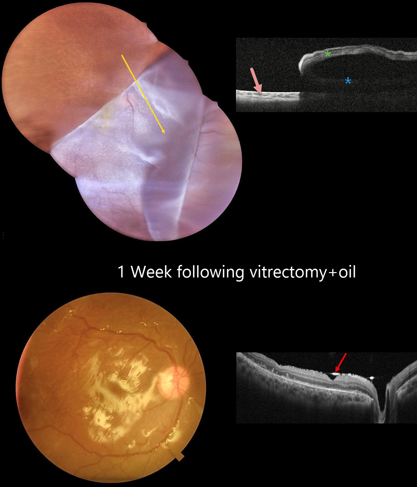
This 12YO boy presented with 3 weeks of vision loss in his right eye immediately following blunt trauma. Vision was hand motion OD and 20/20 in his normal OS.
Color photography shows a superotemporal giant retinal tear that has folded over the optic nerve and macula inferonasally. The yellow macular xanthophyll is visible just inferotemporal to the nerve. OCT scanning shows bare RPE superotemporally, the underlying detachment, and the overlying inverted retina.
Emergent surgical repair with pars plana vitrectomy was scheduled. One week postoperatively, the retina is completely attached. The hyperreflective oil-retinal interface is visible overlying the fovea and optic nerve.
GIANT RETINAL TEAR
Anjana Mirajkar and Manish Nagpal
Originally posted on @retina.rocks November 26, 2024
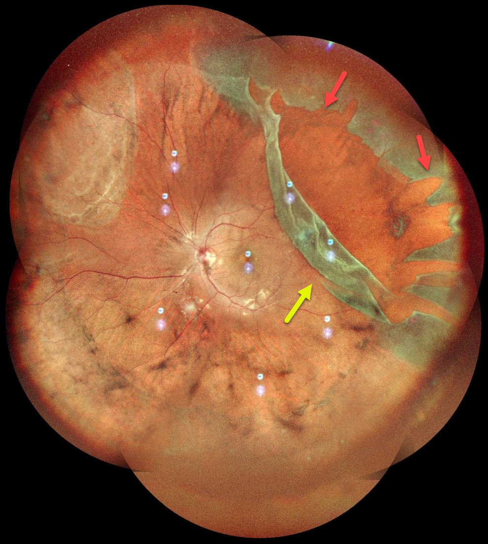
This healthy 24YO male presented with several days of floaters in his left eye. Vision was 20/20 OU.
Pseudocolor SLO imaging shows a superotemporal giant retinal tear (extending over 3 clock hours) and detachment. The anterior portion of the tear has multiple jagged edges, and the posterior flap has folded over onto itself. The macula is attached. The detachment was repaired with emergent pars plana vitrectomy.
GIANT RETINAL TEAR
Originally posted on @retina.rocks September 9, 2022
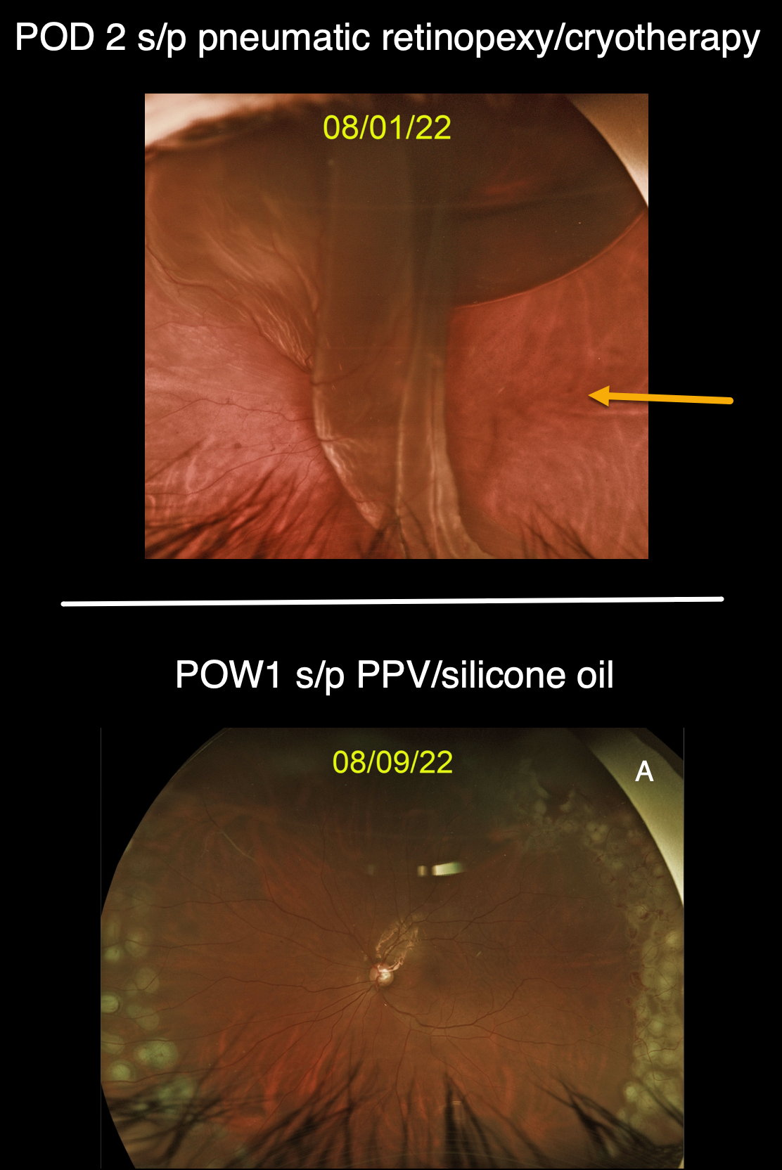
This 50YO male presented with a superior macula-off retinal detachment from a single retinal tear at the 1 o’clock periphery (image not shown), which was treated with a pneumatic retinopexy. Cryotherapy was applied before injecting the gas bubble, confirming this single break.
However, when he returned 2 days later, a new giant retinal tear extended from 12 to 5 o’clock, with subretinal gas migrating beneath the superior retina.
The detachment was repaired with vitrectomy and silicone oil, and the second image shows the 1-week postoperative visit.
GIANT RETINAL TEAR
Originally posted on @retina.rocks March 9, 2022
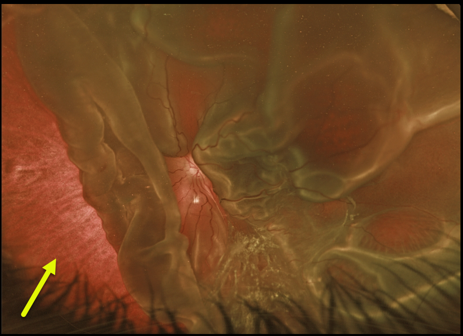
This 6YO boy was found to have this asymptomatic chronic total rhegmatogenous retinal detachment (RRD) in his left eye during a routine eye examination.
There is a large, giant tear nasally from about 6 to 11 o’clock, with an additional giant tear temporally (not shown). There were numerous areas of lattice degeneration with atrophic holes in his right eye (not shown). There was a strong family history of RRD.
He was referred for surgical repair of the left detachment and prophylactic laser in his right eye.
GIANT RETINAL TEAR
Originally posted on @retina.rocks January 27, 2022
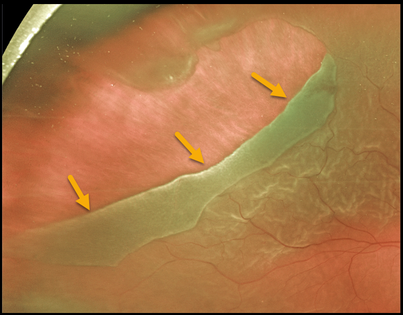
This 57YO male presented with 20/400 vision from a macula-off retinal detachment. The causative giant retinal tear extended from 9 to 12 o’clock.
He underwent successful pars plana vitrectomy surgery using intraoperative perfluorooctane (PFO) liquid to flatten the retina, followed by gas exchange with long-acting C3F8 (perfluoropropane) gas tamponade.
Learning Points:
By definition, giant retinal tears extend at least 3 clock hours. Surgical repair can be greatly facilitated by using Perfluoron (PFO), a heavier-than-water liquid that helps unroll the giant tear and prevents posterior retinal slippage.
GIANT RETINAL TEAR
Originally posted on @retina.rocks December 24, 2021
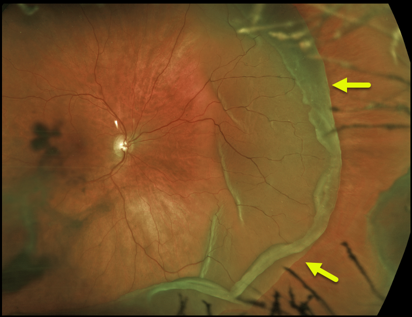
This 54YO female presented with a giant retinal tear with macula-on rhegmatogenous retinal detachment.
At vitrectomy several hours later, the tear was found to extend for 190 degrees.
Learning Points:
By definition, giant retinal tears extend at least 3 clock hours. Surgical repair can be greatly facilitated by using Perfluoron (PFO), a heavier-than-water liquid that helps unroll the giant tear and prevents posterior retinal slippage.
GIANT RETINAL TEAR
Originally posted on @retina.rocks March 1, 2021
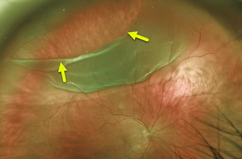
Here is a preoperative Optos photo of a giant retinal tear extending from about 9:30 to 1 o’clock.
This patient underwent successful pars plana vitrectomy surgery with C3F8 (perflouropropane) gas tamponade.
Learning Points:
By definition, giant retinal tears extend at least 3 clock hours.
Surgical repair is greatly facilitated by the use of Perfluoron liquid (PFO, perfluoron octane), which is heavier than water and, intraoperatively, helps unroll the posterior retina and prevent posterior retinal slippage.
The PFO can either be removed at the initial repair or left in place for a week or two before being removed with a secondary procedure.
RHEGMATOGENOUS RETINAL DETACHMENT
Originally posted on @retina.rocks January 25, 2021
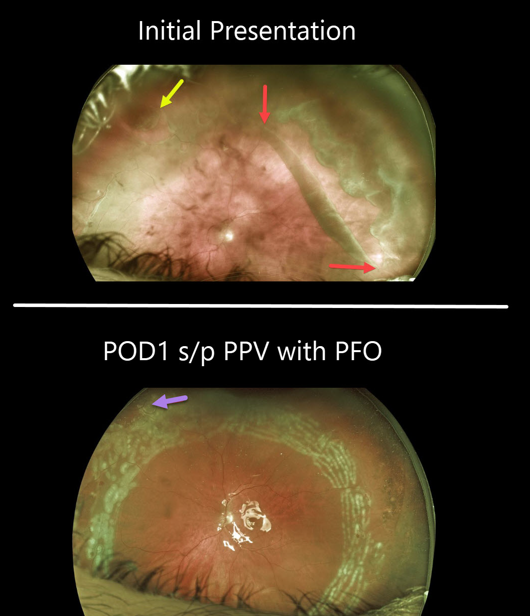
This patient presented with a symptomatic macula-on giant-tear retinal detachment (RD) in his left eye. The giant tear extends from about 12 to 4 o’clock with an additional tear noted superonasally.
One day after vitrectomy with Perfluoron (PFO, perfluoro octane) injection, the retina is completely attached, with fresh white encircling laser scarring. Some PFO bubbles can be seen superonasally.
The PFO liquid was removed two weeks later, and his retina has remained attached.
Learning Points:
By definition, giant retinal tears extend at least 3 clock hours. Surgical repair is greatly facilitated by using PFO liquids, which are heavier than water and help unroll the posterior retina and prevent posterior slippage of the detachment. The PFO can either be removed at the initial repair or left in place for a week or two before being removed with a secondary procedure.
RHEGMATOGENOUS RETINAL DETACHMENT
Originally posted on @retina.rocks June 22, 2020
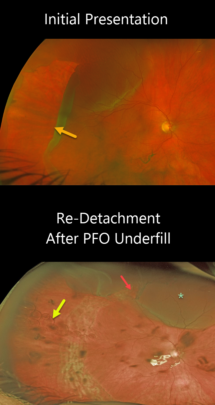
This patient presented with a retinal detachment caused by a giant retinal tear extending from 5 to 11 o’clock.
Unfortunately, the retina re-detached since the eye was inadvertently underfilled with perfluorocarbon (PFO) at the time of vitrectomy. The superior edge of the giant tear is open, resulting in a superior detachment. There are numerous small PFO bubbles floating along the interface between the PFO meniscus below and the liquid vitreous above.
The patient underwent subsequent surgery to remove the Perfluoron and successfully repair the superior detachment.
Learning Points:
PFO is a transparent, heavier-than-water liquid, often used during vitrectomy for these types of detachments. The PFO liquid is injected over the posterior pole, and as it fills the eye, it simultaneously unrolls the giant tear and flattens the retina. Traditionally, the PFO is exchanged for air, then for a long-acting gas. However, this often results in posterior retinal slippage.
Although PFO is not meant as a long-term vitreous substitute, it can be left in the eye for a week or two. Postoperatively, the patient sleeps on their back, allowing the PFO to tamponade the retina while the retinopexy begins to scar. Patients are then taken back to the operating room, where the PFO can be removed and replaced with a gas or silicone oil, without concern about the retina slipping intraoperatively.
GIANT RETINAL TEAR
Originally posted on @retina.rocks March 19, 2020
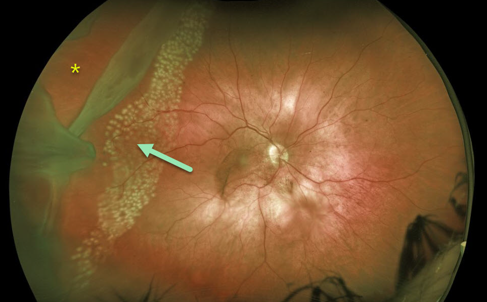
This patient presented with a giant retinal tear. Against medical advice, the patient adamantly refused vitrectomy on multiple occasions, so we decided to demarcate it with laser photocoagulation. As expected, the detachment progressed through the laser scarring with the development of proliferative vitreoretinopathy. He subsequently needed several vitrectomy procedures, including retinectomy and silicone oil to obtain final retinal reattachment.
Learning Points:
Untreated retinal tears have a very high risk for progressing to retinal detachment. Urgent laser photocoagulation or cryotherapy prevents liquid vitreous from leaking thru the break. Patients need to be followed closely postoperatively to be sure no new breaks develop and that chorioretinal scarring surrounds the break.


