

Rhegmatogenous Retinal Detachment (RRD) Loculated Fluid
<< Back to Cases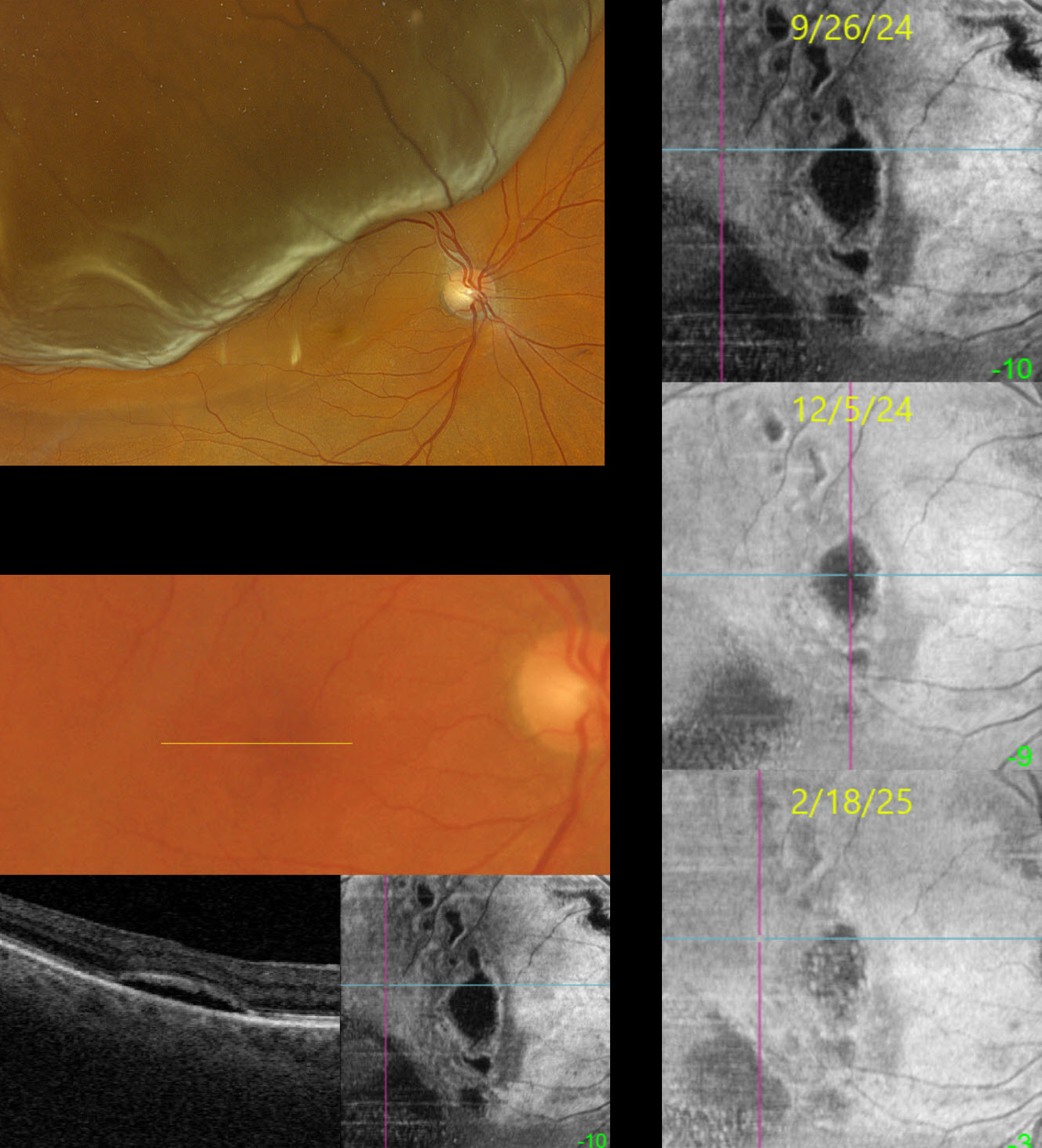
LOCULATED FLUID AFTER SUCCESSFUL PNEUMATIC RETINOPEXY
Originally posted on @retina.rocks October 21, 2025
This 69YO female presented on 4/17/24 with a superior macula-off rhegmatogenous retinal detachment (RRD) and counting fingers vision. Following successful pneumatic retinopexy, the retina reattached, and vision returned to 20/25 one month later.
When examined on 9/26/24, she was subjectively doing well with 20/50 vision. Although the macula looked clinically normal, Triton swept-source OCT B-scan and en face imaging show multifocal pockets of loculated subretinal fluid (SRF). With observation, these lesions continue to shrink and flatten. When last examined on 2/18/25, vision was 20/30, and continued observation was recommended.
Learning Points:
Loculated fluid represents an area of persistent subclinical SRF in the presence of a clinically reattached retina and is seen postoperatively in up to 15% of RRD following vitrectomy (Retina 2020;40:1153-1159) and eventually resolves after many months of observation. In a post hoc analysis of the PIVOT trial, there were no differences in the incidence of loculated fluid between vitrectomy and pneumatic retinopexy (Bansal et al BJO 2023;107:1693-1697). Although subfoveal loculated fluid was associated with reduced visual acuity at 3 months, vision eventually was similar to that of those without fluid at 1 year.
Bansal et al recently described the en face findings for loculated fluid (Ophthalmology Retina 2023;7:496-502). We continue to find that en face imaging often provides a unique and compelling perspective on a variety of pathologies, and we hope other clinicians will add it to their daily clinical routine.
LOCULATED FLUID AFTER RETINAL DETACHMENT REPAIR
Mattie Adams
Originally posted on @retina.rocks July 26, 2023
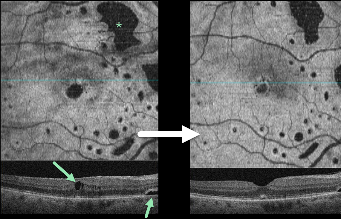
This 66YO female was examined following successful vitrectomy three months earlier for a macula-off, rhegmatogenous retinal detachment (RRD). Vision improved from a preoperative 20/200 to 20/40 postoperatively.
OCT scanning shows multifocal areas of intraretinal and subretinal fluid (SRF). Four months later, vision remains at 20/40, and all areas of the SRF are shrinking.
Learning Points:
Loculated fluid, which represents an area of persistent subclinical SRF in the presence of a clinically reattached retina, is seen postoperatively in up to 15% of RRD following vitrectomy (Retina 2020;40:1153-1159), and eventually resolves after many months of observation.
In a post hoc analysis of the PIVOT trial, there were no differences in the incidence of loculated fluid between vitrectomy and pneumatic retinopexy (BJO 2021-320981, ahead of print). Although subfoveal loculated fluid was associated with reduced visual acuity at 3 months, vision eventually was similar to that of those without fluid at 1 year.
Bansal et al described the en face findings for loculated fluid (Ophthalmology Retina 2023;7:496-502), which prompted us to perform en face imaging in this patient.
In our practice, we continue to find that en face imaging often provides a unique and compelling perspective on a variety of pathologies, and we hope other clinicians will add it to their daily clinical routine.


