

Rhegmatogenous Retinal Detachment (RRD) Outer Retinal Folds/Corrugations
<< Back to Cases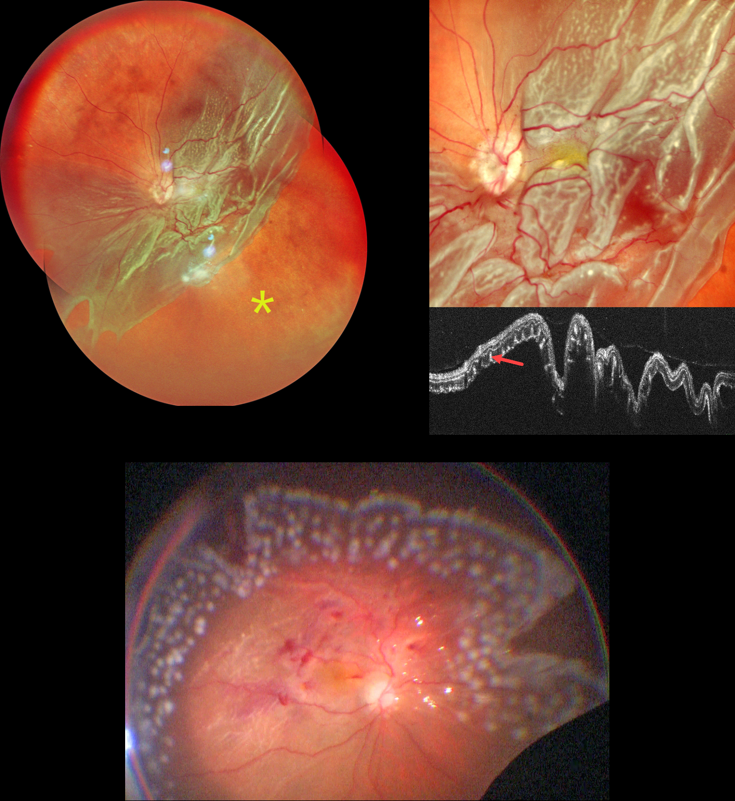
GIANT RETINAL TEAR
Shraddha Raj Shrivastava and Manish Nagpal
Originally posted on @retina.rocks September 1, 2026
This 62YO male presented with 3 months of vision loss in his left eye. Vision was counting fingers.
Pseudocolor SLO imaging shows a giant retinal tear extending from the 2 to 8 o’clock meridians with bare RPE distal to the posterior retina. There are extensive outer retinal corrugations within the posteriorly detached retina. OCT dramatically shows the detached retina, which is redundantly folded onto itself. The outer retinal corrugations appear as numerous hyperreflective outer retinal pillars.
The detachment was emergently repaired with vitrectomy, membrane peeling, endolaser photocoagulation, and silicone oil. The retina was completely attached at the end of the procedure.
Learning Points:
Outer retinal corrugations, also known as hydration outer retinal folds, are one of the key findings that help differentiate a rhegmatogenous retinal detachment from exudative and traction retinal detachments. The cause of these folds remains uncertain but may result from the hydration of glycosaminoglycans in the interphotoreceptor matrix (Dalvin et al, Retinal Cases & Brief Reports 2020;14:355-359).
COMBINED TRACTIONAL-RHEGMATOGENOUS RETINAL DETACHMENT
The European VitreoRetina Society (EVRS) and Shishir Verghese
Originally posted on @retina.rocks September 19, 2025
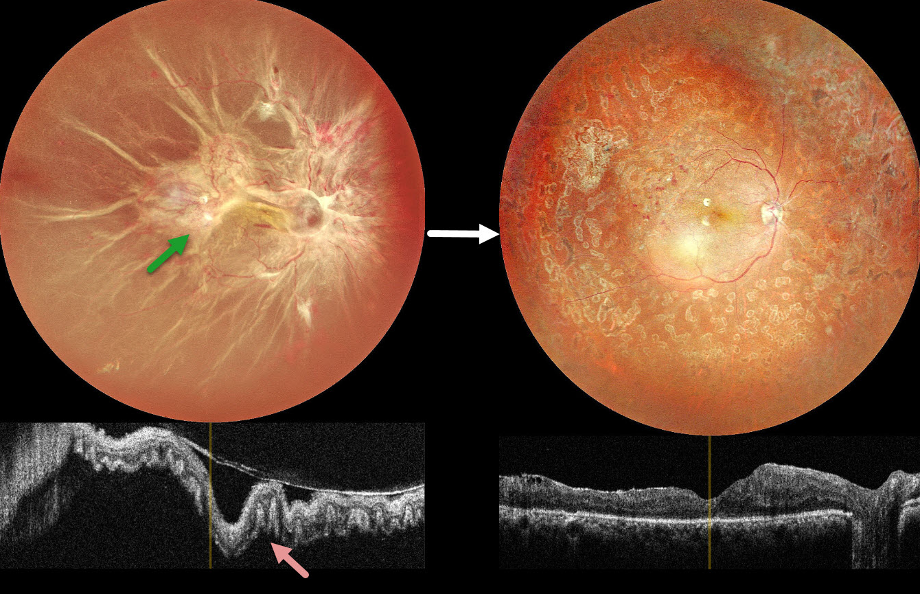
This 40YO female presented with vision loss in her right eye. There was a 15-year history of type 1 diabetes. Vision was hand motion OD.
Color photography shows a total combined traction-rhegmatogenous retinal detachment (RRD). There is a complex pattern of fibrovascular tissue covering the posterior pole with radiating retinal folds extending peripherally. OCT scanning shows a variably undulated detached retina with outer retinal corrugations.
Five days following an intravitreal anti-VEGF injection, she underwent pars plana vitrectomy with membrane peeling, endolaser, and silicone oil injection. Three months later, the silicone oil was removed. Postoperatively, vision improved to 20/80 with complete retinal reattachment.
Learning Points:
Outer retinal corrugations, also known as hydration outer retinal folds, are one of the key findings that help differentiate a RRD from exudative and traction retinal detachments. The cause of these folds remains uncertain but may result from the hydration of glycosaminoglycans in the interphotoreceptor matrix (Dalvin et al, Retinal Cases & Brief Reports 2020;14:355-359). This OCT biomarker confirms the rhegmatogenous component of our patient’s detachment.
OUTER RETINAL FOLDS/CORRUGATIONS
Originally posted on @retina.rocks December 20, 2023
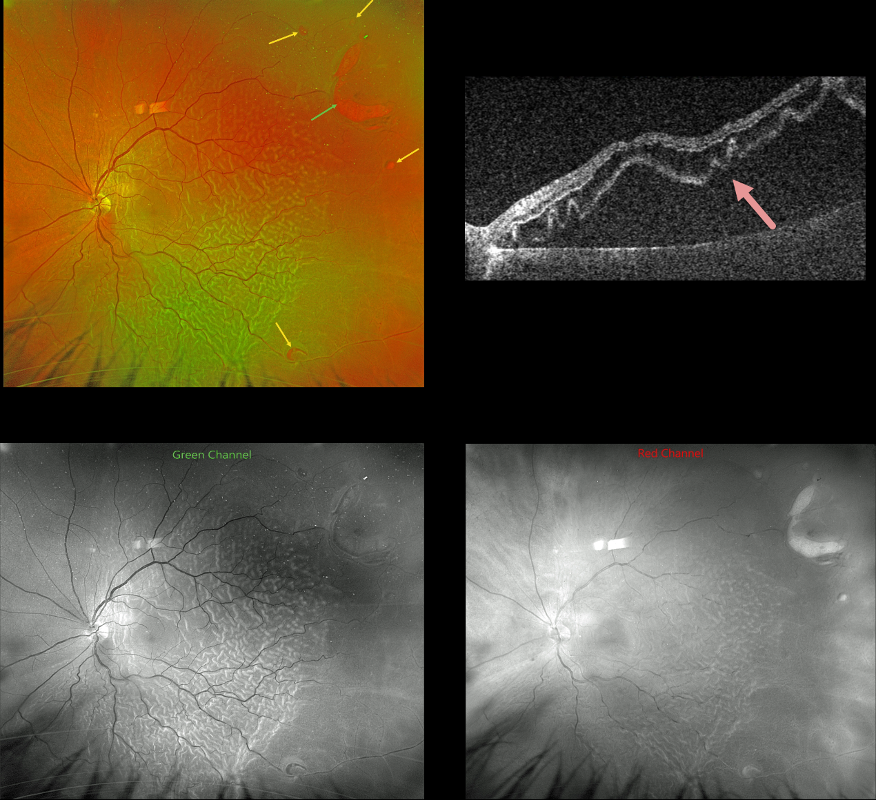
This 61YO male presented with a several-week history of severe vision loss in his left eye. Vision was hand motion.
Optos color RG imaging shows a rhegmatogenous retinal detachment (RRD) with corrugated outer retinal folds. A large horseshoe retinal tear is seen superotemporally, with numerous smaller breaks noted.
The outer retinal folds are best seen with the green channel compared with the red channel. Triton swept-source OCT shows how these findings involve just the outer retina.
Vitrectomy surgery was scheduled.
Learning Points:
Hydration of outer retinal folds is one of the key findings to help differentiate a RRD from exudative and traction retinal detachments. The cause for these folds remains uncertain, but they may form as the result of the hydration of glycosaminoglycans in the interphotoreceptor matrix (Dalvin et al, Retinal Cases & Brief Reports 2020;14:355-359).
OUTER RETINAL FOLDS/CORRUGATIONS
Originally posted on @retina.rocks June 7, 2023
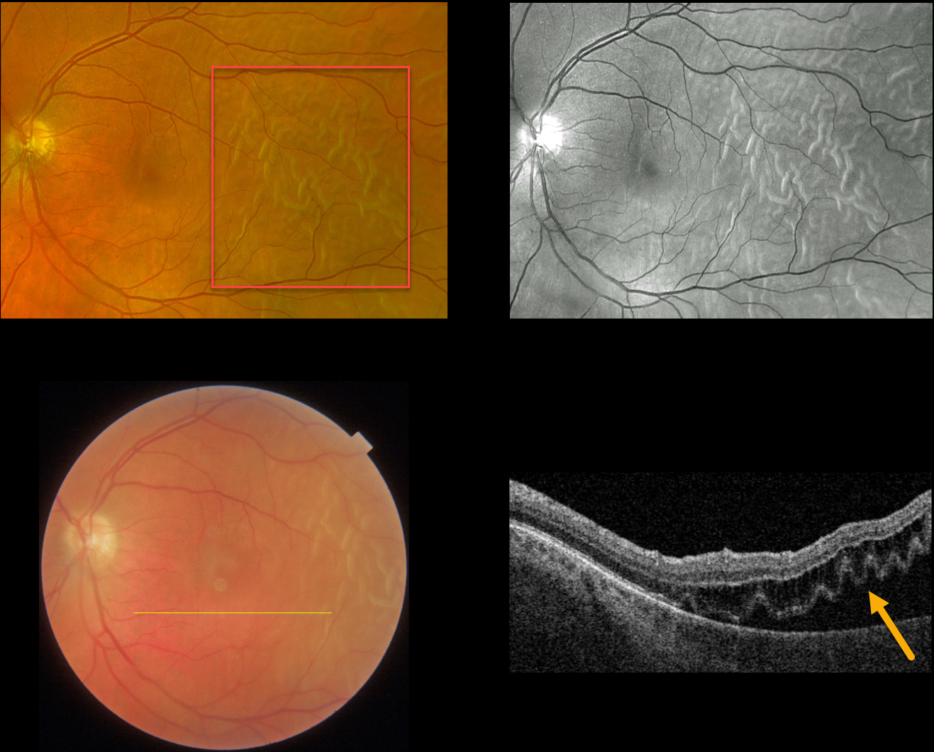
This 51YO male presented with this acute temporal macula-off rhegmatogenous retinal detachment (RRD). Vision was 20/200.
Optos color RGB imaging shows an RRD with corrugated outer retinal folds. These folds are best seen with the green channel. Swept-source OCT shows how these findings involve just the outer retina. The detachment was repaired with vitrectomy the following day.
Learning Points:
Hydration of outer retinal folds are one of the key findings to help differentiate a RRD from exudative and traction retinal detachments.
The cause for these folds remains uncertain, but they may form as the result of hydration of glycosaminoglycans in the interphotoreceptor matrix (Dalvin et al, Retinal Cases & Brief Reports 2020;14;355-359).
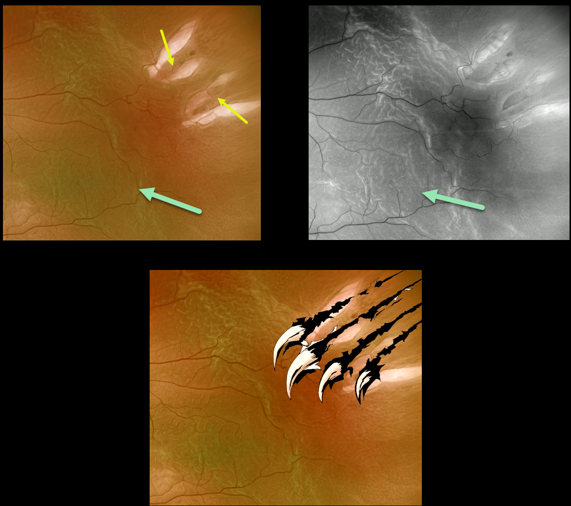
WOLVERINE RETINOPATHY
Originally posted on @retina.rocks May 10, 2022
This 72YO male presented with a macula-off rhegmatogenous retinal detachment (RRD).
Optos imaging gives the illusion of four breaks, although in actuality, there are only two tractional tears. The hydrated outer retinal folds are best visualized on green channel imaging.
Vision was 20/60, and the retina was successfully reattached with emergent pars plana vitrectomy.
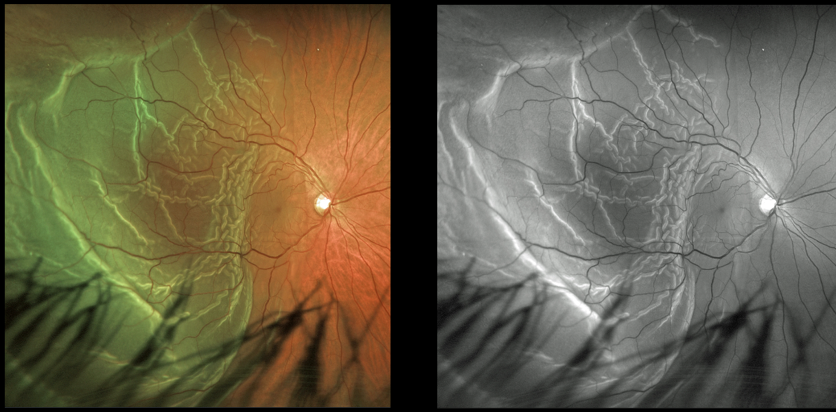
RHEGMATOGENOUS RETINAL DETACHMENT
Originally posted on @retina.rocks December 3, 2021
This 47YO male presented with hand motion vision from a macula-off rhegmatogenous retinal detachment (RRD). The outer retinal folds are dramatically imaged in both the Optos color and green channel images.
Learning Points:
Hydration outer retinal folds are one of the key findings to help differentiate a RRD from exudative and traction retinal detachments.
The cause of these folds remains uncertain but may result from the hydration of glycosaminoglycans in the interphotoreceptor matrix (Dalvin et al, Retinal Cases & Brief Reports 2020;14:355-359).
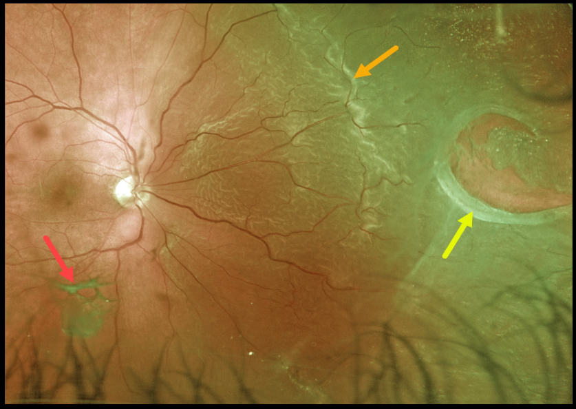
RHEGMATOGENOUS RETINAL DETACHMENT
Originally posted on @retina.rocks November 2, 2021
This patient presented with a macula-on rhegmatogenous retinal detachment (RRD) from a large nasal retinal tear. The tear has a rolled posterior edge, indicating possible early proliferative vitreoretinopathy.
The hydration outer retinal folds at the posterior edge of the detachment are one of the key clinical findings that help differentiate a rhegmatogenous from exudative and traction retinal detachments.
The causative posterior vitreous detachment is seen floating above the retina just inferior to the macula.
Learning Points:
The normal neurosensory retina remains attached to the RPE due to numerous physiological mechanisms, including the inner (retinal vascular endothelium) and outer (tight junctions at the apex of the RPE cells) blood retinal barriers, hyperosmotic choroid passively drawing fluid into the subretinal space, RPE pump, interphotoreceptor matrix, and friction between the RPE microvilli and the photoreceptor outer segments.
Rhegmatogenous retinal detachment (RRD) occurs when liquefied vitreous, entering the subretinal space through a retinal break, overwhelms the forces that favor retinal detachment.
Repairing RRD involves finding and closing all breaks, essentially denying the liquid vitreous access to the subretinal space. The break(s) can be closed internally with a gas bubble with pneumatic retinopexy or vitrectomy surgery, or externally by sewing a piece of silicone material to the eye wall (scleral buckle). This indents, or buckles, the sclera up against the retina break.
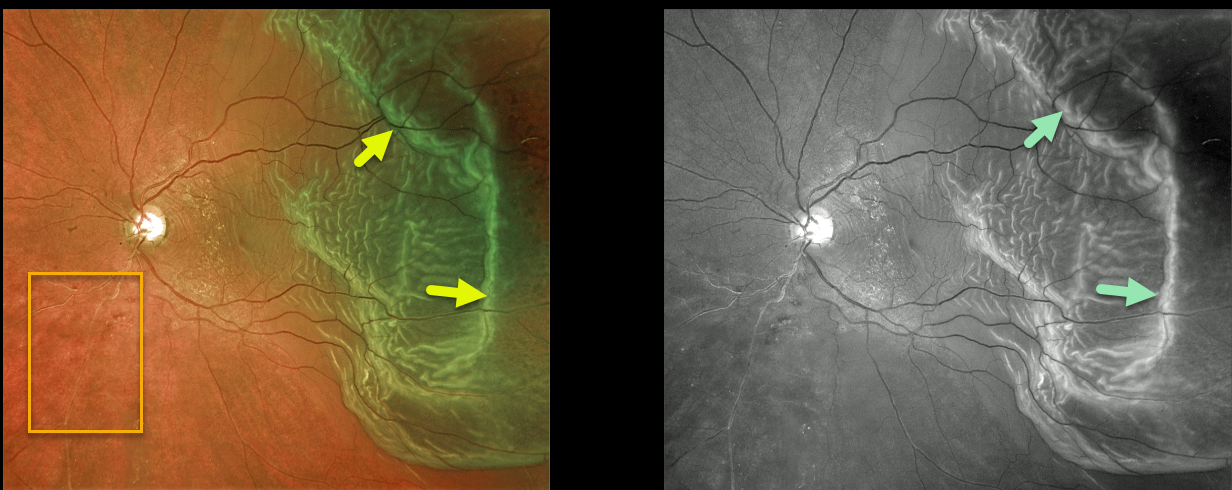
RHEGMATOGENOUS RETINAL DETACHMENT
Originally posted on @retina.rocks August 30, 2021
This 67YO male presented with hand motion vision from a macula-off rhegmatogenous retinal detachment (RRD).
The outer retinal folds are dramatically imaged in both the Optos color and green channel images. An incidental, old asymptomatic branch retinal vein occlusion is also noted inferonasally.
Learning Points:
Hydration outer retinal folds are one of the key findings to help differentiate a RRD from exudative and traction retinal detachments.
The cause of these folds remains uncertain, but they may form as a result of hydration of glycosaminoglycans in the interphotoreceptor matrix (Dalvin et al., Retinal Cases & Brief Reports 2020;14;355-359).


