

Rhegmatogenous Retinal Detachment (RRD) Pneumatic + Gas Bubble
<< Back to Cases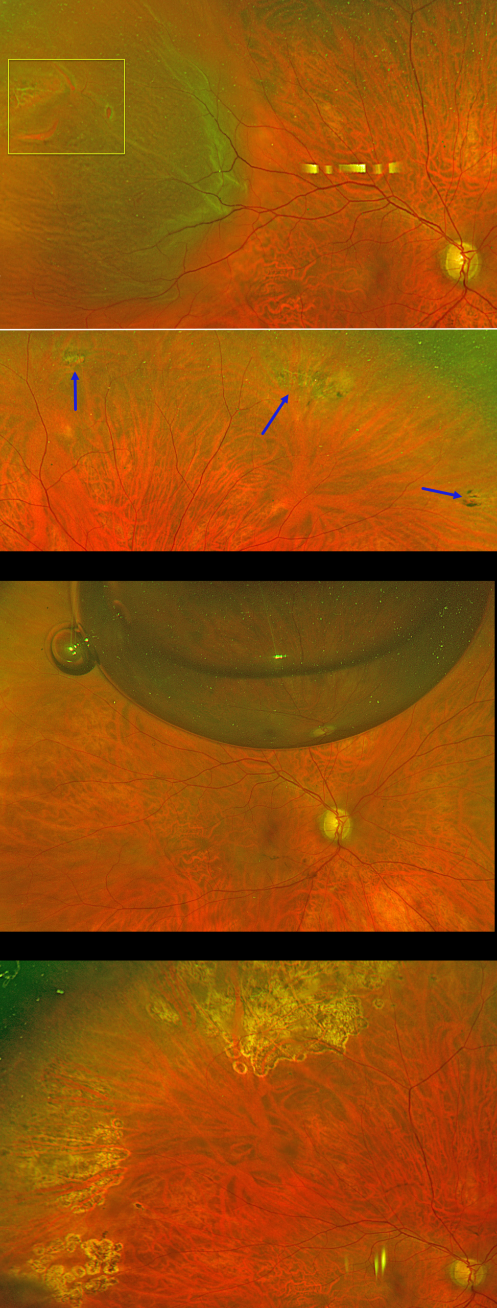
PNEUMATIC RETINOPEXY
Originally posted on @retina.rocks August 13, 2026
This 56YO phakic male presented with floaters and a football-shaped nasal shadow in his right eye for one week. Vision was 20/20 bilaterally.
Optos color RG imaging shows a superotemporal macula-on rhegmatogenous retinal detachment (RRD) from several tiny adjacent breaks within lattice. There were other areas of lattice degeneration superiorly, inferiorly, and inferotemporally within the attached retina. Options for retinal detachment repair were discussed, including pneumatic retinopexy, scleral buckle, and vitrectomy.
The patient chose to undergo pneumatic retinopexy (PnRP) with 0.3cc of pure C3F8. The areas of lattice within the attached retina were lasered just prior to performing the procedure. The retina was completely attached the following day. The causative break was lasered four days later once the detachment flattened. Two months postoperatively, vision was 20/20, and the retina remains attached. Given the diffuse areas of lattice and absence of posterior vitreous detachment in the fellow eye, we performed prophylactic laser retinopexy to the left eye (not shown).
Learning Points:
PnRP was introduced by Hilton and Grizzard in 1986 as an in-office alternative to traditional retinal reattachment surgery (Ophthalmology 1986;93:626-651). It was initially recommended for detachments with one or more breaks within a single clock hour, located within the superior eight clock hours of the fundus.
Forty years after its introduction, the role of PnRP, compared with scleral buckling and vitrectomy, remains controversial. In Canada, where access to operating rooms is more limited than in the US, PnRP is often the initial treatment of choice for appropriate detachments. The Canadian PIVOT trial reported that PnRP offered better final vision, less vertical metamorphopsia, and reduced morbidity compared to vitrectomy (Ophthalmology 2019;126:531-539).
However, in the US, PnRP is not as popular except on a late Friday afternoon (we’re only partially kidding). A recent critical review (Chronopoulos et al, Surv Ophthalmology 2021;66:585-593) argued against PnRP for initial treatment due to the need for “multiple procedures that largely negate its potential cost savings and subjects the patient to prolonged stress and disability…” A recent cost-utility analysis surprisingly found PPV to be the most cost-effective primary procedure (Teja et al, AJO 2023;255:141-154).
Although extensive lattice degeneration is generally considered a contraindication for pneumatic retinopexy, we decided to perform prophylactic treatment. Our patient did well after lasering the areas of lattice prior to performing the pneumatic procedure and was followed closely with scleral depression exams.
PNEUMATIC RETINOPEXY
Originally posted on @retina.rocks March 25, 2026
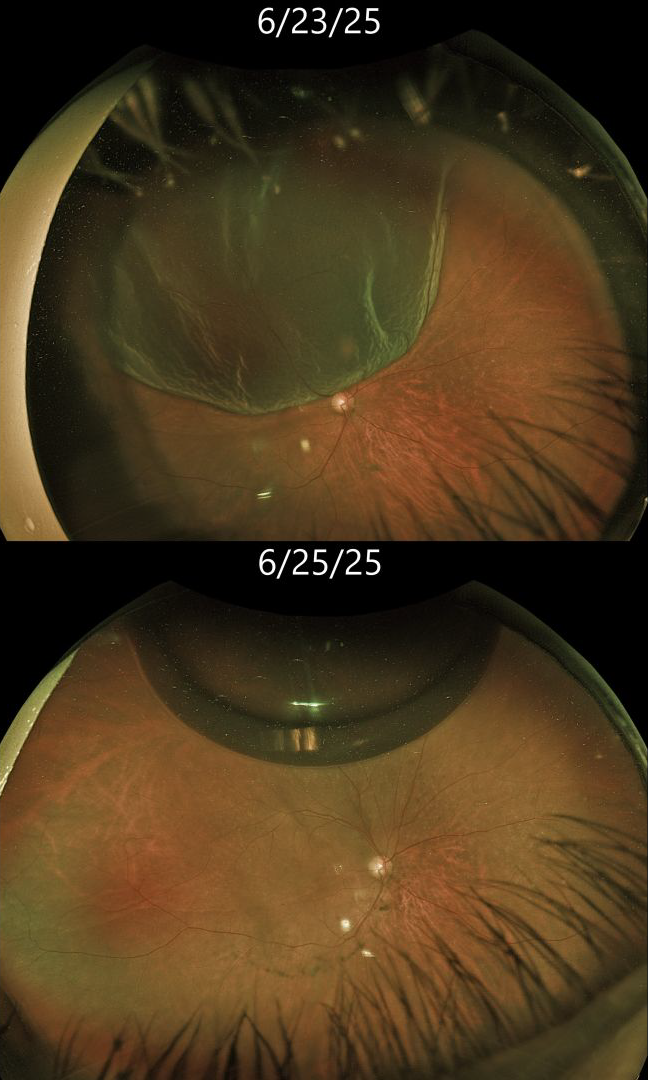
This 76YO female presented with several days of painless vision loss in her right eye. Vision was 20/30.
Optos color RG imaging shows a bullous superotemporal rhegmatogenous retinal detachment (RRD). The macular center was shallowly detached. Scleral depression revealed a single tiny retinal tear anterior to the 11:30 equator. Cryotherapy for this tear, followed by pneumatic retinopexy with 0.2 cc C3F8, was performed. Two days later, vision was 20/30. A single gas bubble is noted superiorly, and the retina is completely attached.
Learning Points:
Pneumatic retinopexy (PnRP) was introduced by Hilton and Grizzard in 1986 as an in-office alternative to traditional retinal reattachment surgery (Ophthalmology 1986;93:626-651). It was initially recommended for detachments with one or more breaks within a single clock hour, located within the superior eight clock hours of the fundus.
Nearly 40 years after its introduction, the role of RRD repair in RRD repair, compared with scleral buckling and vitrectomy, remains controversial. In Canada, where access to operating rooms is more limited than in the US, PnRP is often the initial treatment of choice for appropriate detachments. The Canadian PIVOT trial reported that PnRP offered better final vision, less vertical metamorphopsia, and reduced morbidity compared to vitrectomy (Ophthalmology 2019;126:531-539). However, in the US, PnRP is not as popular except on a late Friday afternoon (we’re only partially kidding). A recent critical review (Chronopoulos et al, Surv Ophthalmology 2021;66:585-593) argued against PnRP for initial treatment due to the need for “multiple procedures that largely negates its potential cost savings and subjects the patient to prolonged stress and disability…” A recent cost-utility analysis surprisingly found PPV to be the most cost-effective primary procedure (Teja et al, AJO 2023;255:141-154).
We personally recommend PnRP for patients with mobile superior RRD with superior breaks. Patients must be able to properly position their head. We must be able to visualize the retinal periphery well to ensure there are no inferior breaks, and ideally, we prefer that the macula be detached in case a failed PnRP converts a macula-on RRD to a macula-off RRD.
We therefore felt that our patient was a perfect candidate for PnRP. The PIVOT trial also supported our decision, with a recent post hoc analysis showing similar single-surgery success with PnRP (88%) vs PPV (91%) for single-break primary RRD (Pecaku et al., Br J Ophthalmol 2025;109:113-118).
PRE-ANTERIOR HYALOID GAS FOLLOWING FAILED PNEUMATIC RETINOPEXY
Arthur Zupelli, Bruno Felipe Oliveira Silva, Leonardo Luis Cassoni Marcelo Brandão Guimarães Rego and Rodrigo Jorge
Originally posted on @retina.rocks January 13, 2026
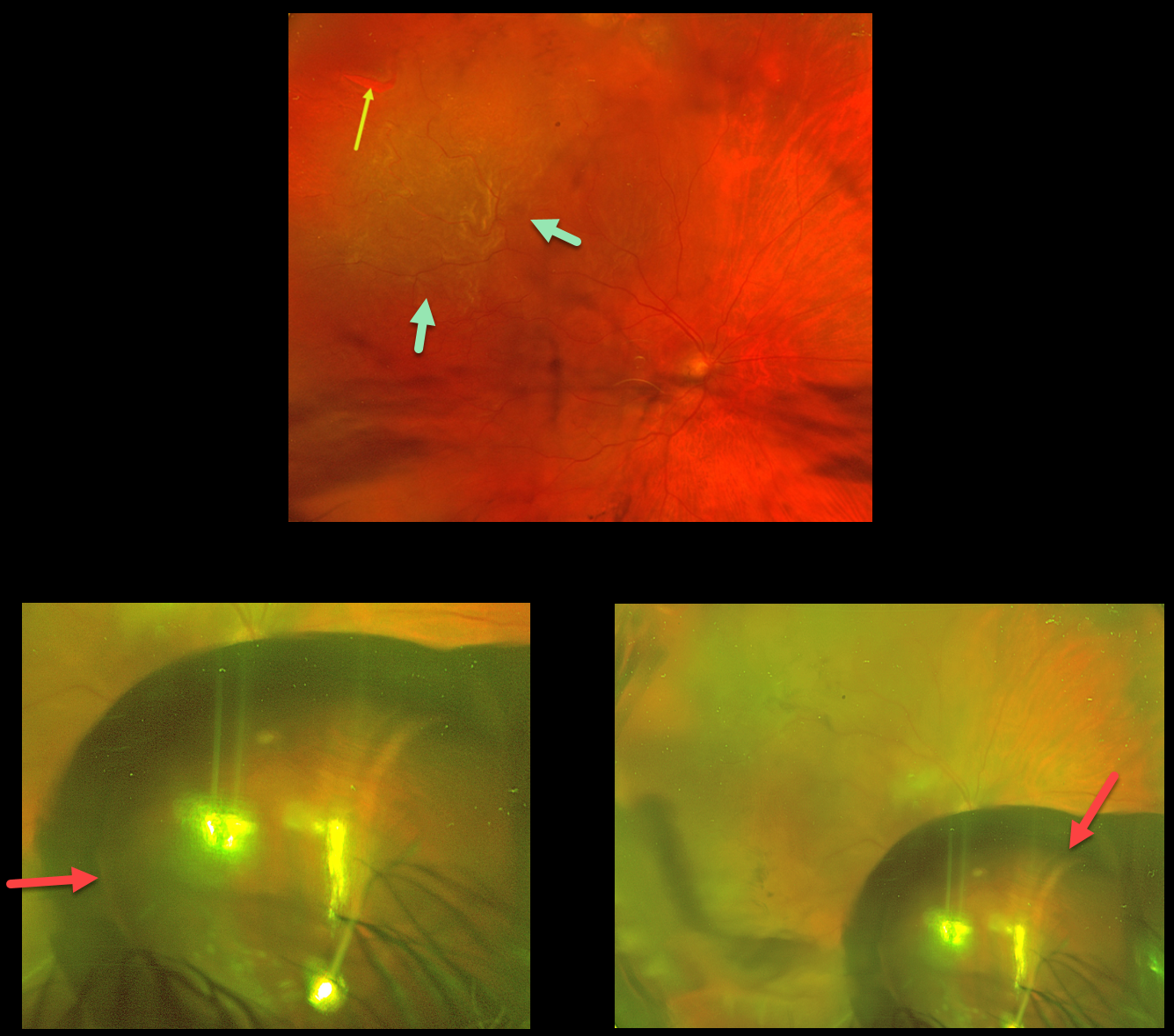
This 71YO male presented with decreased vision in his right eye one month following cataract surgery. Vision was hand motion. Slit lamp examination revealed a well-centered posterior chamber IOL.
Optos color RG imaging shows a superotemporal macula-off rhegmatogenous retinal detachment (RRD) caused by a single break. A pneumatic retinopexy using 0.35cc of pure perfluoropropane gas (C3F8) was performed, followed by intravenous mannitol. One day later, vision was hand motion, and a large gas bubble was trapped in the pre-hyaloid space. This configuration reflected gas sequestration within the potential space bordered by the posterior lens capsule, the anterior hyaloid membrane, and the ciliary body. The appearance was unchanged 2 days later.
We attempted to remove the gas in the office, using a plungerless syringe with the needle inserted into the bubble. This maneuver was unsuccessful, necessitating pars plana vitrectomy. The retina remained attached postoperatively with a final vision of 20/100.
Learning Points:
Gas entrapment in the pre-hyaloid space is an exceedingly rare but visually striking complication of pneumatic retinopexy (Chan et al, Surv Ophthalmol 2008;53:443-478), and most likely is caused by a too shallow needle entry into the vitreous. On slit-lamp or fundus examination, the bubble may appear as a donut- shaped or sausage-like lucency located immediately behind the lens.
LOCULATED FLUID AFTER SUCCESSFUL PNEUMATIC RETINOPEXY
Originally posted on @retina.rocks October 21, 2025
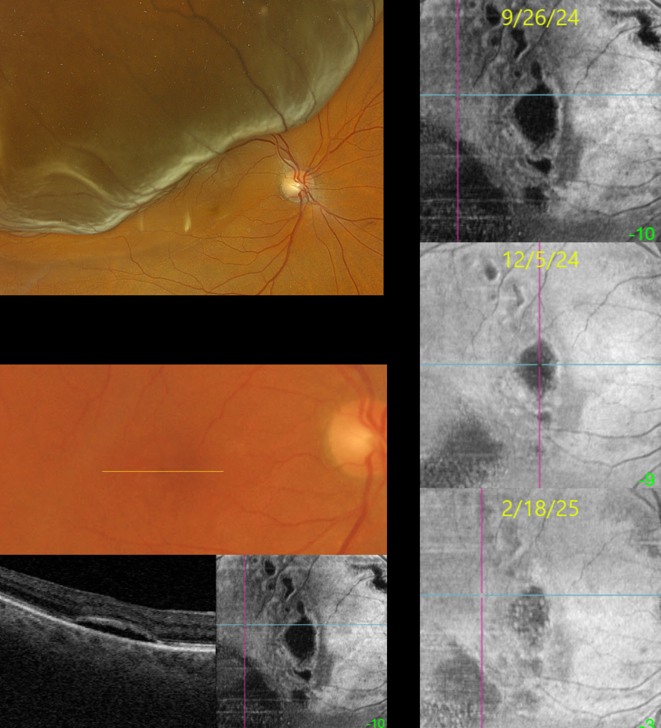
This 69YO female presented on 4/17/24 with a superior macula-off rhegmatogenous retinal detachment (RRD) and counting fingers vision. Following successful pneumatic retinopexy, the retina reattached, and vision returned to 20/25 one month later.
When examined on 9/26/24, she was subjectively doing well with 20/50 vision. Although the macula looked clinically normal, Triton swept-source OCT B-scan and en face imaging show multifocal pockets of loculated subretinal fluid (SRF). With observation, these lesions continue to shrink and flatten. When last examined on 2/18/25, vision was 20/30, and continued observation was recommended.
Learning Points:
Loculated fluid represents an area of persistent subclinical SRF in the presence of a clinically reattached retina and is seen postoperatively in up to 15% of RRD following vitrectomy (Retina 2020;40:1153-1159) and eventually resolves after many months of observation. In a post hoc analysis of the PIVOT trial, there were no differences in the incidence of loculated fluid between vitrectomy and pneumatic retinopexy (Bansal et al BJO 2023;107:1693-1697). Although subfoveal loculated fluid was associated with reduced visual acuity at 3 months, vision eventually was similar to that of those without fluid at 1 year.
Bansal et al recently described the en face findings for loculated fluid (Ophthalmology Retina 2023;7:496-502). We continue to find that en face imaging often provides a unique and compelling perspective on a variety of pathologies, and we hope other clinicians will add it to their daily clinical routine.
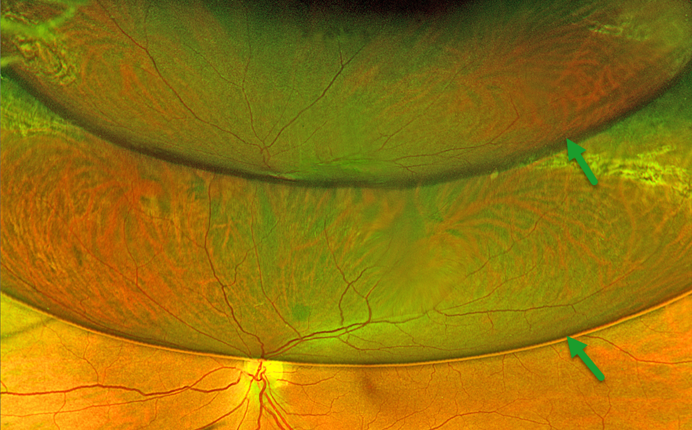
POST-OPERATIVE RD REPAIR WITH GAS
Originally posted on @retina.rocks December 16, 2024
This 56YO female underwent successful vitrectomy surgery for a rhegmatogenous retinal detachment (RRD) 1 month earlier. Optos color RG imaging shows an unusual double-bubble with different reflections of the attached inferior retina.
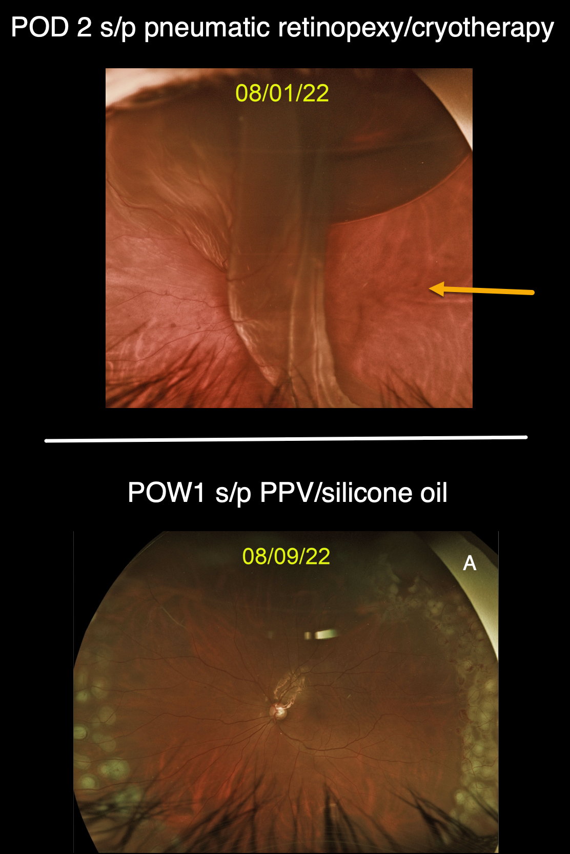
GIANT RETINAL TEAR
Originally posted on @retina.rocks September 9, 2022
This 50YO male presented with a superior macula-off retinal detachment from a single retinal tear at the 1 o’clock periphery (image not shown), which was treated with a pneumatic retinopexy. Cryotherapy was applied before injecting the gas bubble, confirming this single break.
However, when he returned 2 days later, a new giant retinal tear extended from 12 to 5 o’clock, with subretinal gas migrating beneath the superior retina.
The detachment was repaired with vitrectomy and silicone oil, and the second image shows the 1-week postoperative visit.
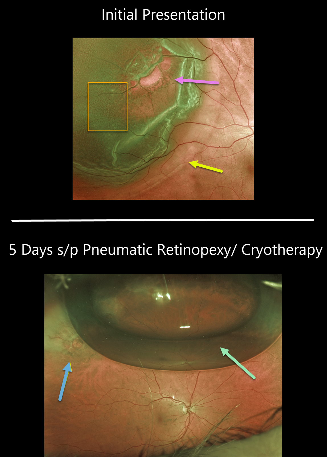
RHEGMATOGENOUS RETINAL DETACHMENT
Originally posted on @retina.rocks February 25, 2022
This 63YO female presented with 20/25 vision and a superotemporal retinal detachment. The posterior edge of the detachment was just outside the macular center. There was diffuse fine microcystic retinal edema within the area of detachment, except for outer retinal hydration folds more posteriorly.
The retina was completely attached 5 days later following successful cryotherapy with pneumatic retinopexy. Early cryotherapy scarring is already visible.
Learning Points:
This detachment could have been repaired with any technique, including vitrectomy, scleral buckling, or pneumatic retinopexy. All techniques yield similar final anatomic success, although the debate continues about the relative merit of one over the other.
See the ___References folder in the Retinal detachment (RD) rhegmatogenous (RRD) folder in the Image Library at www.retinarocks.org for a summary of the key journal articles.
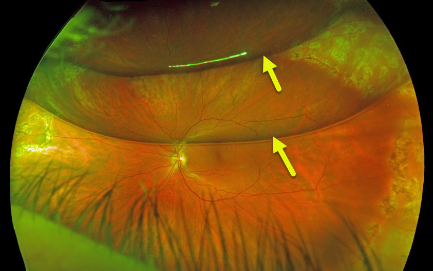
RHEGMATOGENOUS RETINAL DETACHMENT
Originally posted on @retina.rocks February 25, 2020
This patient had a rhegmatogenous retinal detachment that was repaired with pars plana vitrectomy and C3F8 gas.
The patient’s bubble has broken into 2 large bubbles, each with its own beautiful retinal reflection.
Learning Points:
Long-acting intraocular gas is routinely used in vitrectomy for rhegmatogenous retinal detachment repairs. C3F8 and sulfur hexafluoride (SF6) are used due to greater longevity compared to air. C3F8 lasts in the vitreous for 2-3 months, and SF6 lasts for several weeks.


