

Rhegmatogenous Retinal Detachment (RRD) Proliferative Vitreoretinopathy (PVR)
<< Back to Cases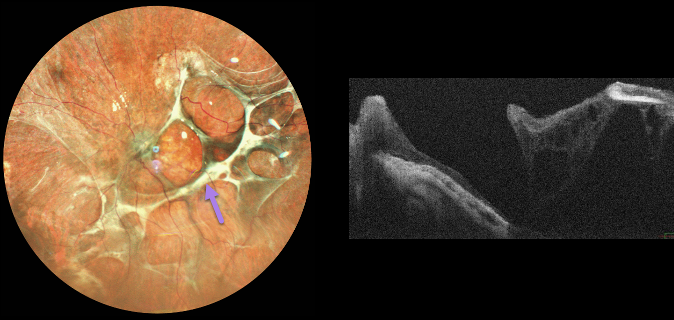
SUBRETINAL AND PRERETINAL PROLIFERATIVE VITREORETIONPATHY
The European VitreoRetinal Society (EVRS), Akansha Sharma, Manish Nagpal and Navneet Mehrotra
Originally posted on @retina.rocks May 29, 2026
This 71YO male presented with one year of poor vision in his left eye following vitrectomy with silicone oil elsewhere for a rhegmatogenous retinal detachment. Vision was light perception.
Pseudocolor SLO imaging shows a complex pattern of interlacing subretinal fibrosis bands. OCT scanning shows areas of disorganized retinal thinning, thickened disorganized retina nasally, and retinal detachment more temporally. Observation was recommended given the poor visual prognosis.
Learning Points:
Proliferative vitreoretinopathy (PVR), which consists of cellular membranes growing on either retinal surface, develops in about 5-10% of RRD. The membranes contract, foreshorten the retina, and likely represent an aberrant wound-healing response. There are no proven pharmacologic therapies to prevent or treat PVR. These complex detachments can be managed with vitrectomy and membrane peeling, retinectomy, scleral buckling, and silicone oil. However, PVR remains the main cause for ultimate retinal reattachment surgical failure.
See Garweg et al Surv Ophthalmol 2013;48:321-329 for a good review of PVR pathophysiology.
PROLIFERATIVE VITREORETINOPATHY
Originally posted on @retina.rocks April 8, 2026
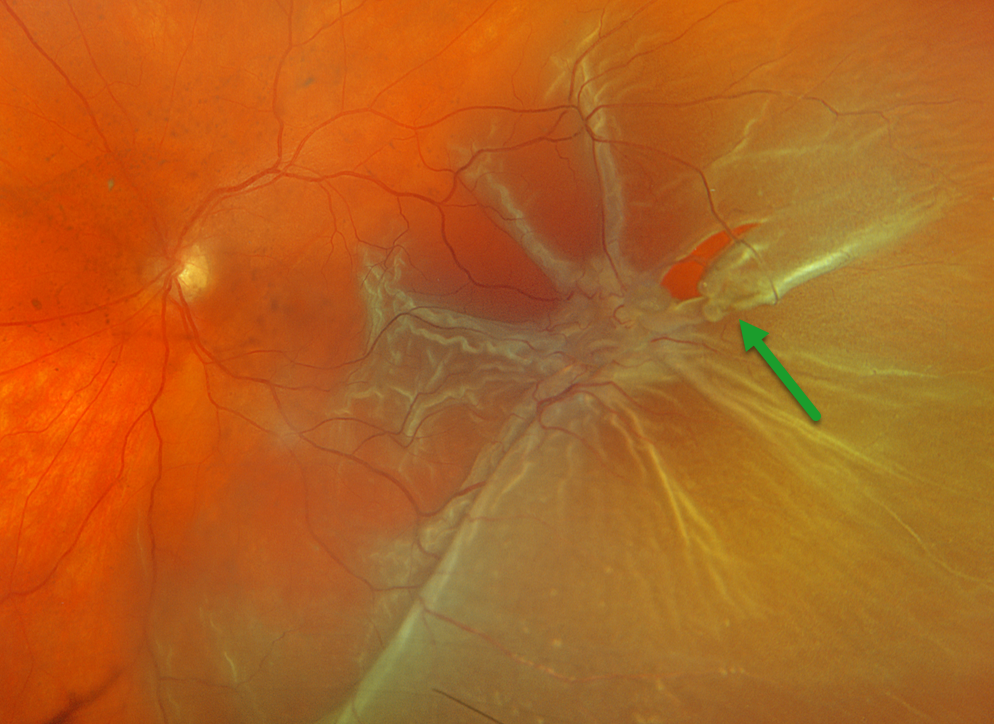
This 63YOF presented with a week of vision loss in her right eye. Optos color RGB imaging shows a temporal macula-off rhegmatogenous retinal detachment (RRD) complicated by proliferative vitreoretinopathy (PVR) with a large star fold just posterior to a retinal tear. She was scheduled for emergent vitrectomy.
Learning Points:
PVR, which consists of cellular membranes growing on either retinal surface, develops in about 5-10% of retinal detachments. RRD with a concurrent non-causal macular hole is found in about 2% detachments (Iros et al, Retina 2023;43:581-584). Although most eyes achieve anatomic success, final vision is usually poor. In our patient, unfortunately, there is no further clinical information, so it is unclear whether the macular hole caused the detachment or was secondary to traction from the star fold.
PROLIFERATIVE VITREORETINOPATHY
The European VitreoRetina Society (EVRS) and Shishir Verghese
Originally posted on @retina.rocks August 8, 2025
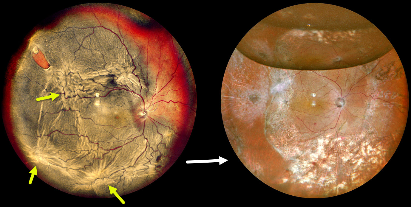
This 75YO male presented with decreased vision in the right eye for four months. Vision was hand motion OD and 20/25 in his normal OS.
Pseudocolor SLO imaging shows a complex total rhegmatogenous retinal detachment with a supertemporal tear and multiple proliferative vitreoretinopathy (PVR) star folds. Following three vitrectomy surgeries, including a relaxing retinectomy, the retina is attached. Vision was 20/120.
GIANT MACULAR HOLE WITH TOTAL RETINAL DETACHMENT
Anand Temkar and Manish Nagpal
Originally posted on @retina.rocks February 10, 2025
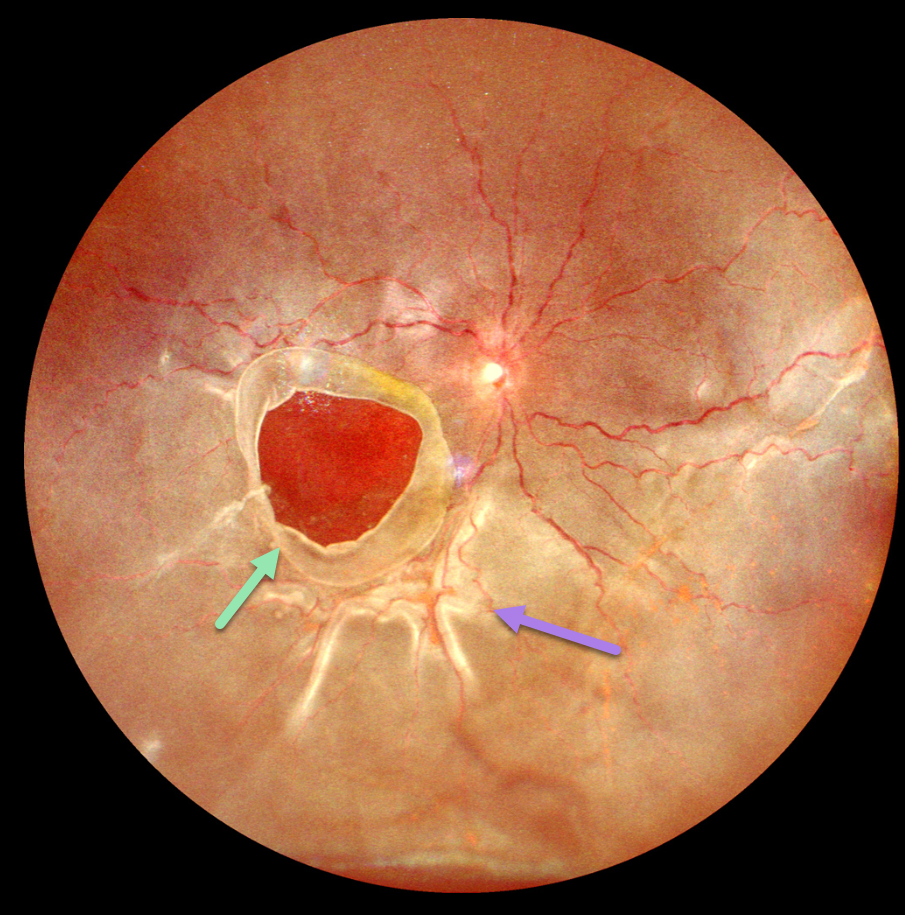
This 10YO boy presented with 1 week of vision loss in his left eye. The parents gave a vague history of trauma with a tennis ball 2 years earlier. Vision was counting fingers OD and 20/20 in his normal OS.
Ultrawidefield imaging shows a giant macular hole occupying nearly the entire macula. The hole’s edges are rolled, and proliferative vitreoretinopathy with star folds is present within the total rhegmatogenous retinal detachment (RRD).
Emergent vitrectomy surgery with silicone oil was scheduled.
Learning Points:
Blunt ocular trauma can result in numerous sequelae, including commotio retina, retinal breaks and detachment, choroidal rupture, and macular hole. A giant macular hole, also reported in Alport syndrome (Shah and Weinberg Ophthalmic Genetics 2010;31(2):93-97), is an exceedingly rare traumatic event (Hernandez-Da Mota Case Rep Ophthalmol 2011;2:283-286).
Traumatic macular holes are thought to result from a vitreous coup contrecoup injury or a jet stream of anterior chamber aqueous humor shooting through Cloquet canal into the premacular bursa.
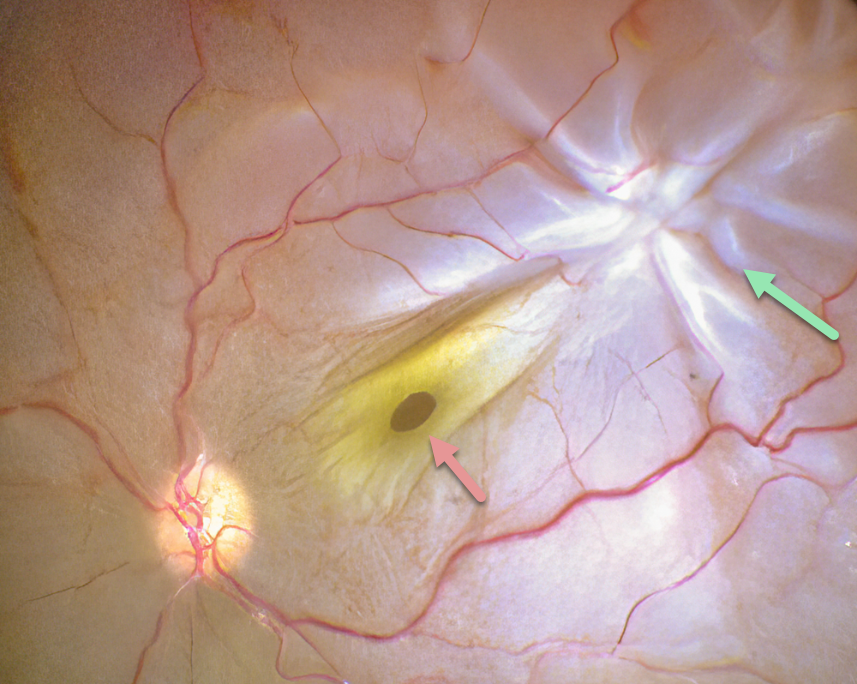
PROLIFERATIVE VITREORETINOPATHY (PVR) WITH MACULAR HOLE
Anjana Mirajkar and Manish Nagpal
Originally posted on @retina.rocks August 14, 2024
This intraoperative photograph shows a rhegmatogenous retinal detachment (RRD) complicated by proliferative vitreoretinopathy (PVR) with a star fold superotemporally. A full-thickness macular hole surrounded by its luteal pigment is dragged towards the PVR. The macular hole and detachment were repaired with vitrectomy.
Learning Points:
PVR, which consists of cellular membranes growing on either retinal surface, develops in about 5-10% of retinal detachments. RRD with a concurrent non-causal macular hole is found in about 2% detachments (Iros et al, Retina 2023;43:581-584). Although most eyes achieve anatomic success, final vision is usually poor. In our patient, unfortunately, there is no further clinical information, so it is unclear whether the macular hole caused the detachment or was secondary to traction from the star fold.
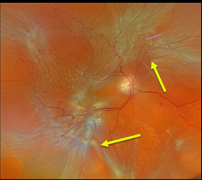
PROLIFERATIVE VITREORETINOPATHY (PVR)
Originally posted on @retina.rocks August 22, 2023
This 76YO male presented with a vague history of recent vision loss in his right eye. Vision was counting fingers.
Optos color RGB imaging shows a total rhegmatogenous retinal detachment (RRD) with diffuse posterior proliferative vitreoretinopathy (PVR). The detachment was successfully repaired with vitrectomy and silicone oil.
Learning Points:
PVR, which consists of cellular membranes growing on either retinal surface, develops in about 5-10% of RRD. The membranes contract and foreshorten the retina, likely as an aberrant wound-healing response.
There are no proven pharmacologic therapies to prevent or treat PVR. These complex detachments can be managed with vitrectomy and membrane peeling, retinectomy, scleral buckling, and silicone oil. However, PVR remains the main cause for ultimate retinal reattachment surgical failure.
See Garweg et al Surv Ophthalmol 2013;48:321-329 for a good review of PVR pathophysiology.
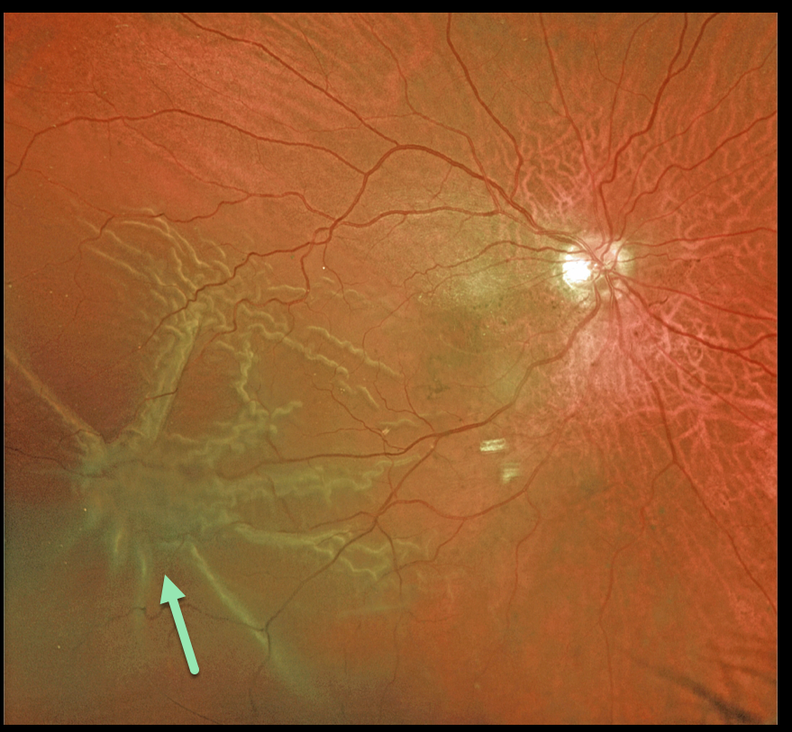
PROLIFERATIVE VITREORETINOPATHY (PVR)
Originally posted on @retina.rocks November 1, 2022
This 72YO male presented with this macula-off rhegmatogenous retinal detachment (RRD).
Optos color imaging shows a prominent star fold inferotemporally. This detachment was repaired with vitrectomy and membrane peeling.
Learning Points:
Proliferative vitreoretinopathy (PVR), which consists of cellular membranes growing on either retinal surface, develops in about 5-10% of retinal detachments.
The membranes contract and foreshorten the retina, likely as an aberrant wound-healing response.
There are no proven pharmacologic therapies to prevent or treat PVR. These complex detachments can be managed with vitrectomy and membrane peeling, retinectomy, scleral buckling, and silicone oil. However, PVR remains the main cause for ultimate retinal reattachment surgical failure.
See Garweg et al Surv Ophthalmol 2013;48:321-329 for a good review of PVR pathophysiology.
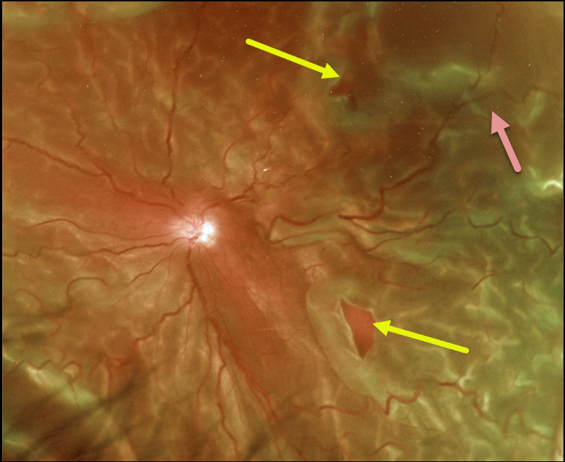
PROLIFERATIVE VITREORETINOPATHY (PVR)
Originally posted on @retina.rocks June 17, 2022
This 65YO male presented with light perception in his left eye secondary to a very chronic total rhegmatogenous retinal detachment (RRD).
Optos color imaging captured the detachment, which was fairly stiff due to diffuse proliferative vitreoretinopathy (PVR). Two posterior retinal breaks are seen.
The retina was successfully reattached with vitrectomy, inferior retinectomy, and silicone oil.
Learning Points:
PVR, which consists of cellular membranes growing on either retinal surface, develops in about 5-10% of retinal detachments. The membranes contract, foreshorten the retina, and likely proliferate as an aberrant wound-healing response. There are no proven pharmacologic therapies to prevent or treat PVR.
These complex detachments can be managed with vitrectomy with membrane peeling, retinectomy, scleral buckling, and silicone oil. However, PVR remains the main cause for ultimate retinal reattachment surgical failure.
See Garweg et al Surv Ophthalmol 2013;48:321-329 for a good review of PVR pathophysiology.
PROLIFERATIVE VITREORETINOPATHY (PVR)
Originally posted on @retina.rocks November 24, 2020
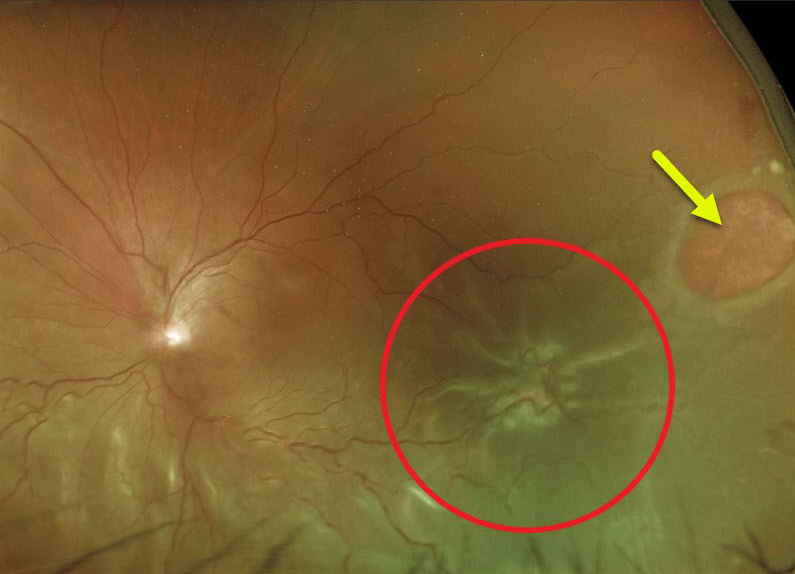
This patient’s Optos image demonstrates a large retinal hole and retinal detachment with proliferative vitreoretinopathy (PVR).
Learning Points:
Proliferative vitreoretinopathy (PVR), consisting of growth of cellular membranes on either retinal surface, develops in about 5-10% of retinal detachments. The membranes contract and foreshorten the retina, likely resulting from an aberrant wound-healing response.
There are no pharmacologic therapies to prevent or treat PVR. These complex detachments can be managed with vitrectomy with membrane peeling, retinectomy, scleral buckling, and silicone oil. However, PVR remains the main cause for ultimate retinal reattachment surgical failure.
See Garweg et al Surv Ophthalmol 2013;48:321-329 for a good review of PVR pathophysiology.
PROLIFERATIVE VITREORETINOPATHY
Originally posted on @retina.rocks December 31, 2019
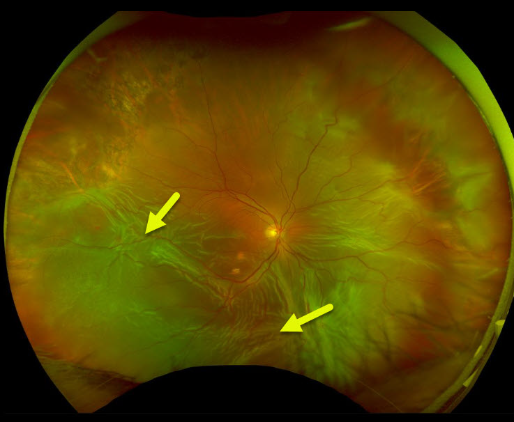
This eye has a total retinal detachment, with numerous foci of fibrotic retina in the inferior hemisphere.
Learning Points:
PVR, or proliferative vitreoretinopathy, consists of fibrotic membranes growing on the retina surface.
It is the primary cause of retinal reattachment surgical failure and occurs in 5-10% of detachments.


