

Rhegmatogenous Retinal Detachment (RRD)
<< Back to Cases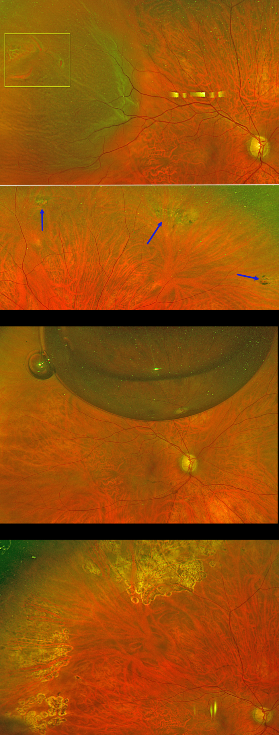
PNEUMATIC RETINOPEXY
Originally posted on @retina.rocks August 13, 2026
This 56YO phakic male presented with floaters and a football-shaped nasal shadow in his right eye for one week. Vision was 20/20 bilaterally.
Optos color RG imaging shows a superotemporal macula-on rhegmatogenous retinal detachment (RRD) from several tiny adjacent breaks within lattice. There were other areas of lattice degeneration superiorly, inferiorly, and inferotemporally within the attached retina. Options for retinal detachment repair were discussed, including pneumatic retinopexy, scleral buckle, and vitrectomy.
The patient chose to undergo pneumatic retinopexy (PnRP) with 0.3cc of pure C3F8. The areas of lattice within the attached retina were lasered just prior to performing the procedure. The retina was completely attached the following day. The causative break was lasered four days later once the detachment flattened. Two months postoperatively, vision was 20/20, and the retina remains attached. Given the diffuse areas of lattice and absence of posterior vitreous detachment in the fellow eye, we performed prophylactic laser retinopexy to the left eye (not shown).
Learning Points:
PnRP was introduced by Hilton and Grizzard in 1986 as an in-office alternative to traditional retinal reattachment surgery (Ophthalmology 1986;93:626-651). It was initially recommended for detachments with one or more breaks within a single clock hour, located within the superior eight clock hours of the fundus.
Forty years after its introduction, the role of PnRP, compared with scleral buckling and vitrectomy, remains controversial. In Canada, where access to operating rooms is more limited than in the US, PnRP is often the initial treatment of choice for appropriate detachments. The Canadian PIVOT trial reported that PnRP offered better final vision, less vertical metamorphopsia, and reduced morbidity compared to vitrectomy (Ophthalmology 2019;126:531-539).
However, in the US, PnRP is not as popular except on a late Friday afternoon (we’re only partially kidding). A recent critical review (Chronopoulos et al, Surv Ophthalmology 2021;66:585-593) argued against PnRP for initial treatment due to the need for “multiple procedures that largely negate its potential cost savings and subjects the patient to prolonged stress and disability…” A recent cost-utility analysis surprisingly found PPV to be the most cost-effective primary procedure (Teja et al, AJO 2023;255:141-154).
Although extensive lattice degeneration is generally considered a contraindication for pneumatic retinopexy, we decided to perform prophylactic treatment. Our patient did well after lasering the areas of lattice prior to performing the pneumatic procedure and was followed closely with scleral depression exams.
COMBINED RETINOSCHISIS-RHEGMATOGENOUS RETINAL DETACHMENT
Originally posted on @retina.rocks August 12, 2026
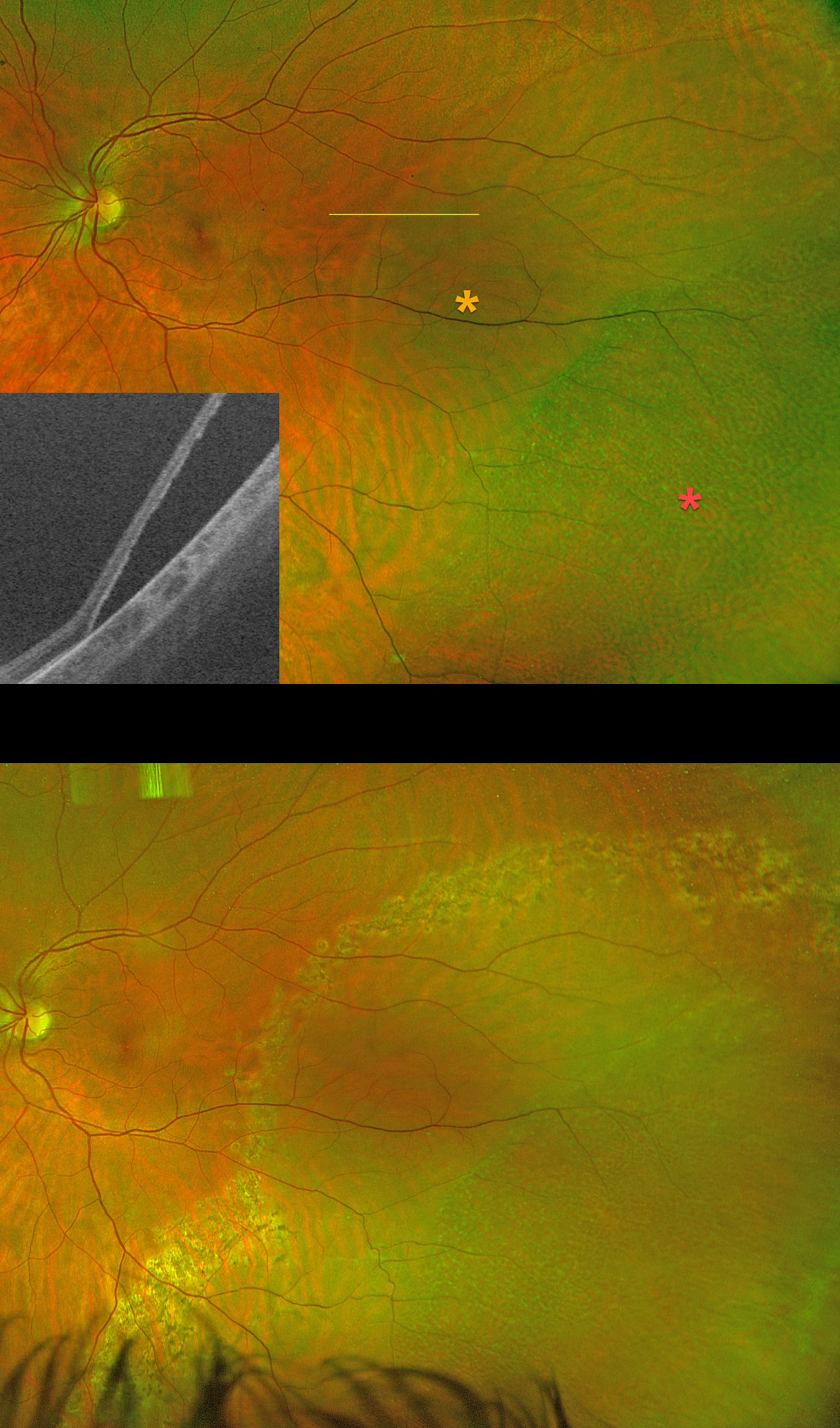
This 35YO pregnant female presented with 3 days of flashes in her left eye. She had a family history of retinal tears in her father and rhegmatogenous retinal detachment (RRD) in her maternal uncle. Vision was 20/20 OS.
Silverstone color RG imaging shows a macula-on combined retinoschisis-RRD. There was no posterior vitreous detachment. Schisis is noted more peripherally, with the rhegmatogenous fluid noted more posteriorly. Swept-source OCT confirms the presence of a full-thickness retinal detachment. Treatment options were discussed, including laser demarcation, primary scleral buckle with cryotherapy, and pars plana vitrectomy. She elected for laser retinopexy, which was performed that day.
She delivered a healthy baby, and her retina has remained stable over the past 9 months as of her last visit.
Learning Points:
Isolated degenerative retinoschisis should not be treated. However, it can progress to a combined schisis-RRD in the presence of breaks in both the inner and outer layers. Asymptomatic combined detachments can be demarcated, as in our patient. Symptomatic detachments are usually treated with pars plana vitrectomy.
SELF-SEALING RHEGMATOGENOUS RD WITH HEAVY ENCIRCLING DEMARCATION LINE
Anand Gandhi and Manish Nagpal
Originally posted on @retina.rocks August 11, 2026
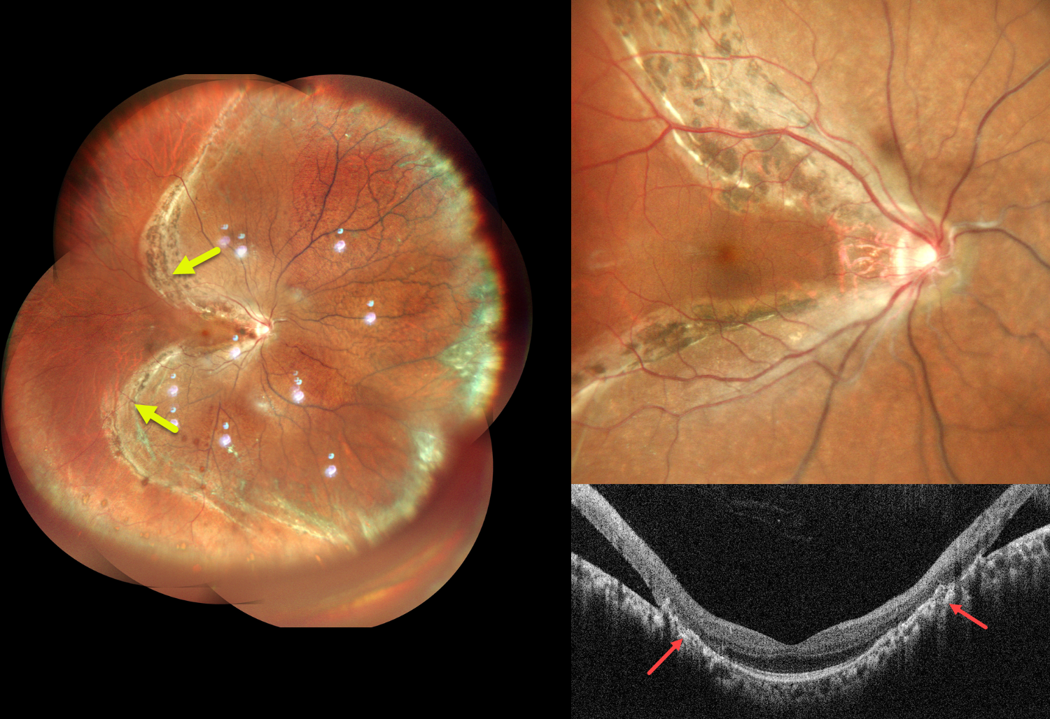
This healthy 31YO male presented with blurred vision in his right eye for 2 years. Best-corrected visual acuity was 20/20 OU.
Pseudocolor SLO imaging shows a chronic rhegmatogenous retinal detachment (RRD) involving the superior, nasal, and inferior quadrants, sparing the central and temporal macula. A prominent pigmented demarcation encircles the RRD. Vertical OCT scanning shows a normal macular center with broad chorioretinal scarring superiorly and inferiorly bordering the attached and detached retina.
Learning Points:
Acute RRDs are virtually always symptomatic, requiring emergent surgery. However, asymptomatic detachments often have a low risk of progression and can usually be safely observed (Cohen et al, AJO 2005;139;777-779; Sengillo et al, Ophthalmology Retina 2023;7:215-220) or demarcated (Vrabec and Baumal, Ophthalmology 2000;107:1063-1067).
In the present case, although the retinal detachment was symptomatic at onset, it was not diagnosed until several years later. The heavy demarcation line indicates chronicity with likely stability. Given the preserved visual acuity, macular sparing, and chronic nature of the detachment, close observation was recommended.
RHEGMATOGENOUS RD WITH LASER DEMARCATION
Originally posted on @retina.rocks July 29, 2026
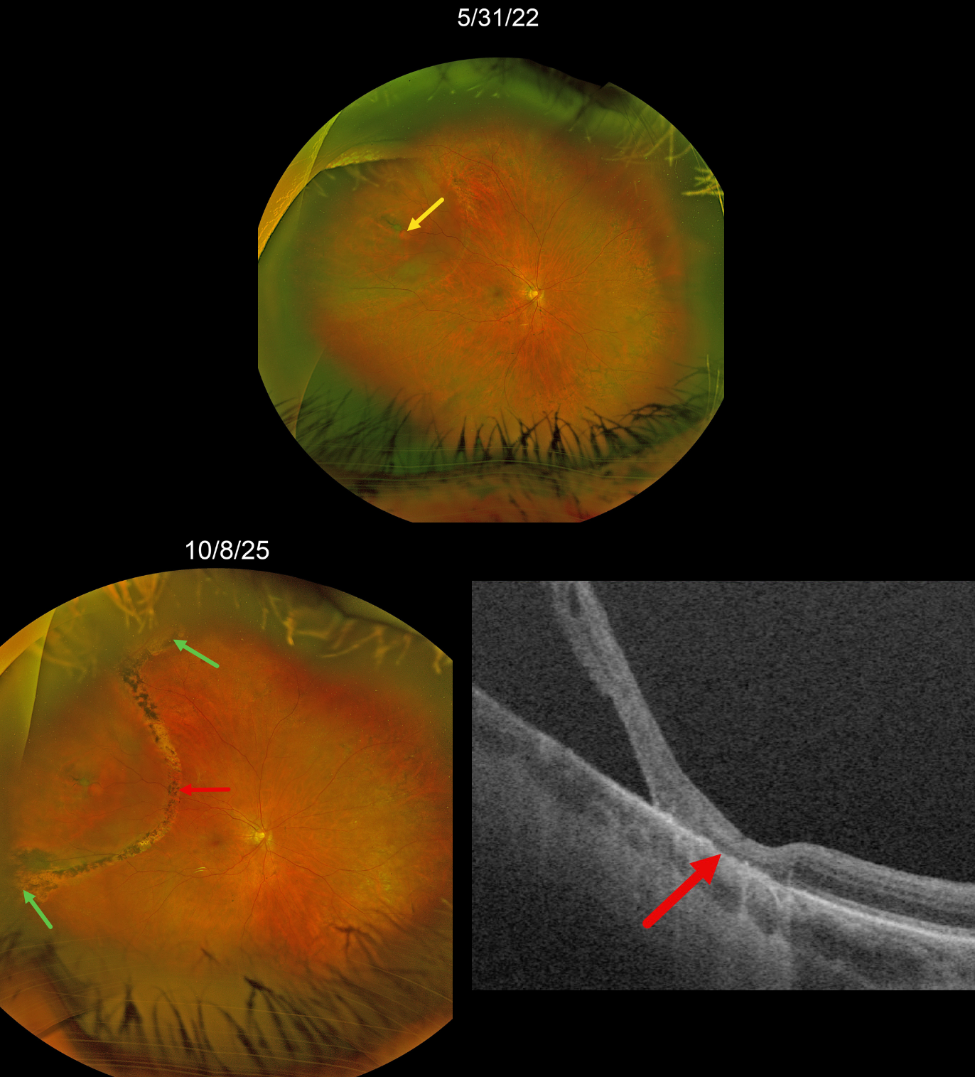
This 74YO female was referred on 5/31/22 for an asymptomatic rhegmatogenous retinal detachment (RRD) in her right eye.
Optos color RG imaging shows a chronic appearing superotemporal macula-on RRD with a single retinal break. A faint demarcation line is noted along the detachment’s posterior edge. The detachment was subsequently demarcated with thermal laser photocoagulation posteriorly and cryotherapy to its anterior edges.
We have been examining her twice yearly since. When last examined on 10/8/25, the detachment remained well-demarcated and without spread, with scarring from laser and cryotherapy. Optos Silverstone RGB swept-source OCT shows a broad area of outer retinal atrophy within the area of retinopexy.
Learning Points:
Demarcation, rather than reattachment, of the retina is a valid option for select cases of RRD. When demarcating these detachments, it is critical that the retinopexy completely surrounds the subretinal fluid and that treatment extends to the ora; otherwise, the detachment can spread through the untreated retina. Patients need to be followed postoperatively, as detachment can occasionally progress despite prior retinopexy.
See Vrabec and Baumal, Ophthalmology 2000;107:1063-1067 for an excellent review regarding demarcation laser photocoagulation for selected macula-sparing retinal detachments.
RHEGMATOGENOUS RETINAL DETACHMENT
Abhishek Karra and Dinesh Garg
Originally posted on @retina.rocks July 1, 2026
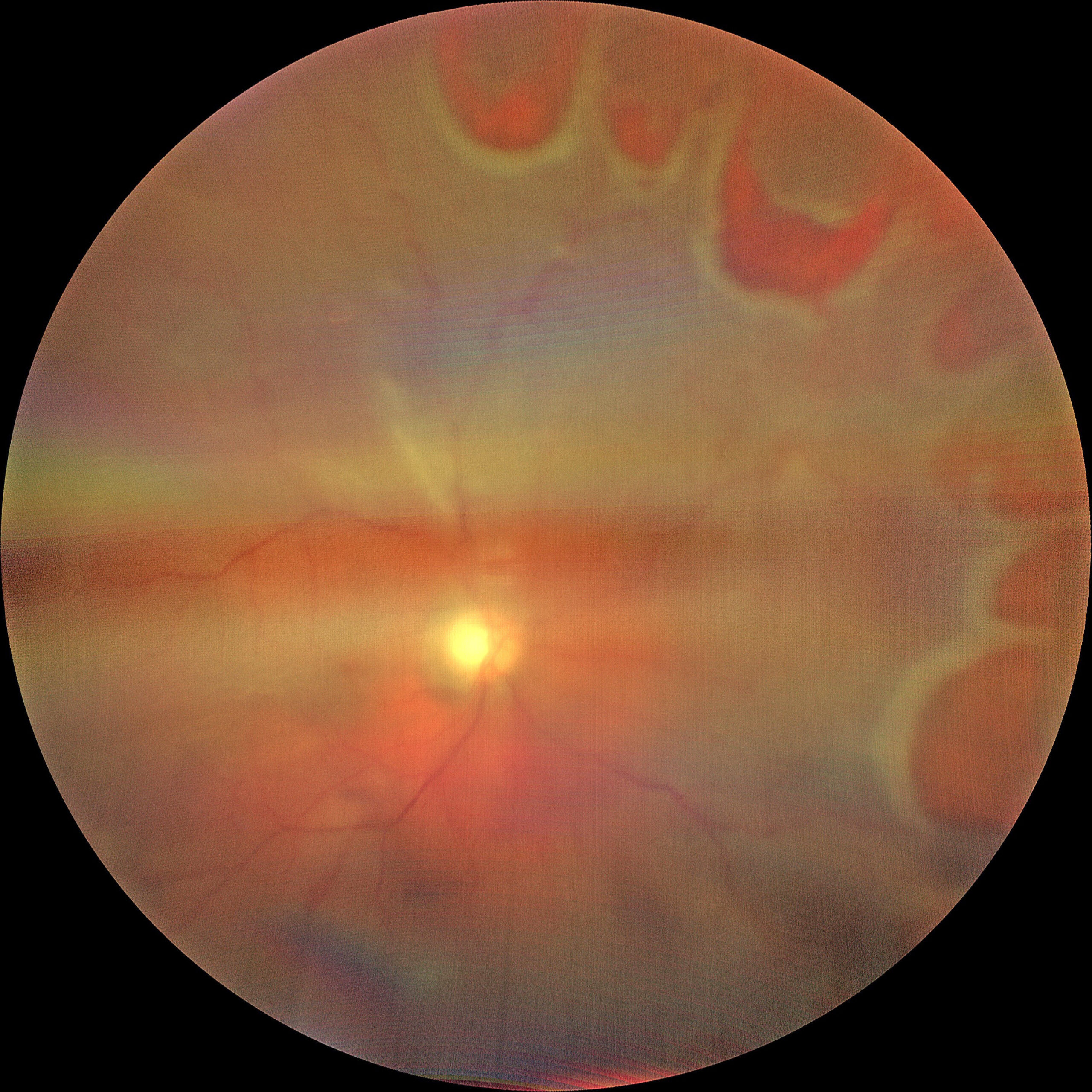
This 53YO male presented with 1 week of sudden vision loss in his right eye. Vision was counting fingers.
Fundus photography shows multiple retinal tears extending from 12 to 4 o’clock. There is a macula-off superior and nasal rhegmatogenous retinal detachment (RRD). The image is hazy due to an overlying vitreous hemorrhage. Emergency vitrectomy was scheduled.
INTRAOPERATIVE HEART DURING TRIAMCINOLONE-ASSISTED MEMBRANE PEELING
The European VitreoRetinal Society (EVRS), Claudio Brancato and Gregorio Lo Giudice
Originally posted on @retina.rocks February 13, 2026
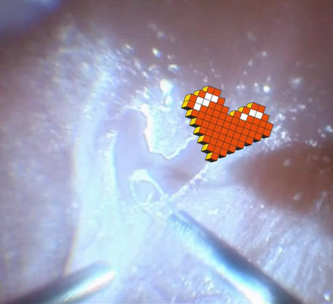
This 75YO male presented with hand motion vision in his right eye from a rhegmatogenous retinal detachment (RRD). Intraoperatively, triamcinolone was injected to stain the posterior hyaloid. During FINESSE Flex Loop-assisted membrane peeling, some love came back at us from the patient’s retina. The retina remained attached postoperatively, and vision improved to 20/50.
Learning Points:
Valentine’s Day traces its origins to a blend of ancient Roman customs, early Christian tradition, and the imaginative influence of medieval literature. What began as mid-February festivals gradually became associated with figures named Valentine, remembered for acts of compassion and steadfast conviction. Over time, poets such as Chaucer helped link the date with courtly love, giving rise to the exchange of affectionate notes and tokens. By the 18th and 19th centuries, these gestures evolved into the expensive cards, flowers, and symbols of romance that define the holiday today, transforming a complex historical tapestry into a widely celebrated expression of affection.
So, for you retinal surgeons out there, what could be a more romantic Valentine’s Day gift than offering to vitrectomize your loved one?

BRILLIANT BLUE DYE G-STAINED PERFLUOROCARBON LIQUID BUBBLES
Shishir Verghese
Originally posted on @retina.rocks January 28, 2026
This intraoperative image was obtained from a 55YO female undergoing pars plana vitrectomy for a total rhegmatogenous retinal detachment. Brilliant Blue G (BBG) dye was injected to facilitate membrane peeling, followed by the injection of perfluorocarbon liquid (PFCL) to flatten the posterior retina.
The PFCL was introduced while residual BBG dye was still present within the vitreous cavity. During the injection, a transient increase in infusion pressure caused mild intraoperative turbulence, resulting in fragmentation of the PFCL into multiple microbubbles that unexpectedly became infused with BBG dye.
Intraoperative photography captured this striking appearance of mid-vitreous BBG-stained PFCL microbubbles. The posterior retina is attached beneath a larger, unstained PFCL bubble, while the retina remains detached in the more anterior regions.
CNV AT INTERNAL DRAINAGE SITE FOLLOWING VITRECTOMY FOR RHEGMATOGENOUS RD
Originally posted on @retina.rocks November 10, 2025
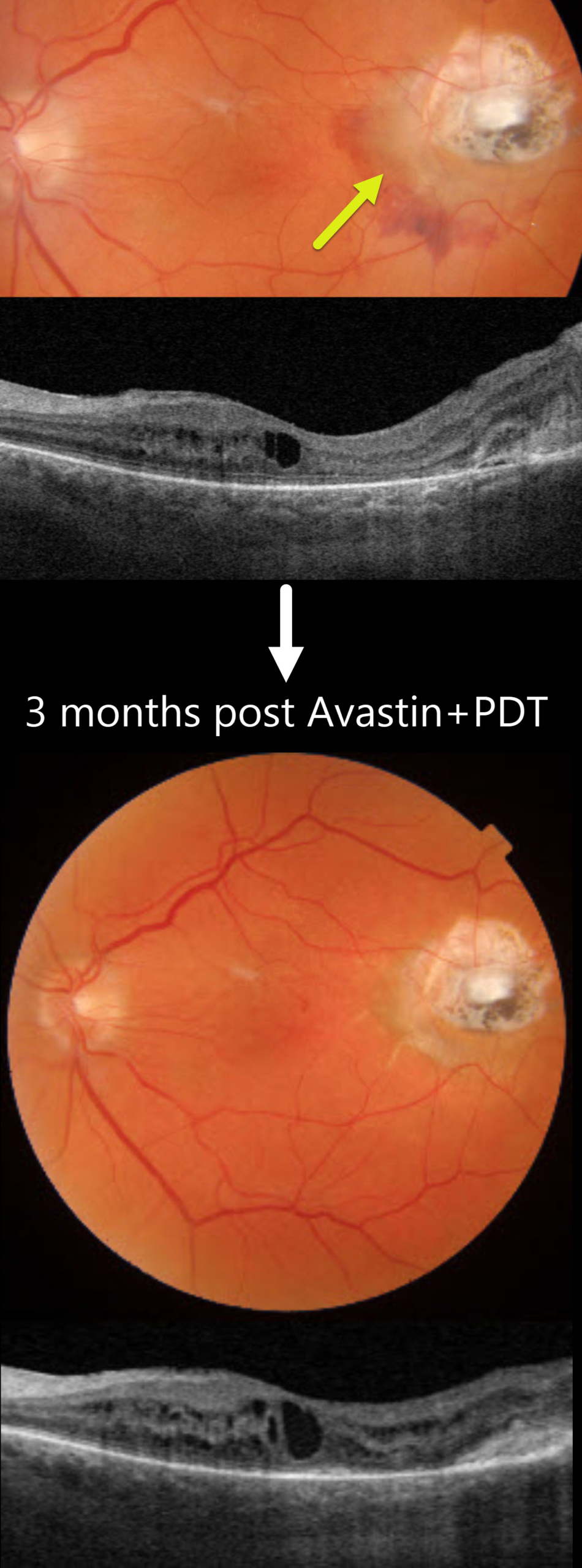
This 75YO male underwent successful vitrectomy for a macula-off rhegmatogenous retinal detachment (RRD). Vision improved from a preoperative hand motion vision to counting vision 7 months following surgery. However, he returned 4 months later with new distortion and blurriness. Vision was 20/400.
Triton color imaging shows an active choroidal neovascularization (CNV) with associated subretinal blood along the temporal and inferior edge of the drainage retinotomy scar. Swept-source OCT shows mild macular cystic edema arising from the temporal type 2 (above the RPE) CNV. The peripheral retina remained attached.
The lesion failed to respond to several intravitreal Avastin injections, necessitating combined Avastin and photodynamic therapy. Exudation completely resolved 3 months following this treatment.
Learning Points:
Invented by Steve Charles in the early days of vitrectomy, the drainage retinotomy has become an integral part of retinal reattachment surgery (Ramamurthy et al, Surv Ophthalmology 2023;68:1038-1049). Following the adage that ‘every time a hole is made in the retina, some vision leaks out,’ it is always preferable to drain subretinal fluid through a pre-existing break whenever possible. When required, the site should ideally be located superiorly and well away from the macula. Drainage retinotomies are associated with an increased risk of macular pucker and cystoid macular edema (McKay et al, AJO 2022;241:227-237). CNV is an exceedingly rare complication, which may be caused by inadvertent RPE-choroidal touch by the drainage needle or thermal-induced damage.
RHEGMATOGENOUS RD WITH RETINAL INCARCERATION FOLLOWING FAILED SCLERAL BUCKLING
Malvika Singh and Manish Nagpal
Originally posted on @retina.rocks November 4, 2025
PRIOR CHRONIC ICSC WITH NEW TRACTIONAL MACULAR HOLE-RRD WITH POSTOPERATIVE MENISCUS MICROPYON
Originally posted on @retina.rocks October 29, 2025
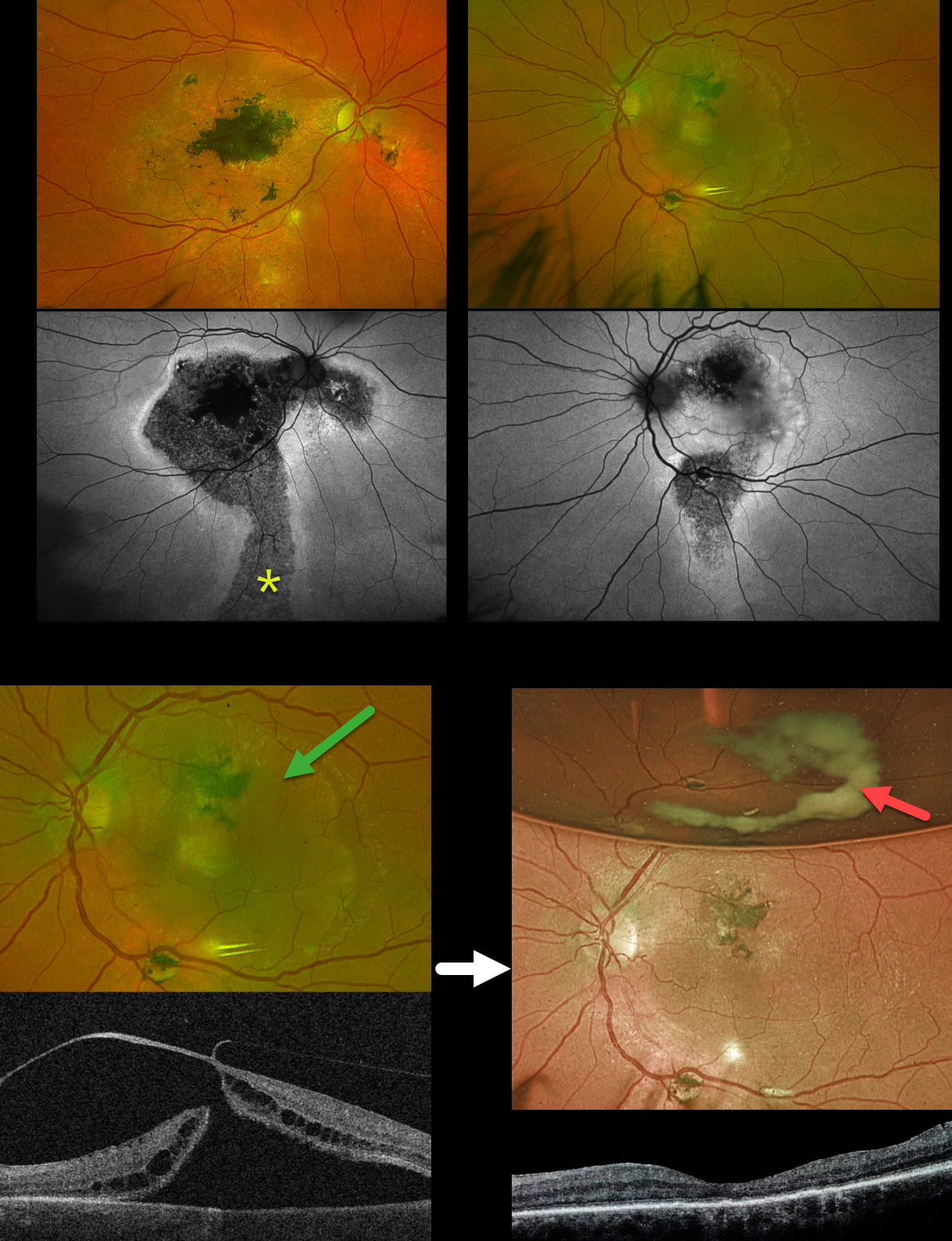
This 58YO male was referred for a retinal detachment in his left eye. He has a long history of idiopathic central serous chorioretinopathy (ICSC). He was previously treated elsewhere for neovascular AMD OD. For many years, he has been receiving intravitreal Avastin OS every 1-2 months. Vision was counting fingers OD and 20/400 OS.
Optos color RG imaging shows bilateral macular scarring with confluent hyperpigmented foveal changes and intraretinal pigment migration, especially in his right eye. Inferior hypopigmented gutters are most obvious on fundus autofluorescence (FAF) as hypo-FAF with surrounding hyper-FAF margins. A rhegmatogenous retinal detachment (RRD) occupies the entire left macula. Triton swept-source OCT shows a full-thickness macula hole, active vitreoretinal traction on the temporal edge of the hole, and a macular detachment.
The tractional macular hole with secondary RRD in our patient’s left eye is completely unrelated to his poorly treated ICSC. One month following vitrectomy, vision improved to 20/100. The retina was completely attached, and the macular hole was closed. A meniscus micropyon is adherent to the inferior meniscus of the gas bubble.
Learning Points:
Photodynamic therapy (PDT) is currently the standard of care for ICSC with symptomatic acute or chronic subretinal fluid. Unfortunately, Visudyne is rarely available outside the US, which has led to the adoption of unproven therapies, including mineralocorticoid receptor antagonists (eplerenone), beta blockers, and intravitreal anti-VEGF agents. Recent meta-analyses and systematic reviews confirm that these alternative therapies are ineffective (Felipe et al Int J Retina Vitreous 2022;8;34 and Kim et al Ophthalmology 2025;132:343-353). Despite the clear lack of efficacy in the literature, specialists both within and outside the US continue to use these drugs (Vankatesh et al Int J Retina Vitreous 2023;9;61). Our patient’s macular findings are a testament to what can happen when proven therapies are ignored.
Russell et al recently reported a unique ophthalmoscopic sign, a meniscus micropyon, that follows PPV with gas tamponade for all causes (Retina 2024;44:1766-1776). Features they noted that distinguish this sterile micropyon from post-PPV fibrin include a delayed appearance, hyperautofluorescence, and absence of anterior chamber or vitreous fibrinous strands/sheets. Vitreous biopsy in two of their cases revealed predominantly white blood cells. The micropyon appearance was associated with epiretinal proliferation (80%). Recurrent RRD from proliferative vitreoretinopathy (PVR) developed 33% of eyes, 12% re-detached without frank PVR, and 18% developed worsening epiretinal membranes.
Our patient’s meniscus micropyon disappeared as the bubble resolved. Three months postoperatively, vision was 20/60, and the retina remained completely reattached without PVR.
COMBINED TRACTIONAL-RHEGMATOGENOUS RETINAL DETACHMENT WITH MACULAR HOLE
Sucheta Kulkarani, Anita Gaikwad and Anjana Mirajkar
Originally posted on @retina.rocks July 24, 2025
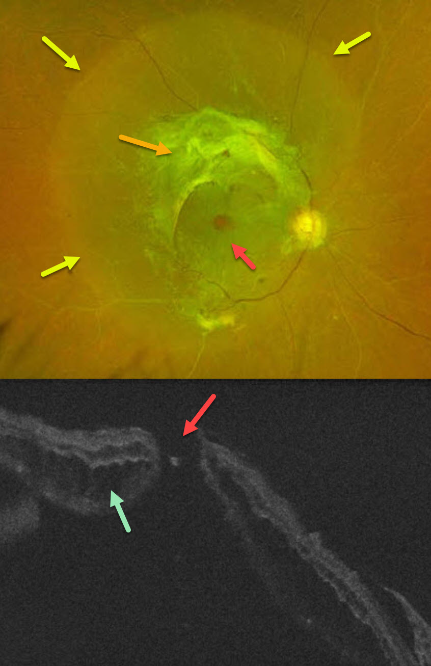
This 49YO male with type 2 diabetes noticed decreased vision in his right eye for 4 months. A single anti-VEGF injection was given elsewhere in his right eye around the time of his vision loss. Vision was 20/400 OD and 20/30 OS.
Optos color RG imaging OD shows a combined tractional-rhegmatogenous retinal detachment from dense fibrovascular proliferation and a full-thickness macular hole. OCT shows diffuse cystic edema within the detached retina, predominantly in the outer nuclear layer, and a macular hole.
Learning Points:
Tractional macular holes as a cause of combined tractional-rhegmatogenous diabetic retinal detachments are a rare event in our experience. Unfortunately, our patient was lost to follow-up immediately. We expect him to lose central vision in his left eye soon if he does not seek care.
RHEGMATOGENOUS RD WITH INADEQUATE LASER DEMARCATION
Originally posted on @retina.rocks July 9, 2025
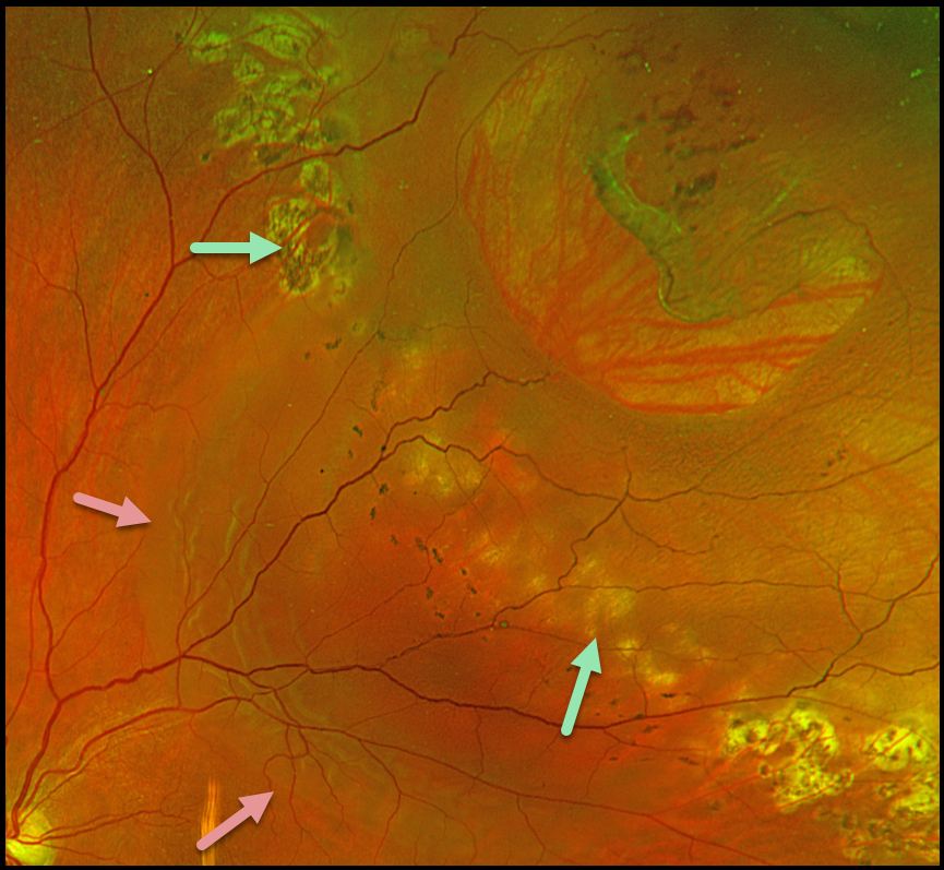
This 56YO male presented with several days of decreased vision in his left eye. He received prior treatment 1.5 years ago elsewhere for a retinal detachment. Vision was 20/30.
Optos color RG imaging shows a large lattice tear superotemporally with a surrounding rhegmatogenous retinal detachment (RRD). Several rows of appropriate and nearly confluent laser photocoagulation scarring are noted along the superonasal and inferotemporal edges of the detachment. However, barely visible depigmented laser scarring is noted more posteriorly, along with multiple tiny, 50-150 micron hyperpigmented laser scars. The detachment has broken through this region of inadequate laser.
Learning Points:
Demarcating rather than reattaching the retina is a valid option in select cases of RRD. When demarcating these detachments, it is critical that the retinopexy completely surrounds the subretinal fluid and extends to the ora; otherwise, the detachment can spread through the untreated retina, as occurred in this patient. Treatment should consist of several rows of nearly confluent, 300-500 micron photocoagulation burns. Cryotherapy or treatment with the laser indirect ophthalmoscope is often needed for the anterior treatment.
Assuming appropriate initial treatment, patients need to be followed postoperatively since the detachment can occasionally progress through the prior retinopexy. See Vrabec and Baumal, Ophthalmology 2000;107:1063-1067 for an excellent review regarding demarcation laser photocoagulation for selected macula-sparing retinal detachments.
LINCOFF RULES TO LOCATE BREAKS IN RHEGMATOGENOUS RETINAL DETACHMENT
Nilesh Kumar
Originally posted on @retina.rocks July 1, 2025
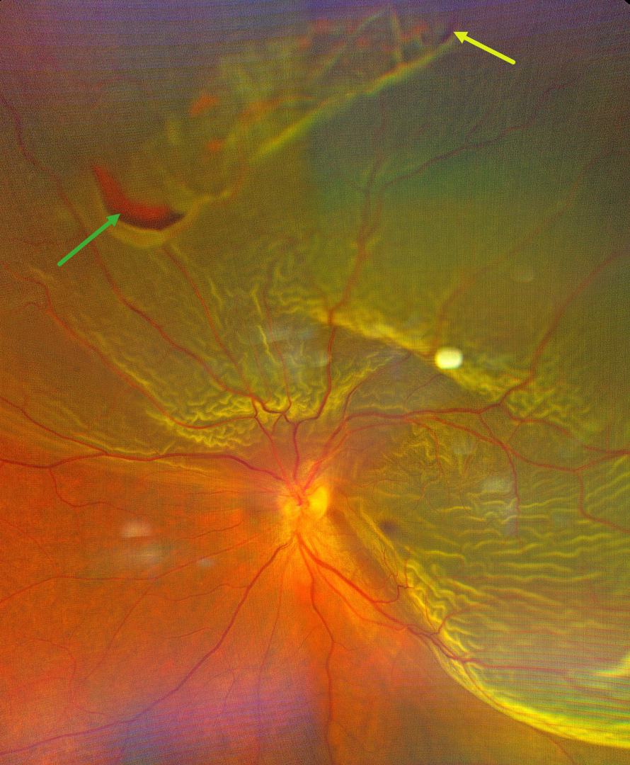
This 44YO male presented with sudden, painless, and progressive central and inferior peripheral vision loss. Color photography shows a superior and temporal macula-off rhegmatogenous retinal detachment (RRD). There is a superior patch of lattice degeneration with retinal tears at either edge of the lattice at 10:30 and 12:30.
Learning Points:
RRD occurs when liquid vitreous accesses the subretinal space through at least one full-thickness break in the neurosensory retina. A key principle for surgical success is identifying and closing all breaks. In 1971, Harvey Lincoff, in his classic paper ‘Finding the Retinal Hole,’ defined 4 findings based on RRD morphology that guide clinicians in determining where the causative breaks should be found (Lincoff and Gieser, Arch Ophthalmology 1971;85:565-569). These have since become known as the ‘Lincoff Rules.’ Simply put, gravity helps explain a detachment’s configuration and the location of the causative breaks.
Lincoff Rule 2 specifically addresses superior detachments crossing the 12 o’clock midline. It states that the primary break lies within a triangular zone with an apex at 12 o’clock, spanning 1.5 clock hours on either side (10:30-1:30 positions). This rule applies to 93% of such cases, as gravity causes subretinal fluid to spread symmetrically downward from the break.
Our patient’s detachment extends more inferiorly on the temporal edge. The two identified breaks in our image probably do not fully explain our RRD’s configuration, since the inferior edge of the detachment should fall on the nasal, not the temporal edge. At surgery, we would therefore search extra hard for at least one additional break along the temporal side.
MENISCUS MICROPYON
Mattie Adams
Originally posted on @retina.rocks April 3, 2025
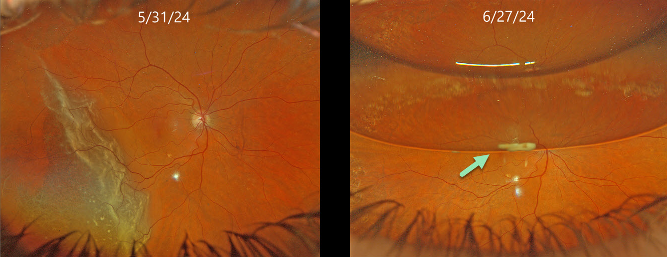
This 72YO female presented with one day of peripheral vision loss in her right eye. Vision was 20/25 OD and 20/25 in her healthy OS.
Optos color RG imaging (image 2, top) shows a temporal macula-on rhegmatogenous retinal detachment (RRD). Pars plana vitrectomy (PPV) with a C3F8 bubble was performed later that day. On her 1-month postoperative visit, vision was 20/50 with about a 50% gas bubble. A greyish globular deposit was adherent to the inferior edge of the gas meniscus.
Learning Points:
Russell et al recently reported a unique ophthalmoscopic sign, a meniscus micropyon, that follows PPV with gas tamponade for all causes (Retina 2024;44:1766-1776). Features they noted that distinguish this sterile micropyon from post-PPV fibrin include delayed appearance, hyperautofluorescence, and absence of fibrinous strands or sheets in the anterior chamber or vitreous. Vitreous biopsy in two of their cases revealed predominantly white blood cells. The micropyon appearance was associated with epiretinal proliferation (80%). Recurrent RRD from proliferative vitreoretinopathy (PVR) developed in 33% of eyes, 12% re-detached without frank PVR, and 18% developed worsening epiretinal membranes.
Fortunately, our patient’s postoperative course was uneventful. The micropyon resolved at the following visit and 4 months postoperatively, vision was 20/20 and the retina remained completely attached without epiretinal proliferation (not shown). Since the publication of Russell’s article, we have seen several patients with this biomarker, so we suspect it to be a not uncommon finding.
RHEGMATOGENOUS RETINAL DETACHMENT WITH SUB-ILM HEMORRHAGE
Originally posted on @retina.rocks February 12, 2025
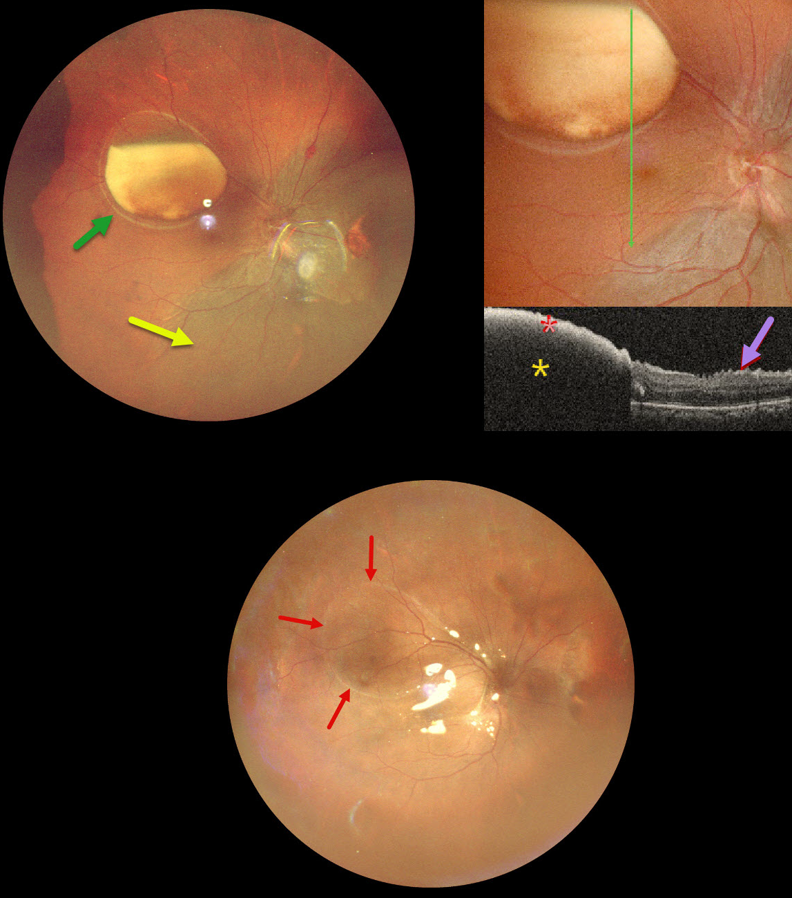
This 39YO male underwent vitrectomy OD elsewhere for a vitreous hemorrhage one month earlier. The treating surgeon noticed a sub-internal limiting membrane (ILM) hemorrhage at a postoperative visit and referred him to us for further evaluation. Vision was 20/30 OD and 20/20 in his normal OS.
Ultrawidefield pseudocolor SLO imaging shows a large partially layered sub-ILM hemorrhage with devitalized yellow blood in the superotemporal midperiphery. A retinal detachment is noted inferiorly and nasally. Inner retinal folds are visible at higher magnification. The blood is highly reflective on OCT scanning with total posterior shadowing. The inner retinal folds are noted inferiorly.
We don’t have a good explanation for our patient’s sub-ILM hemorrhage following vitrectomy for a vitreous hemorrhage. The yellow devitalized blood must have been present preoperatively, even though the initial surgeon told us he just noticed it one month postoperatively. Possibly our patient developed a breakthrough vitreous hemorrhage from a Valsalva maneuver, but without further history, this is just speculative.
We repaired the detachment with vitrectomy and silicone oil, and drained the sub-ILM blood intraoperatively. The retina was completely attached 2 weeks later. A high-water mark defines the margins of the prior ILM detachment.
RHEGMATOGENOUS RETINAL DETACHMENT
Fraser McKay
Originally posted on @retina.rocks February 3, 2025
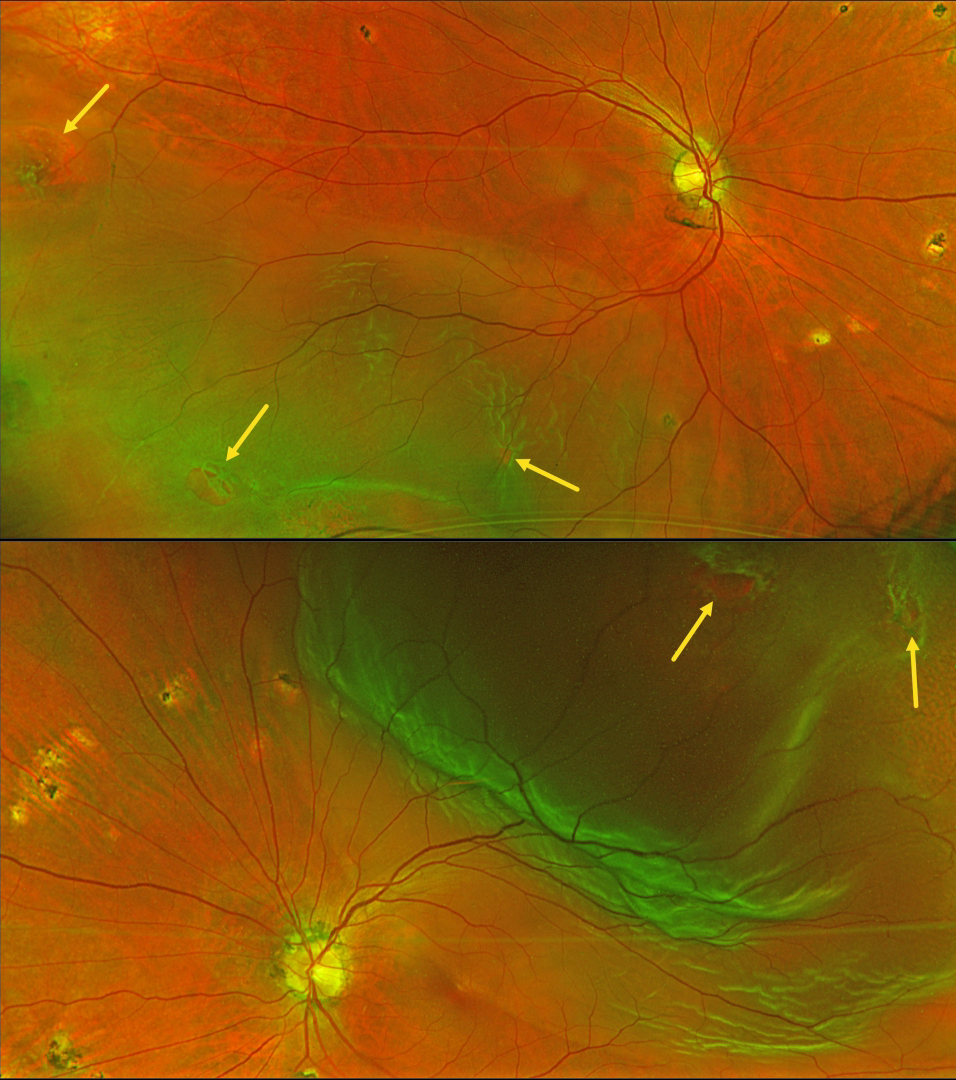
This 60YO male presented with 2 weeks of floaters in his left eye. Vision was 20/25 OD and 20/30 OS. Optos color RG imaging shows bilateral rhegmatogenous retinal detachments (RRD) with numerous breaks. The asymptomatic right detachment extended into the inferotemporal macula and split the left fovea. Scattered ocular histoplasmosis scars are present bilaterally.
Learning Points:
Patients with a RRD have a lifelong 10% risk of detachment in their fellow eye. Only a very small percentage will present with simultaneous detachments (Singh et al, Retina 2019;39:1504-1509).
Although the surgical success for each eye should be no different from that in patients presenting with unilateral detachments, treating the patient (not the eye) is more complicated. If one chooses pars plana vitrectomy (PPV), the postoperative gas bubble can cause severe, temporary vision loss. If both eyes receive PPV close together in time, the patient will be functionally blind for several weeks to months. We therefore find it helpful in these cases to treat the first eye with either silicone oil (with postoperative glasses) or perfluoro-octane liquid (followed by surgical removal a few weeks later). We can then repair the second eye within a few weeks of the initial eye. If possible, scleral buckling is also a good choice since no gas bubble is needed.
Emergent pars plana vitrectomy surgery was scheduled for our patient’s left detachment. We will follow the right eye closely perioperatively and will be forced to operate sooner if the detachment spreads towards the macular center. Otherwise, the right detachment will be repaired once the intraocular bubble is less than 50% in his left eye.
RETINAL TEAR WITH BRIDGING VESSEL
Originally posted on @retina.rocks January 9, 2025
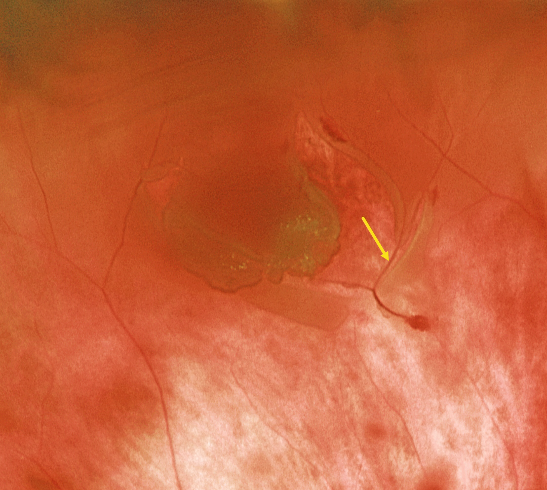
This patient presented with a macula-on rhegmatogenous retinal detachment (RRD). Optos color RG imaging shows the causative retinal tear with a bridging vessel. The RRD was successfully repaired with vitrectomy.
Learning Points:
The overall risk of an acute PVD causing a retinal break or detachment is about 5% (Seider et al, Ophthalmology 2022;129:67-72), and this risk is increased in the presence of vitreous pigment or blood. The reason for this is that, as in our patient, the retina often tears along a blood vessel. An occult retinal break is often the cause of an unexplained vitreous hemorrhage, which is why early vitrectomy is often recommended for these eyes, even when a break cannot be found on office examination (Flores-Sanchez et al, Eye 2023;37:3191-3196).
The bridging vessels in retinal breaks can also cause recurrent vitreous hemorrhage, a condition known as the avulsed retinal vessel syndrome (Robertson et al, Arch Ophthalmology 1971;85:669-672). The traction can spontaneously release, or vitrectomy may be required for sputtering hemorrhages or non-clearing blood.
RHEGMATOGENOUS RETINAL DETACHMENT
Yuenpang Cheung
Originally posted on @retina.rocks December 19, 2024
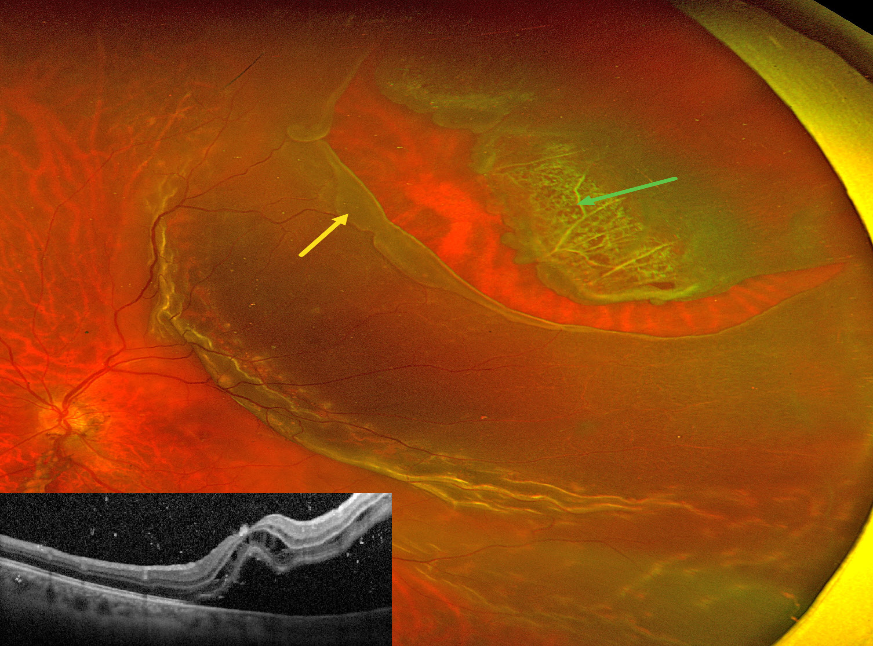
This 46YO male presented with recent vision loss in his left eye. Vision was counting fingers.
Optos color RG imaging shows a superotemporal macula-off rhegmatogenous retinal detachment (RRD). The anterior flap of the causative tear encompasses an entire lattice lesion. The posterior edge of the tear has a posteriorly-rolled edge, indicating early Grade B proliferative vitreoretinopathy (The Retina Society Terminology Committee, Ophthalmology 1983;90:121-125).
OCT shows cystic retinal edema mostly in the outer nuclear layer, along with hyperreflective vitreous and subretinal cells. The patient was scheduled for emergent vitrectomy.
CHOROIDAL DETACHMENT
Anand Temkar and Manish Nagpal
Originally posted on @retina.rocks October 31, 2024
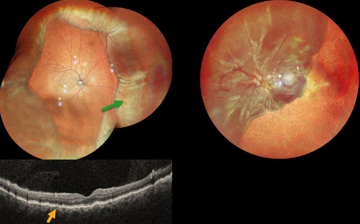
This 52YO male presented with 2 weeks of vision loss OD. He had a complicated ocular history of multiple prior surgeries elsewhere, including cataract surgery OD 2 months earlier and multiple retinal detachment surgeries OS. Vision was 20/30 OD and LP OS. Pressure was 8mmHg with no wound leaks on slit lamp OD.
MultiColor imaging shows encircling peripheral choroidal detachments OD, and OCT shows shallow macular chorioretinal folds. A total retinal detachment is noted OS with bare choroid nasally and inferiorly.
Learning Points:
A choroidal detachment is defined by the abnormal presence of fluid or blood in the suprachoroidal space. Serous choroidal detachments, also known as choroidal effusions, are a frequent complication of glaucoma surgery or following any intraocular procedure. Frank wound leaks usually require surgical repair. Otherwise, conservative management, often with topical/systemic steroids, is recommended.
Since our patient’s surgical wound was secure and the anterior chamber was formed, topical and oral steroids were prescribed.
CMV RETINITIS WITH RETINAL DETACHMENT
Originally posted on @retina.rocks September 2, 2024
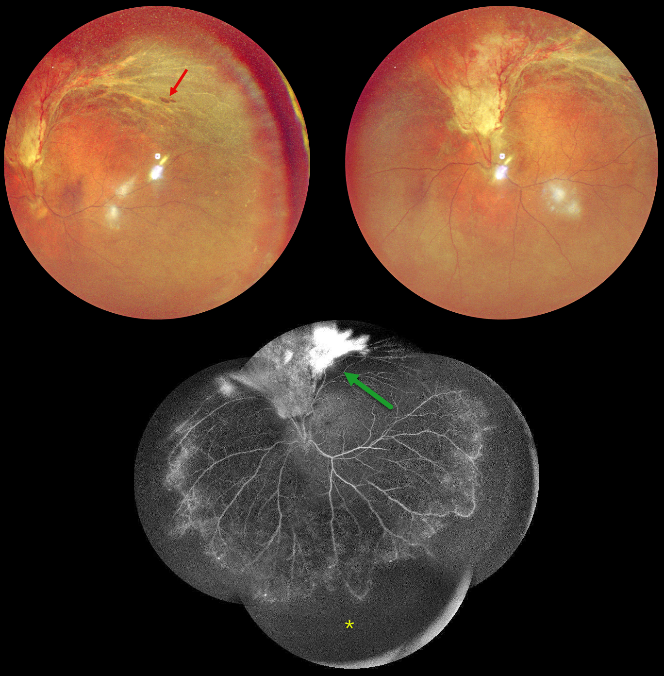
This 48YO HIV positive male on highly active antiretroviral therapy (HAART) presented with 2 months of vision loss in his left eye. His last CD4 count was 81. Vision was 20/20 OD and counting fingers OS.
Multicolor imaging shows active cytomegalovirus (CMV) retinitis with retinal detachment extending superiorly from the optic nerve into the superotemporal midperiphery. A few atrophic retinal breaks are noted. Fluorescein angiography of his left eye shows variable leakage within the area of retinitis superiorly and severe encircling peripheral ischemia. Vitrectomy with silicone oil was scheduled to repair the detachment in his left eye.
Learning Points:
CMV retinitis develops as a reactivation of latent CMV in immunosuppressed individuals. Before effective antiretroviral treatment emerged in the mid to late 1990’s, CMV retinitis developed in up to 40% of HIV/AIDS patients, often within the last 6 months of life. Treatment includes a combination of intravenous and intravitreal medications, including ganciclovir, foscarnet and cidofovir. Rhegmatogenous retinal detachment is a common late sequela and requires vitrectomy with silicone oil.
COMBINED TRACTIONAL-RHEGMATOGENOUS RETINAL DETACHMENT
Originally posted on @retina.rocks March 27, 2024
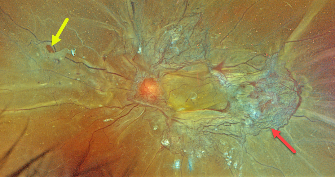
This 46YO female with type 2 diabetes presented with recent bilateral vision loss. Vision was 20/200 OD and hand motion OS.
Optos color RGB imaging of her left eye shows a combined tractional-rhegmatogenous total retinal detachment with fibrovascular tissue encircling the macula. Temporally radial outer retinal folds are dragged toward the more succulent, red neovascularization. A retinal tear is noted temporally.
Vitrectomy surgery was scheduled.
Proliferative diabetic retinopathy with disc and peripheral neovascularization was noted in her right eye (not shown). Panretinal photocoagulation was performed in her left eye and surgery was scheduled for her left eye.
JACK O- LANTERN DETACHMENT
Sandeep Kumar
Originally posted on @retina.rocks October 31, 2023
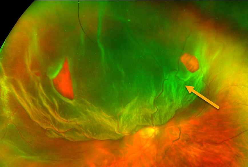
This 45YO female presented with a superior Jack-O-Lantern macula-off retinal detachment in her right eye. The detachment was successfully repaired with vitrectomy and C3F8 tamponade.
RHEGMATOGENOUS RETINAL DETACHMENT
Originally posted on @retina.rocks June 26, 2023
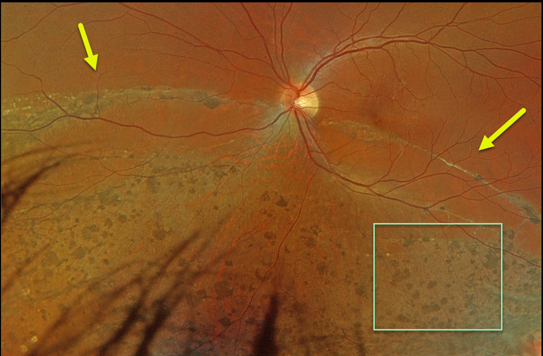
This 36YO male was referred for asymptomatic unilateral fundus findings. He had a history of retinal reattachment surgery 13 years earlier. Vision was 20/40.
Optos color RGB imaging shows chorioretinal scarring with diffuse intraretinal pigment migration within the area of prior detachment, giving the appearance of pseudo-sector retinitis pigmentosa (RP). A demarcation line courses through the inferior macula. This just spares the central macula, thankfully leaving him with good central acuity.
Learning Points:
Photoreceptor loss from numerous causes, including blunt trauma, retinal detachment, macular telangiectasia, and RP, can allow for intraretinal RPE migration with this shared phenotype.
Unilateral RP is exceedingly rare and requires normal electrophysiologic findings and long-term observation to ensure delayed findings in the fellow eye.
RHEGMATOGENOUS RETINAL DETACHMENT
Originally posted on @retina.rocks May 29, 2023
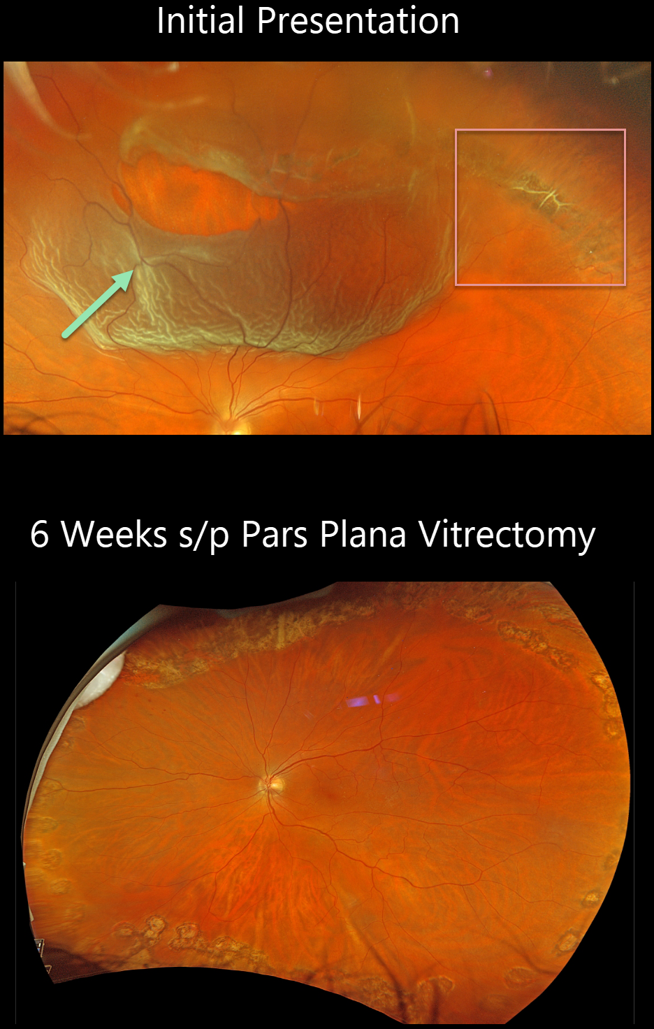
This 55YO male presented with this acute superior macula-on rhegmatogenous retinal detachment (RRD). Vision was 20/30.
Optos color RGB imaging shows a large patch of equatorial lattice degeneration extending from 11 to 2 o’clock. Nasally, the lattice lesion has torn along its lateral and posterior edges, causing the detachment. Several intact bridging retinal vessels are noted within the tear.
The detachment was repaired with vitrectomy the following day and remained attached 6 weeks later, following complete absorption of the intraocular gas bubble.
RHEGMATOGENOUS RETINAL DETACHMENT
Priyanak Gupta
Originally posted on @retina.rocks April 11, 2023
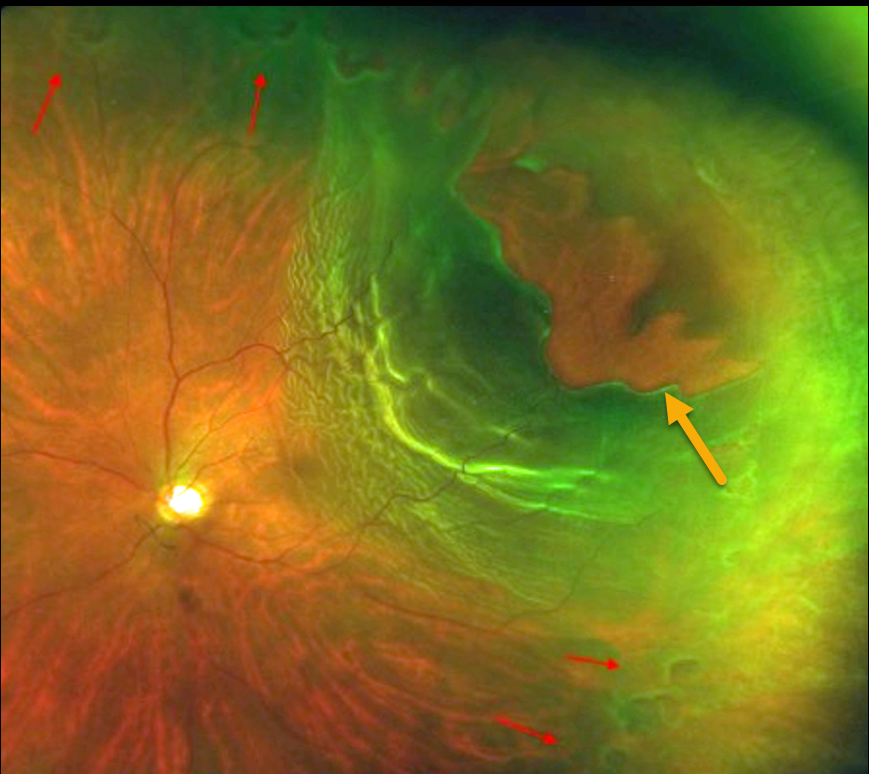
This patient presented with sudden vision loss from this rhegmatogenous retinal detachment (RRD).
Optos imaging shows a superotemporal macula-off RRD with an equatorial ring of numerous small retinal tears, along with a large ragged tear simulating stalactites. The patient subsequently underwent successful retinal reattachment surgery with vitrectomy.
RETINAL TEAR
Originally posted on @retina.rocks November 17, 2022
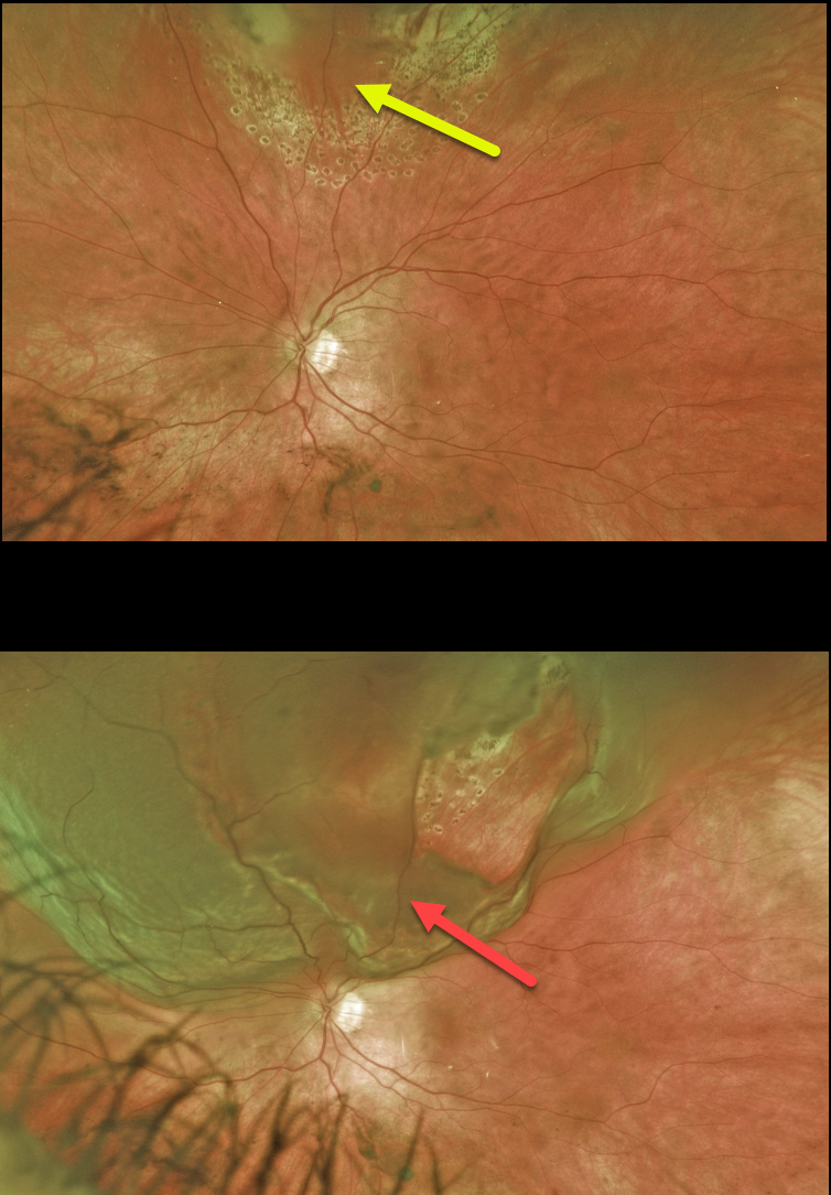
This 37YO female presented with a symptomatic superior retinal tear in her left eye. The posterior and lateral edges of the break were surrounded with thermal laser. The anterior edges could not be reached with the laser, so these were surrounded with cryotherapy. One week later, pigmented scarring surrounds the treated tear.
Three weeks later, she presented with a few days of inferior visual field loss. She now has a superior macula-on rhegmatogenous retinal detachment (RRD) due to lifting off the originally treated tear. Vitrectomy surgery was performed the following day.
Learning Points:
Untreated symptomatic retinal tears have a high risk for RRD. Retinopexy, either with thermal laser or cryotherapy, must completely surround the break and, for more anterior lesions, ideally extend to the ora.
Patients must be followed closely, especially during the first month, due to the risk of additional breaks or retinal detachment. A firm chorioretinal adhesion takes several weeks to form (Yoon and Marmor, Ophthalmology 1988;95:1385-1388). Patients must always be cautioned to return immediately for new flashes, floaters, or vision loss.
RHEGMATOGENOUS RETINAL DETACHMENT
Originally posted on @retina.rocks October 13, 2022
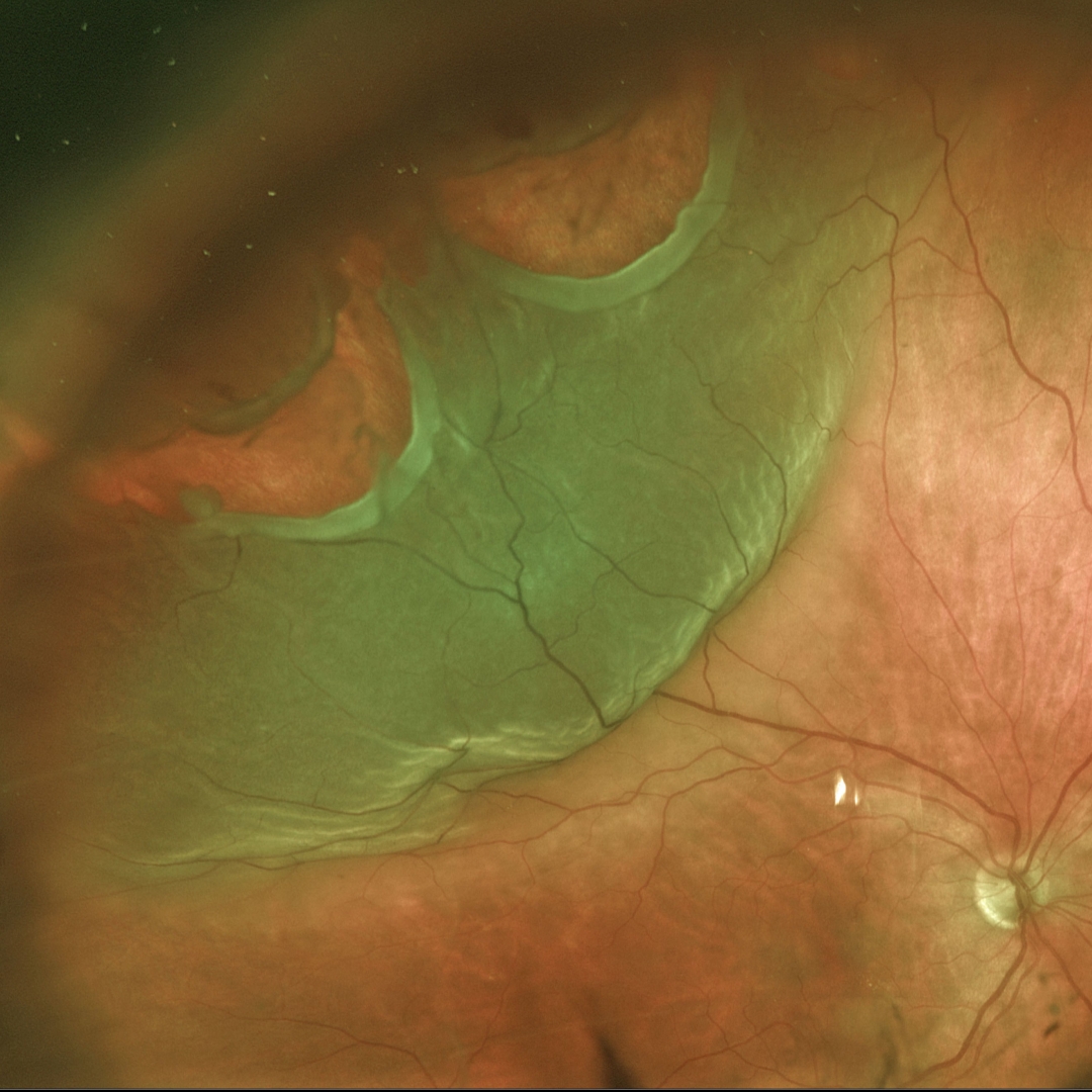
This 60YO female was referred for a symptomatic superotemporal rhegmatogenous retinal detachment (RRD) in her right eye.
Two large causative retinal tears are seen, both with posteriorly rolled edges.
The patient underwent emergent successful pars plana vitrectomy.
CYSTICERCOSIS WITH RHEGMATOGENOUS RETINAL DETACHMENT
Chhaya Bharti
Originally posted on @retina.rocks September 14, 2022
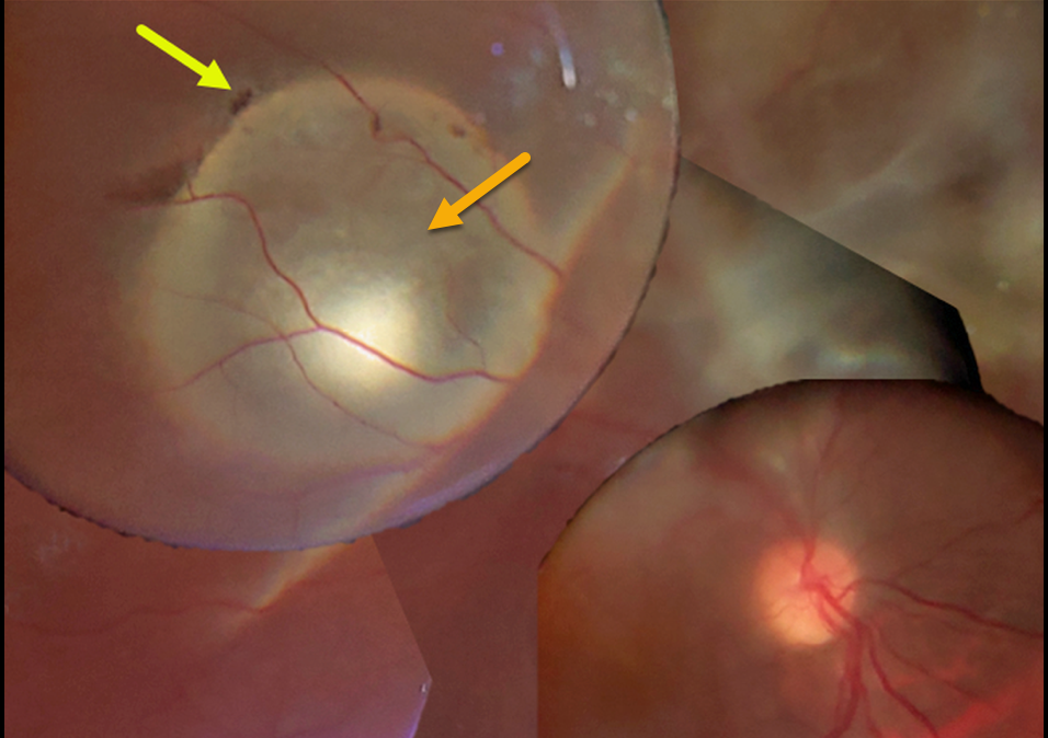
This 15YO boy presented with 20/200 vision. A large subretinal cysticercosis cyst is noted in the right distal superotemporal macula.
There is an overlying rhegmatogenous retinal detachment caused by a small retinal break. Unfortunately, the patient was subsequently lost to follow-up.
Learning Points:
Ocular cysticercosis is a rare parasitic eye infection caused by a pork tapeworm (larval form of the cestode, Taenia solium).
The ocular adnexa are most commonly affected, but the encysted organism can appear within the eye, including the anterior segment, vitreous cavity, and the subretinal space, as in this patient.
Vitrectomy with complete removal of the subretinal cyst is required since the dead organism can cause a severe inflammatory response.
For a great recent cysticercosis in ophthalmology review article, see Pujari et al, Surv Ophthalmology 2022;67:544-569.
PROLIFERATIVE VITREORETINOPATHY (PVR)
Originally posted on @retina.rocks June 17, 2022
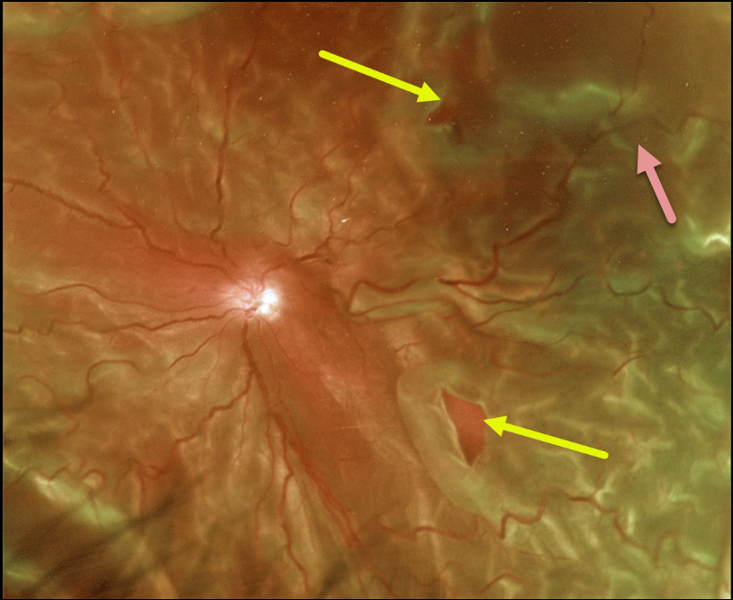
This 65YO male presented with light perception in his left eye secondary to a very chronic total rhegmatogenous retinal detachment (RRD).
Optos color imaging captured the detachment, which was fairly stiff due to diffuse proliferative vitreoretinopathy (PVR). Two posterior retinal breaks are seen.
The retina was successfully reattached with vitrectomy, inferior retinectomy, and silicone oil.
Learning Points:
PVR, which consists of cellular membranes growing on either retinal surface, develops in about 5-10% of retinal detachments. The membranes contract, foreshorten the retina, and likely proliferate as an aberrant wound-healing response. There are no proven pharmacologic therapies to prevent or treat PVR.
These complex detachments can be managed with vitrectomy with membrane peeling, retinectomy, scleral buckling, and silicone oil. However, PVR remains the main cause for ultimate retinal reattachment surgical failure.
See Garweg et al Surv Ophthalmol 2013;48:321-329 for a good review of PVR pathophysiology.
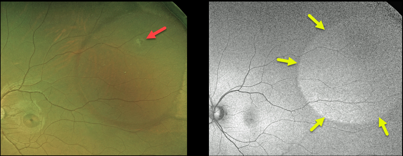
RHEGMATOGENOUS RETINAL DETACHMENT
Originally posted on @retina.rocks June 8, 2022
This 25YO female was referred for an asymptomatic rhegmatogenous retinal detachment (RRD) in her left eye. The detachment had an unusual oval shape that did not reach the ora, with a small causative break.
Fundus autofluorescence (FAF) shows variable hyper-FAF throughout the detachment, likely due to unmasking of the underlying RPE by dysfunctional photoreceptors.
Various treatment options were discussed, and the patient opted for thermal laser demarcation.
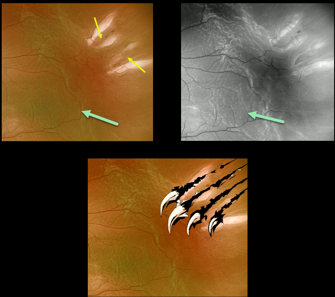
WOLVERINE RETINOPATHY
Originally posted on @retina.rocks May 10, 2022
This 72YO male presented with a macula-off rhegmatogenous retinal detachment (RRD).
Optos imaging gives the illusion of four breaks, although in actuality, there are only two tractional tears. The hydrated outer retinal folds are best visualized on green channel imaging.
Vision was 20/60, and the retina was successfully reattached with emergent pars plana vitrectomy.
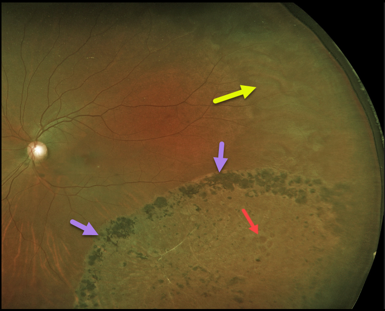
RHEGMATOGENOUS RETINAL DETACHMENT
Originally posted on @retina.rocks April 15, 2022
Years earlier, this 44YO female underwent laser demarcation of an asymptomatic inferotemporal rhegmatogenous retinal detachment (RRD). The prior detachment remains well demarcated, but the detachment itself has completely flattened.
Some of the causative lattice-related atrophic holes are seen within the area of prior detachment. White without pressure is also noted in the temporal periphery.
Learning Points:
Demarcation, rather than reattachment, of the retina is a valid option for select cases of RRD. When demarcating these detachments, it is critical that the retinopexy completely surrounds the subretinal fluid and that treatment extends to the ora; otherwise, the detachment can spread through the untreated retina.
Patients need to be followed postoperatively for life since the detachment can occasionally progress through the prior retinopexy.
Interestingly, we have seen that some of these detachments, as occurred in this case, spontaneously flatten following demarcation.
See Vrabec and Baumal, Ophthalmology 2000;107:1063-1067 for an excellent review regarding demarcation laser photocoagulation for selected macula-sparing retinal detachments.
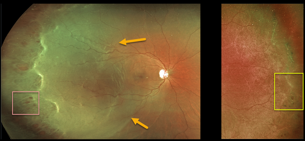
RHEGMATOGENOUS RETINAL DETACHMENT
Originally posted on @retina.rocks April 1, 2022
This 32YO female presented with a macula-off rhegmatogenous retinal detachment (RRD) in her right eye. There were innumerable temporal retinal holes.
The detachment was successfully repaired with vitrectomy surgery.
Her asymptomatic left eye showed extensive temporal lattice lesions with atrophic holes, and prophylactic laser was recommended.
RHEGMATOGENOUS RETINAL DETACHMENT
Originally posted on @retina.rocks February 25, 2022
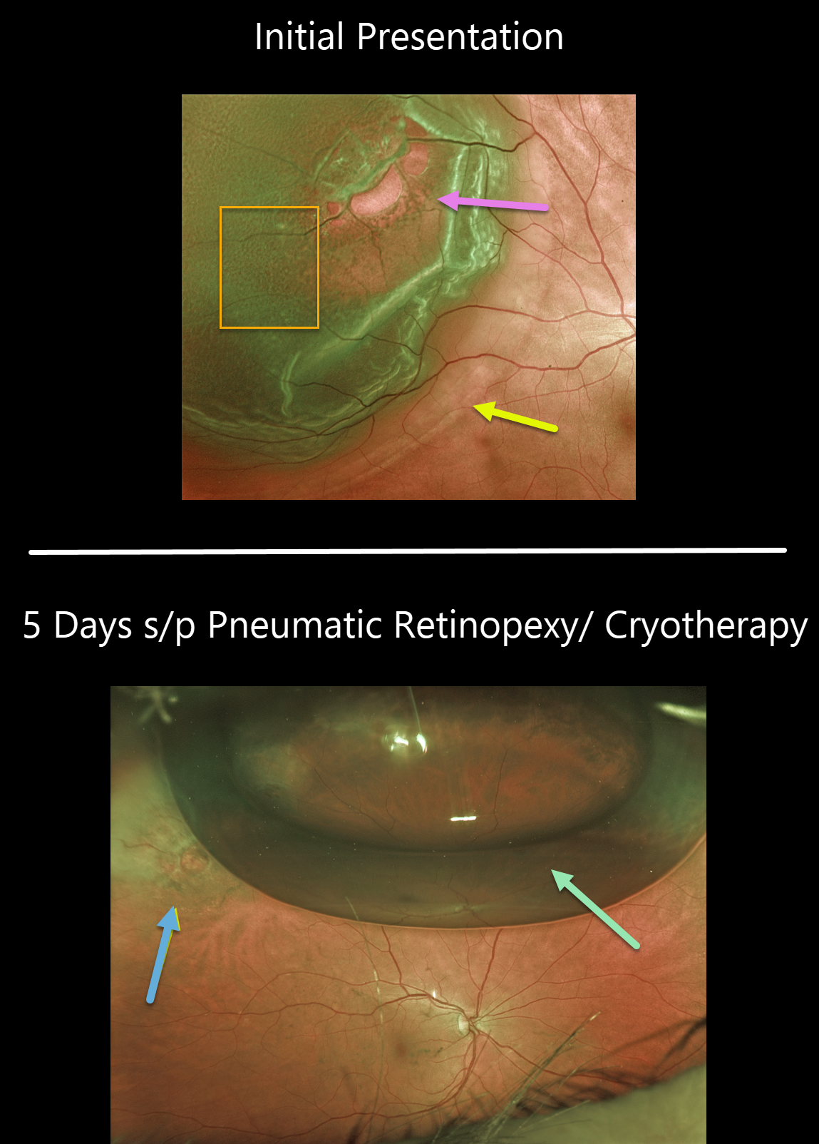
This 63YO female presented with 20/25 vision and a superotemporal retinal detachment. The posterior edge of the detachment was just outside the macular center. There was diffuse fine microcystic retinal edema within the area of detachment, except for outer retinal hydration folds more posteriorly.
The retina was completely attached 5 days later following successful cryotherapy with pneumatic retinopexy. Early cryotherapy scarring is already visible.
Learning Points:
This detachment could have been repaired with any technique, including vitrectomy, scleral buckling, or pneumatic retinopexy. All techniques yield similar final anatomic success, although the debate continues about the relative merit of one over the other.
See the ___References folder in the Retinal detachment (RD) rhegmatogenous (RRD) folder in the Image Library at www.retinarocks.org for a summary of the key journal articles.
RHEGMATOGENOUS RETINAL DETACHMENT
Originally posted on @retina.rocks December 17, 2021
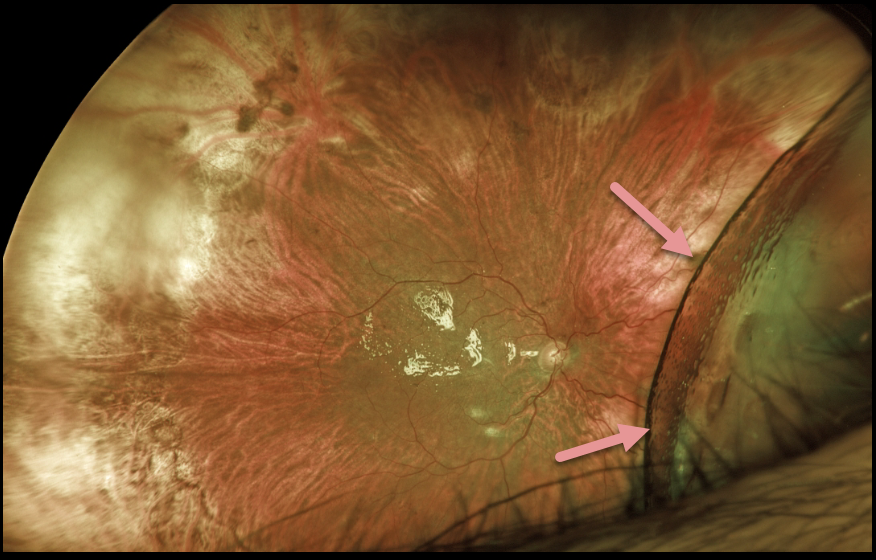
This patient presented with light perception vision following a 1-year-old total retinal detachment with proliferative vitreoretinopathy. At vitrectomy, all membranes were peeled, and the anterior loop traction was relieved (or so we thought). The retina was mobile and completely flattened with fluid-air exchange. 1000 centistoke silicone oil was infused.
At the one-day and one-week postoperative visits, vision remained light perception. The retina was completely attached except for the nasal periphery, which was locally detached with subretinal silicone oil.
Since the surgical goal was to prevent total blindness and possible phthisis, we have decided to observe for now as long as the residual detachment remains localized.
What would your management be at this point?
RHEGMATOGENOUS RETINAL DETACHMENT
Originally posted on @retina.rocks December 3, 2021
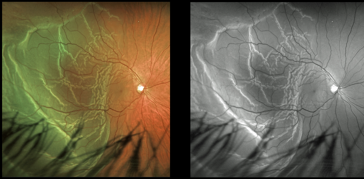
This 47YO male presented with hand motion vision from a macula-off rhegmatogenous retinal detachment (RRD). The outer retinal folds are dramatically imaged in both the Optos color and green channel images.
Learning Points:
Hydration outer retinal folds are one of the key findings to help differentiate a RRD from exudative and traction retinal detachments.
The cause of these folds remains uncertain but may result from the hydration of glycosaminoglycans in the interphotoreceptor matrix (Dalvin et al, Retinal Cases & Brief Reports 2020;14:355-359).
RHEGMATOGENOUS RETINAL DETACHMENT
Originally posted on @retina.rocks November 2, 2021
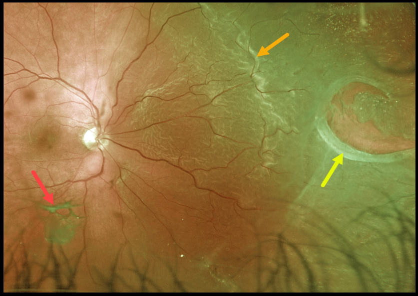
This patient presented with a macula-on rhegmatogenous retinal detachment (RRD) from a large nasal retinal tear. The tear has a rolled posterior edge, indicating possible early proliferative vitreoretinopathy.
The hydration outer retinal folds at the posterior edge of the detachment are one of the key clinical findings that help differentiate a rhegmatogenous from exudative and traction retinal detachments.
The causative posterior vitreous detachment is seen floating above the retina just inferior to the macula.
Learning Points:
The normal neurosensory retina remains attached to the RPE due to numerous physiological mechanisms, including the inner (retinal vascular endothelium) and outer (tight junctions at the apex of the RPE cells) blood retinal barriers, hyperosmotic choroid passively drawing fluid into the subretinal space, RPE pump, interphotoreceptor matrix, and friction between the RPE microvilli and the photoreceptor outer segments.
Rhegmatogenous retinal detachment (RRD) occurs when liquefied vitreous, entering the subretinal space through a retinal break, overwhelms the forces that favor retinal detachment.
Repairing RRD involves finding and closing all breaks, essentially denying the liquid vitreous access to the subretinal space. The break(s) can be closed internally with a gas bubble with pneumatic retinopexy or vitrectomy surgery, or externally by sewing a piece of silicone material to the eye wall (scleral buckle). This indents, or buckles, the sclera up against the retina break.
RHEGMATOGENOUS RETINAL DETACHMENT
Originally posted on @retina.rocks September 22, 2021
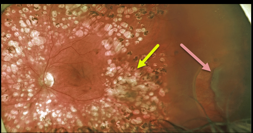
This 50YO male presented for a routine diabetic follow-up. A new asymptomatic peripheral retinal tear causing a rhegmatogenous retinal detachment was noted anterior to full panretinal photocoagulation (PRP) scarring.
We will often demarcate asymptomatic retinal detachments with laser photocoagulation (Vrabec and Baumel, Ophthalmology 2000;107:1063-1067). This patient essentially had laser demarcation before his retinal detachment, but the end result was the same, with his detachment being contained by the laser scarring.
Learning Points:
We opted to treat this eye with vitrectomy, but one could argue that this detachment could also be closely followed since it was contained by the prior laser.
CHOROIDAL COLOBOMA
Originally posted on @retina.rocks September 10, 2021
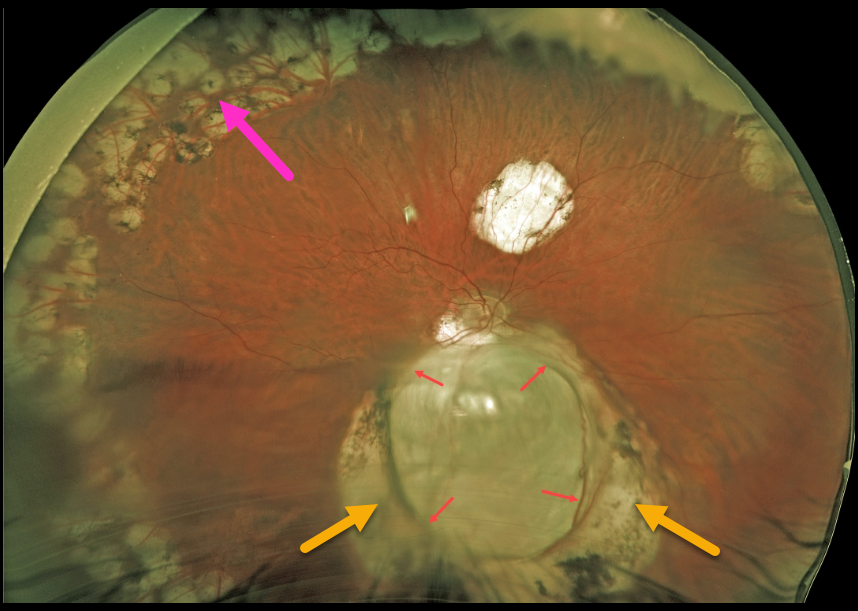
This 57YO male underwent successful vitrectomy surgery 8 years earlier for rhegmatogenous retinal detachment (RRD). Vision is now 20/40.
The detachment was caused by a retinal break associated with an inferior choroidal coloboma. The margins of the coloboma are surrounded by confluent depigmented scarring from prior laser photocoagulation. Prophylactic peripheral laser was also applied intraoperatively.
Learning Points:
Choroidal colobomas arise from incomplete closure of the embryonic fissure during the second month of embryonic development.
Patients are at lifelong risk of rhegmatogenous retinal detachment due to breaks in the overlying thinned retina.
RHEGMATOGENOUS RETINAL DETACHMENT
Originally posted on @retina.rocks August 30, 2021
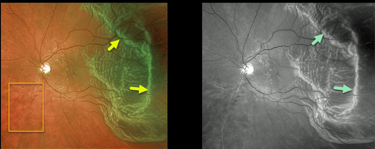
This 67YO male presented with hand motion vision from a macula-off rhegmatogenous retinal detachment (RRD).
The outer retinal folds are dramatically imaged in both the Optos color and green channel images. An incidental, old asymptomatic branch retinal vein occlusion is also noted inferonasally.
Learning Points:
Hydration outer retinal folds are one of the key findings to help differentiate a RRD from exudative and traction retinal detachments.
The cause of these folds remains uncertain, but they may form as a result of hydration of glycosaminoglycans in the interphotoreceptor matrix (Dalvin et al., Retinal Cases & Brief Reports 2020;14;355-359).
RHEGMATOGENOUS RETINAL DETACHMENT
Originally posted on @retina.rocks June 10, 2021
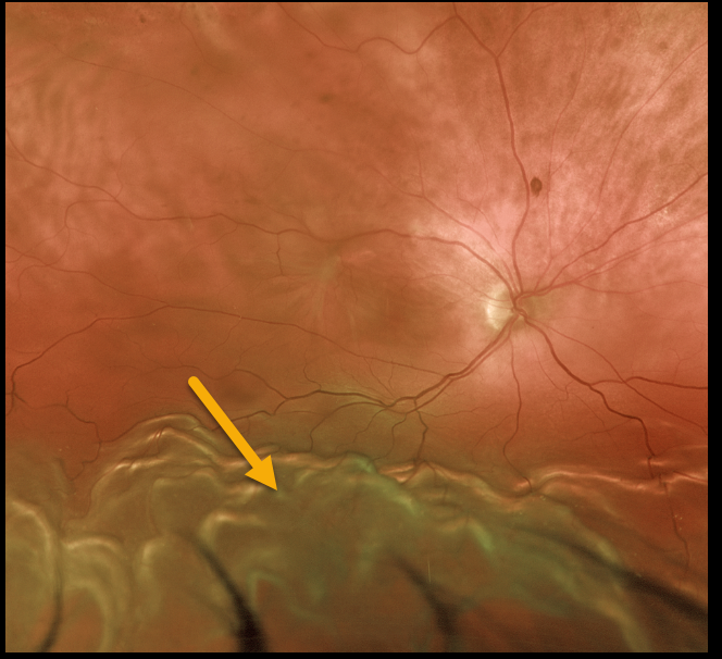
This 77YO male presented with 20/400 vision from a chronic macula-off rhegmatogenous retinal detachment (RRD).
Demarcation lines are common in these cases (not present here). The billowing, corrugated convex appearance of the inferior retina is typical of RRD. The retina is mobile without proliferative vitreoretinopathy.
Learning Points:
The normal neurosensory retina remains attached to the RPE due to numerous physiologic mechanisms, including the inner (retinal vascular endothelium) and outer (tight junctions at the apex of the RPE cells) blood retinal barriers, hyperosmotic choroid passively drawing fluid into the subretinal space, RPE pump, interphotoreceptor matrix, and friction between the RPE microvilli and the photoreceptor outer segments.
Rhegmatogenous retinal detachment (RRD) occurs when liquid vitreous, entering the subretinal space through a retinal break, overwhelms these forces, favoring retinal detachment.
RHEGMATOGENOUS RETINAL DETACHMENT
Originally posted on @retina.rocks May 3, 2021
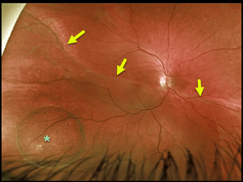
This 34YO male presented with 20/80 vision from a chronic macula-off rhegmatogenous retinal detachment (RRD).
A demarcation line extends along the entire posterior extent of the detachment. There is a retinal macrocyst within the detachment inferotemporally.
Learning Points:
Chronic retinal detachments rarely develop these macrocysts, which are located within the outer plexiform layer (see Marcus and Aaberg, Arch Ophthalmology 1979;97;1275-1279).
They do not need to be treated during RRD surgery unless they prevent closure of the retinal break(s). They promptly flatten following retinal reattachment.
RHEGMATOGENOUS RETINAL DETACHMENT
Originally posted on @retina.rocks April 19, 2021
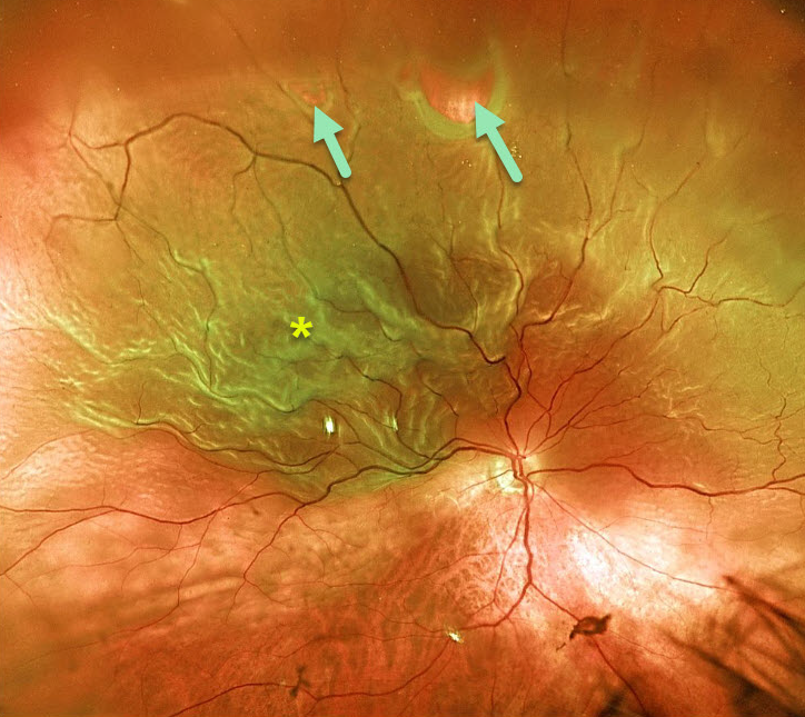
This 77YO patient presented with 20/200 vision from a superior macula-off retinal detachment. Optos imaging beautifully captures the detachment that was caused by several superior retinal tears.
Learning Points:
The normal neurosensory retina remains attached to the RPE due to numerous physiologic mechanisms, including the inner (retinal vascular endothelium) and outer (tight junctions at the apex of the RPE cells) blood retinal barriers, hyperosmotic choroid passively drawing fluid into the subretinal space, RPE pump, interphotoreceptor matrix, and friction between the RPE microvilli and the photoreceptor outer segments.
Rhegmatogenous retinal detachment (RRD) occurs when liquid vitreous, entering the subretinal space through a retinal break, overwhelms these forces, favoring retinal detachment.
RHEGMATOGENOUS RETINAL DETACHMENT
Originally posted on @retina.rocks April 9, 2021
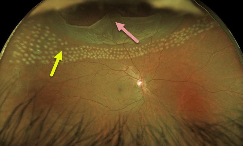
This 51YO male presented with a completely asymptomatic superior rhegmatogenous retinal detachment. There was a history of blunt trauma 1 year earlier.
We discussed the various surgical options, and he decided to proceed with laser demarcation due to the lack of symptoms and remote history of the causative injury. The immediate post-laser Optos image shows the barrier laser treatment.
We were unable to reach the ora with the slit lamp delivery system, so the anterior edges were subsequently treated with cryotherapy.
Learning Points:
When demarcating these detachments, it is critical that the retinopexy completely surrounds the subretinal fluid and that treatment extends to the ora; otherwise, the detachment can spread through the untreated retina.
Patients need to be followed postoperatively, as detachment can occasionally progress despite prior retinopexy.
See Vrabec and Baumal, Ophthalmology 2000;107:1063-1067 for an excellent review regarding demarcation laser photocoagulation for selected macula-sparing retinal detachments.
DEMENTOR DETACHMENT
Originally posted on @retina.rocks March 22, 2021
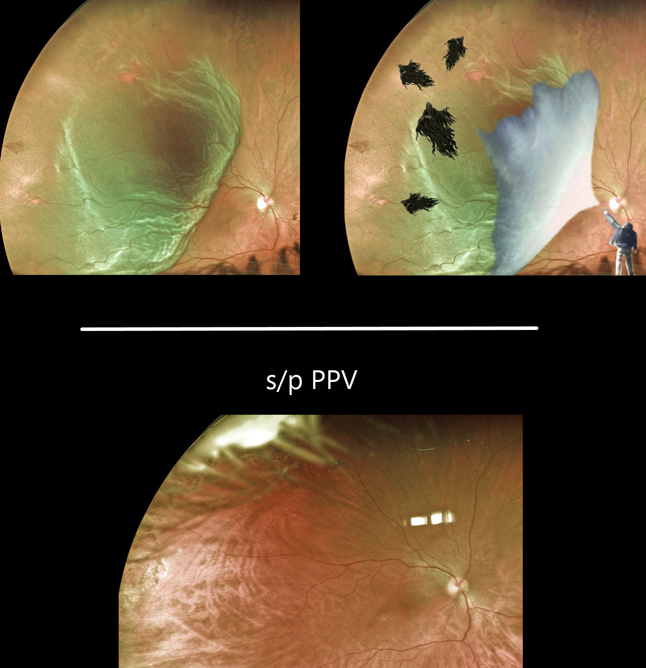
Multiple temporal retinal tears are visible in this superotemporal macula-on rhegmatogenous retinal detachment. Yet it appears to be held off like Harry Potter’s Dementors.
We happen to have a powerful wizard, too.
Just look at the beautifully attached retina after a successful pars plana vitrectomy!
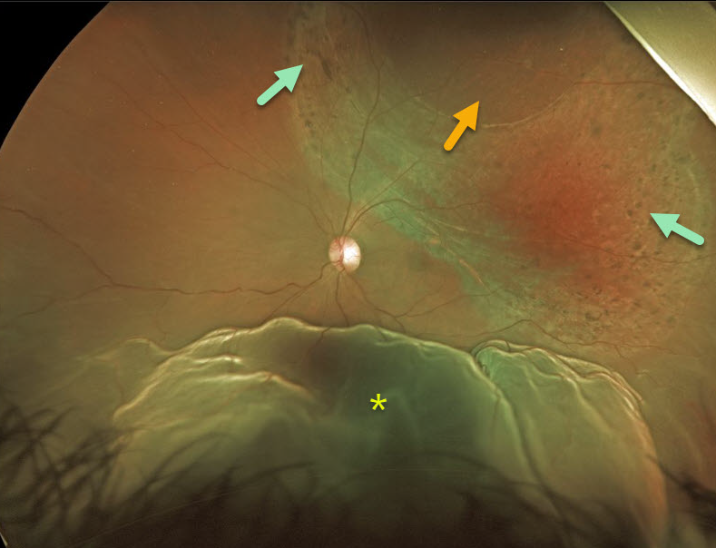
COMBINED RETINOSCHISIS-RHEGMATOGENOUS RETINAL DETACHMENT
Originally posted on @retina.rocks December 7, 2020
This 33YO patient presented with a prominent inferior retinal detachment associated with superotemporal retinoschisis with a large outer layer hole (OLH). The OLH was surrounded by laser-like chorioretinal scarring, although the patient denied prior laser surgery.
At vitrectomy, a superonasal gutter of peripheral detachment extended to an equatorial tear at 10:30 (not visualized in these images). The OLH and schisis were not detached.
Learning Points:
Patients with degenerative retinoschisis and associated inner- and outer-layer holes are at risk of rhegmatogenous retinal detachment.
Despite our patient probably undergoing prior prophylactic laser photocoagulation around the outer layer hole, he still developed a retinal detachment from a presumed new retinal tear outside the area of schisis.
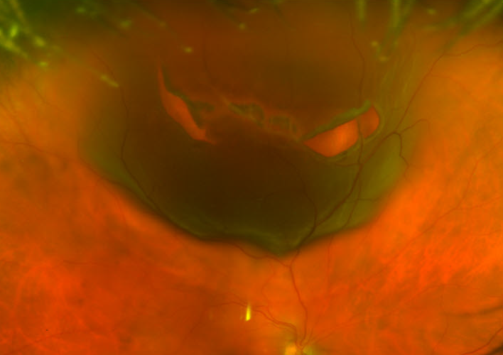
HALLOWEEN RETINOPATHY
Originally posted on @retina.rocks October 30, 2020
This patient presented with a jack-o’-lantern retinal detachment 2 weeks before Halloween.
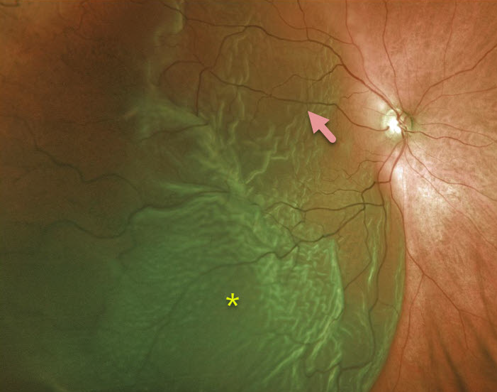
RHEGMATOGENOUS RETINAL DETACHMENT
Originally posted on @retina.rocks June 10, 2020
This patient has an acute, macula-off retinal detachment with a large cilioretinal artery.
Learning Points:
Cilioretinal arteries arise from the choroidal circulation. This results in cilioretinal sparing with central retinal artery occlusion.
Unfortunately, this vessel offers no visual protection against retinal detachment!
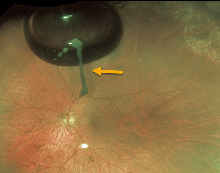
TRIAMCINOLONE-MOXIFLOXACIN (TriMoxi)
Originally posted on @retina.rocks May 22, 2020
Six weeks prior to this retinal detachment repair, the patient underwent uncomplicated cataract surgery. This image shows TriMoxi (triamcinolone acetonide/moxifloxacin) medication literally “hanging on” the absorbing gas bubble!
Learning Points:
During cataract extraction, a commonly used combination of triamcinolone (15 mg/mL) and moxifloxacin (1mg/mL) is injected intracamerally. The medication eventually absorbs completely.
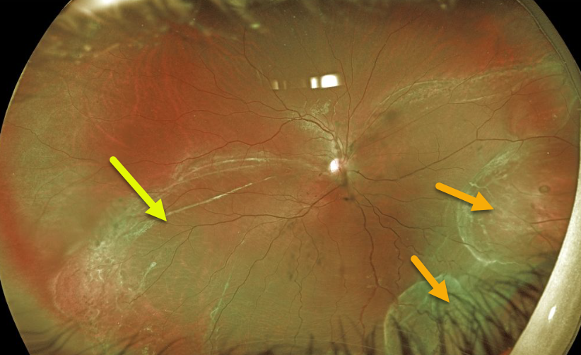
RHEGMATOGENOUS RETINAL DETACHMENT
Originally posted on @retina.rocks May 6, 2020
This 16YO presented with a chronic inferior macula-off rhegmatogenous retinal detachment (RRD). Vision was 20/200.
What’s most unique about this detachment is the rare appearance of intraretinal macrocysts inferonasally.
These cysts are a sign of very long-standing retinal detachments. The multiple demarcation lines also indicate chronicity.
Learning Points:
During RRD repair, the cysts require no special modifications and resolve following retinal reattachment.
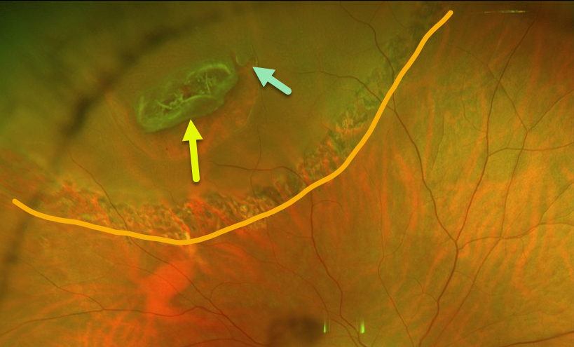
LATTICE DEGENERATION
Originally posted on @retina.rocks April 23, 2020
This patient had an asymptomatic peripheral lattice-related retinal detachment. Note that the entire lattice lesion represents the flap of the tear, with an adjacent secondary smaller tear.
The detachment was demarcated with laser photocoagulation and has remained stable since.
Learning Points:
Retinal detachment occurs in 1 in 10,000 people per year. Lattice is found in 10% of the general population, especially in high myopes.
Since about one-third of patients with retinal detachment have lattice as the cause, the question always arises, especially in patients like this: should prophylactic retinopexy be performed? The short answer is that asymptomatic lattice generally should not be treated.
However, this patient had lattice-related retinal detachment and thus underwent laser demarcation.
An especially relevant article for this topic is by Vrabec and Baumal, Ophthalmology 2000;107;1063-1067.
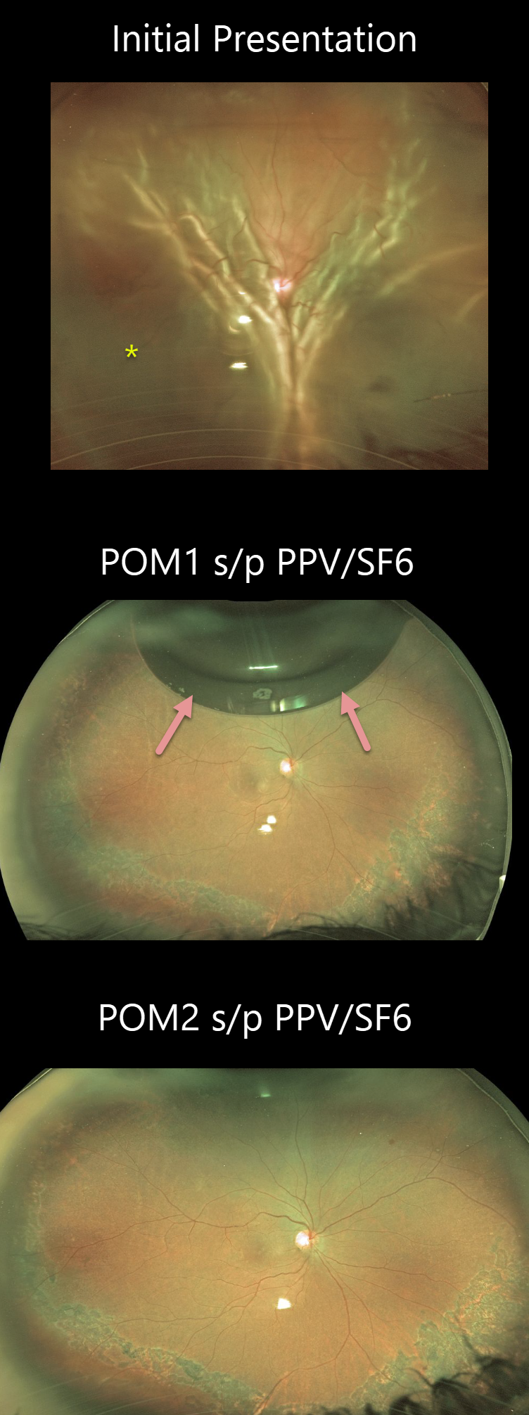
RHEGMATOGENOUS RETINAL DETACHMENT
Originally posted on @retina.rocks March 25, 2020
This patient presented with a total rhegmatogenous retinal detachment in the right eye.
The patient underwent repair with pars plana vitrectomy and SF6 gas. The Optos images show a small residual gas bubble at 1-month post-vitrectomy. Two months post-op, the patient’s vision is 20/200.
Learning Points:
Long-acting intraocular gas is routinely used in vitrectomy for rhegmatogenous retinal detachment repairs. C3F8 and sulfur hexafluoride (SF6) are used due to greater longevity compared to air. C3F8 lasts in the vitreous for 2-3 months, and SF6 lasts for several weeks.
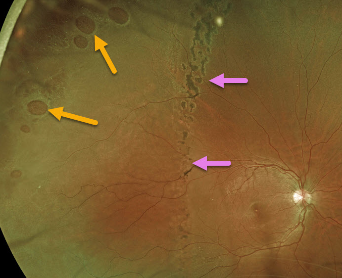
RHEGMATOGENOUS RETINAL DETACHMENT
Originally posted on @retina.rocks March 4, 2020
This patient with multiple retinal holes has a well demarcated retinal detachment.
Learning Points:
The normal neurosensory retina remains attached to the RPE due to numerous physiologic mechanisms including the inner (retinal vascular endothelium) and outer (tight junctions at the apex of the RPE cells) blood retinal barriers, hyperosmotic choroid passively drawing fluid into the subretinal space, RPE pump, interphotoreceptor matrix, and friction between the RPE microvilli and the photoreceptor outer segments.
Rhegmatogenous retinal detachment (RRD) occurs when liquid vitreous, entering the subretinal space through a retinal break, overwhelms these forces favoring retinal detachment.
Pigmented demarcation lines can form in a chronic RRD and limit the spread of subretinal fluid.
RHEGMATOGENOUS RETINAL DETACHMENT
Originally posted on @retina.rocks February 25, 2020
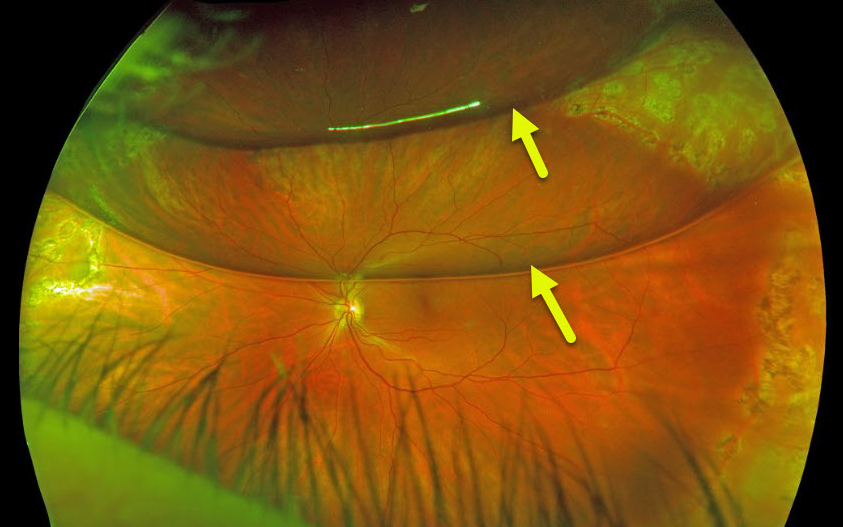
This patient had a rhegmatogenous retinal detachment that was repaired with pars plana vitrectomy and C3F8 gas.
The patient’s bubble has broken into 2 large bubbles, each with its own beautiful retinal reflection.
Learning Points:
Long-acting intraocular gas is routinely used in vitrectomy for rhegmatogenous retinal detachment repairs. C3F8 and sulfur hexafluoride (SF6) are used due to greater longevity compared to air. C3F8 lasts in the vitreous for 2-3 months, and SF6 lasts for several weeks.
RHEGMATOGENOUS RETINAL DETACHMENT
Originally posted on @retina.rocks February 4, 2020
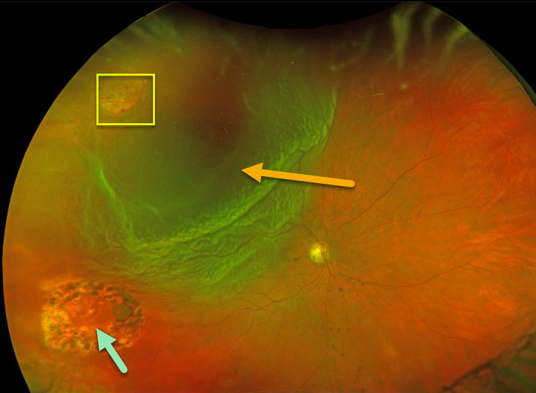
This patient presented with a new retinal tear causing retinal detachment in an eye with previously lasered operculated hole.
Learning Points:
The normal neurosensory retina remains attached to the RPE due to numerous physiologic mechanisms including the the inner (retinal vascular endothelium) and outer (tight junctions at the apex of the RPE cells) blood retinal barriers, hyperosmotic choroid passively drawing fluid into the subretinal space, RPE pump, interphotoreceptor matrix, and friction between the RPE microvilli and the photoreceptor outer segments.
Rhegmatogenous retinal detachment (RRD) occurs when liquid vitreous, entering the subretinal space through a retinal break, overwhelms these forces favoring retinal detachment.
Repairing RRD involves finding and closing all breaks, essentially denying the liquid vitreous access to the subretinal space. The break(s) can be closed internally with a gas bubble with pneumatic retinopexy or vitrectomy surgery, or externally by sewing a piece of silicone material to the eye wall (scleral buckle). This indents, or buckles, the sclera up against the retinal break.
RHEGMATOGENOUS RETINAL DETACHMENT
Originally posted on @retina.rocks January 8, 2020
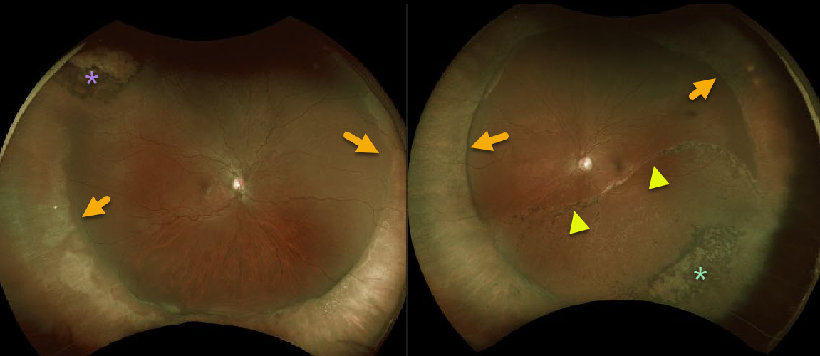
This patient has bilateral scleral buckles following prior rhegmatogenous retinal detachment surgeries.
Cryotherapy scarring is present superotemporally OD and laser scarring is present inferotemporally OS around a few tiny retinal holes that caused the initial detachment.
There is also a large demarcation line OS indicating that the left detachment was present for some time before being repaired.
Learning Points:
Primary scleral buckle can be used to repair certain types of rhegmatogenous retinal detachments.


