

RPE Hypertrophy + CHRPE + Bear Tracks
<< Back to Cases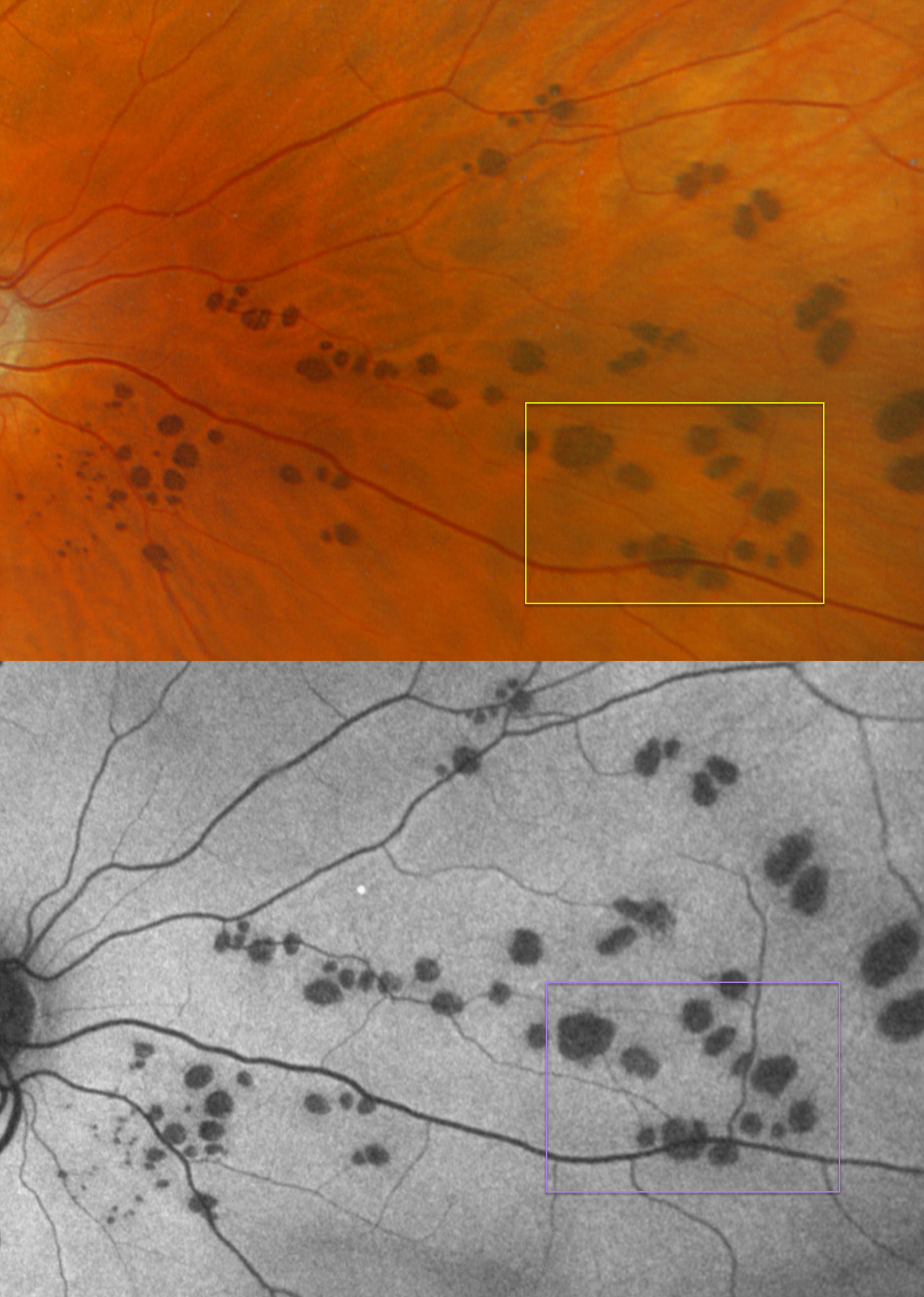
BEAR TRACKS
Originally posted on @retina.rocks September 14, 2026
This healthy 59YO male was referred for asymptomatic lesions in his right eye.
Optos color RGB imaging shows multiple variably round-to-oval subretinal pigmented lesions scattered throughout the nasal right fundus. These lesions are hypo-autofluorescent.
Learning Points:
Group-type congenital pigmented nevi of the RPE (bear tracks) is a relatively rare congenital condition characterized by well-demarcated, hyperpigmented, flat, variably sized RPE lesions that resemble bear footprints. Histopathologically, these lesions resemble congenital hypertrophy of the RPE (CHRPE), with increased numbers of pigment granules in normal-sized RPE cells. These lesions are benign and cause no visual symptoms.
The major fluorophore in fundus autofluorescence (FAF) is lipofuscin, which is intraliposomal RPE material generated as byproducts of outer segment metabolism. The outer retina overlying these bear track lesions shows varying degrees of atrophy. This leads to less lipofuscin production with hypo-FAF.
Although bear tracks and the typical CHRPE lesions (large, round, unilateral, with depigmented lacunae with age) are not associated with systemic abnormalities, the multifocal, often bilateral and small comet-shaped lesions with FAP are associated with colonic polyps and cancer (Romania et al, Ophthalmology 1992;99:911-913).
FAMILIAL ADENOMATOUS POLYPOSIS (FAP)
Joe Wilson
Originally posted on @retina.rocks June 12, 2025
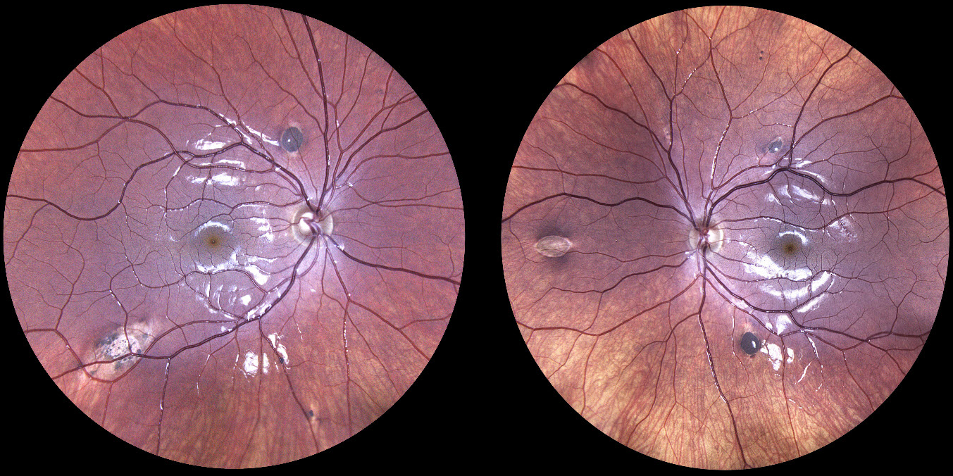
This healthy 13YO girl’s mother has a known history of familial adenomatous polyposis (FAP). She was referred to us for asymptomatic retinal findings. Fundus imaging shows multiple, bilateral, variably comet-shaped, variably pigmented jet-black subretinal lesions.
Learning Points:
Familial adenomatous polyposis (FAP) is an autosomal dominant syndrome associated with colonic polyps and colorectal cancer. Although typical CHRPE lesions (large, round, unilateral, developing depigmented lacunae with age) are not associated with systemic abnormalities, the FAP multifocal, often bilateral, small, comet-shaped lesions, as seen in our patient, are associated with colonic polyps and cancer (Romania et al, Ophthalmology 1992;99:911-913). The likely diagnosis of FAP was discussed with our patient’s parents, and she was referred to her PCP for further management and genetic testing.
BENIGN LOBULAR INNER NUCLEAR LAYER PROLIFERATIONS (BLIP) + BEAR TRACKS
Sharat Hegde
Originally posted on @retina.rocks April 30, 2025
CONGENITAL HYPERTROPHY OF THE RPE (CHRPE)
Leslie Cecil
Originally posted on @retina.rocks December 10, 2024
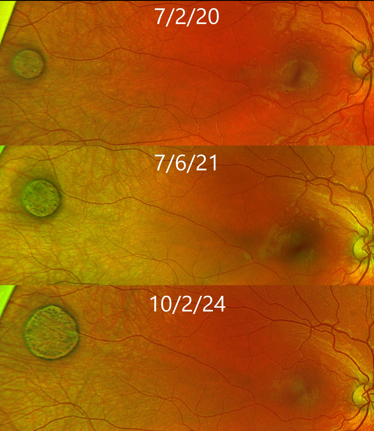
This healthy 7YO girl has been followed since she was 3YO for a benign congenital hypertrophy of the RPE (CHRPE) lesion. The CHRPE has shown subtle yet definite concentric enlargement, as well as redistribution of the lesion’s central pigmentation. Continued observation was recommended since these lesions have virtually no malignant potential.
Learning Point:
Although historically CHRPE lesions were thought not to grow, subtle growth actually occurs in the vast majority of eyes (see Shields et al, Ophthalmology 2003;110:1968-1973).
BEAR TRACKS WITH BENIGN LOBULAR INNER NUCLEAR LAYER PROLIFERATIONS
Sharat Hegde
Originally posted on @retina.rocks November 13, 2024
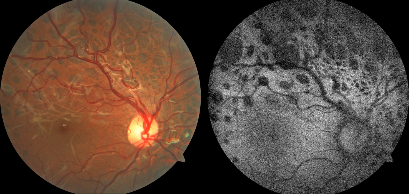
This healthy 10YO female presented with these asymptomatic bilateral lesions. Vision was 20/20 OU.
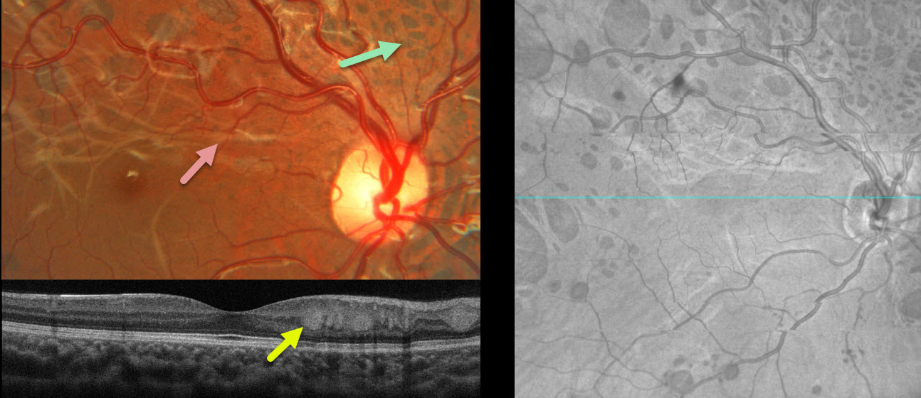
Fundus photography of her right eye shows multiple bear track lesions scattered throughout the posterior pole. An interlacing pattern of whitish deep retinal lines courses through the macula. The pigmented lesions are hypo-autofluorescent. Similar findings were noted in the left eye (not shown).
Learning Points:
Group-type congenital pigmented nevi of the RPE (bear tracks) is a relatively rare congenital condition characterized by well-demarcated, hyperpigmented, flat, variably sized RPE lesions that resemble bear footprints. Histopathologically, these lesions are similar to congenital hypertrophy of the RPE (CHRPE), with increased numbers of pigment granules in normal-sized RPE cells. These lesions are benign and cause no visual symptoms.
The major fluorophore in fundus autofluorescence (FAF) is lipofuscin, which is intraliposomal RPE material generated as byproducts of outer segment metabolism. The outer retina overlying these bear track lesions shows varying degrees of atrophy. This leads to less lipofuscin production with hypo-FAF.
Although bear tracks and the typical CHRPE lesions (large, round, unilateral, with depigmented lacunae with age) are not associated with systemic abnormalities, the multifocal, often bilateral, and small comet-shaped lesions with familial adenomatosis are associated with colonic polyps and cancer (Romania et al Ophthalmology 1992;99:911-913).
Benign lobular inner nuclear layer proliferations were described by Sanfilippo et al in 2023 as a unique benign lesion often associated with CHRPE lesions (Ophthalmology 2023;130:265-273). They appear as white, multifocal, often lobular tumors centered in the posterior pole. On OCT, they appear in the inner nuclear layer at the border of the outer plexiform layer.
CONGENITAL HYPERTROPHY OF THE RPE (CHRPE)
Originally posted on @retina.rocks January 11, 2024
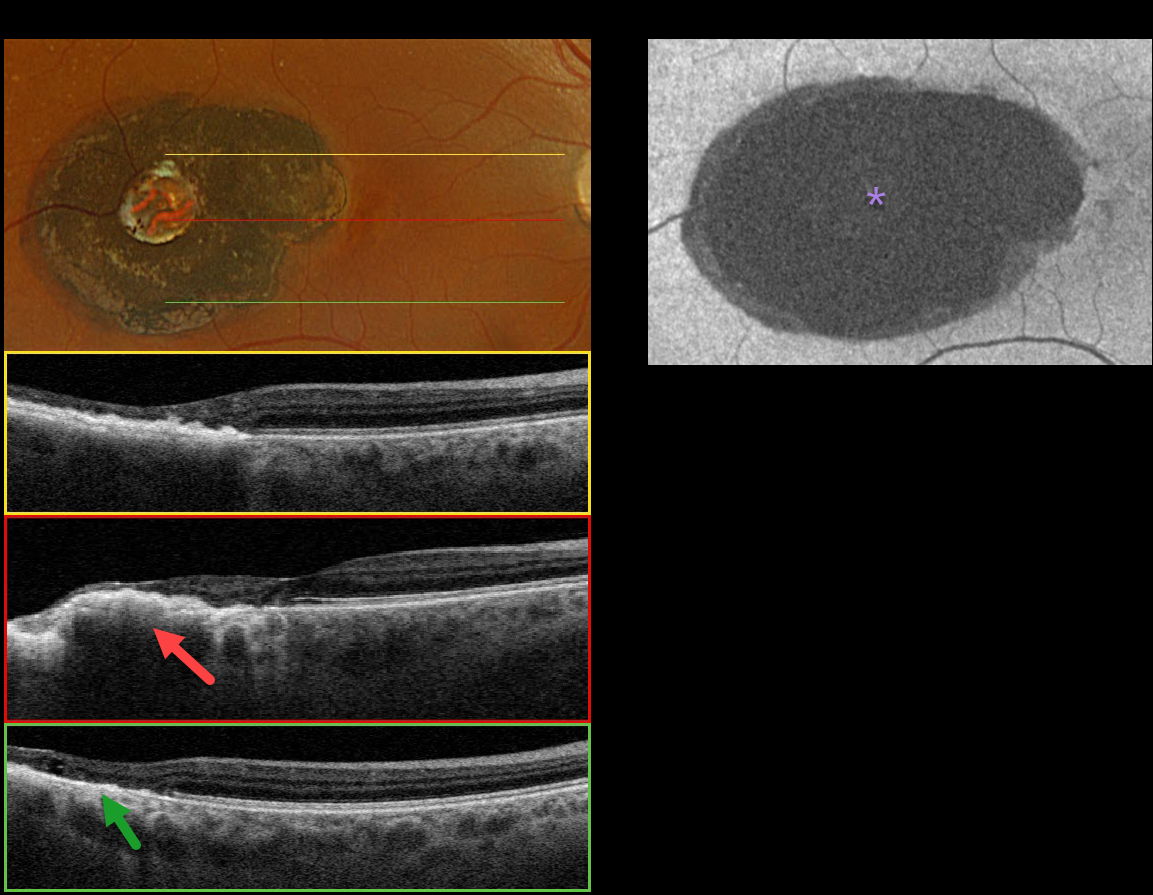
This 57YO female was referred for an asymptomatic pigmented lesion in her right macula. On further questioning, she was told there was something in the back of her eye since childhood due to congenital toxoplasmosis. Vision was 20/20 in her right eye and 20/20 in her left eye.
Optos color RGB imaging shows a well-defined, jet-black lesion in the temporal macula. It has a well-defined hyper/hypopigmented ring along with a central round area of depigmentation.
Triton swept-source OCT shows an irregularly thickened, elevated, and hyperreflective RPE with variable overlying outer retinal atrophy. There is no coloboma. Fundus autofluorescence (FAF) shows uniform hypo-FAF. Observation was recommended.
Learning Points:
Our patient had previously been diagnosed with toxoplasmosis scarring. However, we believe a CHRPE lesion is more likely, given the smooth, well-demarcated hyper/hypopigmented rim typical of CHRPE, the central lacuna, and the lack of colobomatous changes often found in congenital toxoplasmosis lesions (Kumar et al, AJO 2019;200:47-56).
Although CHRPE lesions can enlarge with time (Shields et al, Ophthalmology 2003;110:1968-1973), they carry virtually no malignant potential. Histologic RPE hypertrophy can present with several clinical manifestations.
The typical isolated CHPRE lesions (this case) and bear tracks are not associated with intestinal polyps or cancer, and thus require no systemic evaluation. Only the multiple, small comet-shaped lesions are part of the familial polyposis spectrum.
BEAR TRACKS
Yuenpang Cheung, Stephanie Choi, and Royce Chen
Originally posted on @retina.rocks November 21, 2023
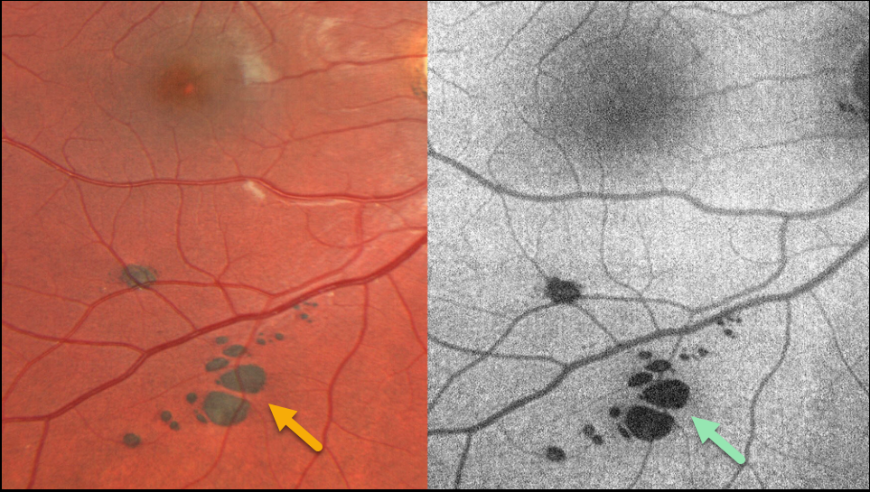
This healthy 19YO male was seen for routine eye examination. Fundus imaging shows bear-track lesions (grouped congenital hypertrophy of the RPE) inferiorly in his right eye. These lesions hypo-autofluoresce.
He had a family history of an aunt with colon cancer. He underwent genetic testing as a child and was fortunately negative for the familial adenomatous polyposis (FAP) gene.
Learning Points:
Group-type congenital pigmented nevi of the RPE (bear tracks) is a relatively rare congenital condition characterized by well-demarcated, flat, hyperpigmented, variably sized RPE lesions that resemble bear tracks. Histopathologically, these lesions are similar to congenital hypertrophy of the RPE (CHRPE), with increased numbers of pigment granules in normal-sized RPE cells. These lesions are benign and cause no visual symptoms.
The major fluorophore in fundus autofluorescence (FAF) is lipofuscin, which is intraliposomal RPE material generated as byproducts of outer segment metabolism. The outer retina overlying these bear track lesions shows varying degrees of atrophy. This leads to less lipofuscin production with hypo-FAF.
Although bear tracks and the typical CHRPE lesions (large, round, unilateral, with depigmented lacunae with age) are not associated with systemic abnormalities, the multifocal, often bilateral, and small comet-shaped lesions with FAP are associated with colonic polyps and cancer (see Romania et al Ophthalmology 1992;99:911-913).
BEAR TRACKS
Joe Yuenpang Cheung
Originally posted on @retina.rocks May 23, 2023
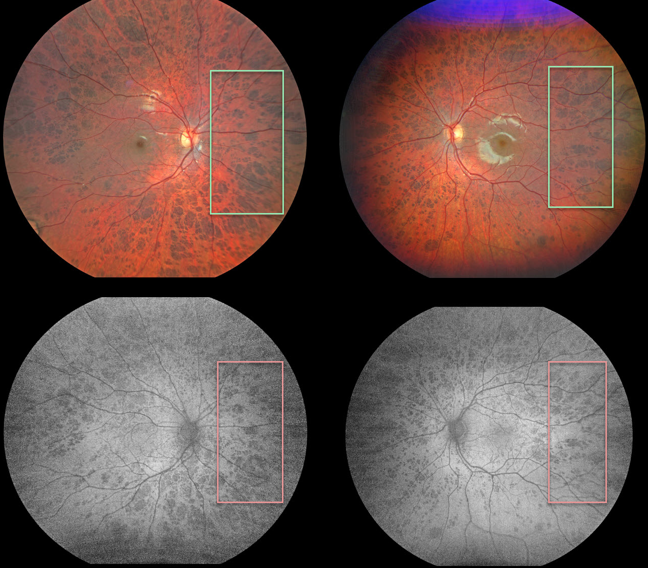
This healthy 8YO was seen for a routine eye examination. Vision was 20/50 OU. Fundus imaging shows extensive bilateral bear track lesions, which are hypoautofluorescent.
Learning Points:
Group-type congenital pigmented nevi of the RPE (bear tracks) is a relatively rare congenital condition characterized by well-demarcated, hyperpigmented, flat, variably sized RPE lesions that resemble bear footprints.
Histopathologically, these lesions are similar to congenital hypertrophy of the RPE (CHRPE), with increased numbers of pigment granules in normal-sized RPE cells. These lesions are benign, cause no visual symptoms, and are not associated with familial polyposis.
The major fluorophore with fundus autofluorescence (FAF) is lipofuscin, which is intraliposomal RPE material generated as byproducts of outer segment metabolism. The outer retina overlying these bear track lesions shows varying degrees of atrophy. This leads to less lipofuscin production with hypo-FAF.
POLAR BEAR TRACKS
Will Gibson
Originally posted on @retina.rocks March 15, 2023
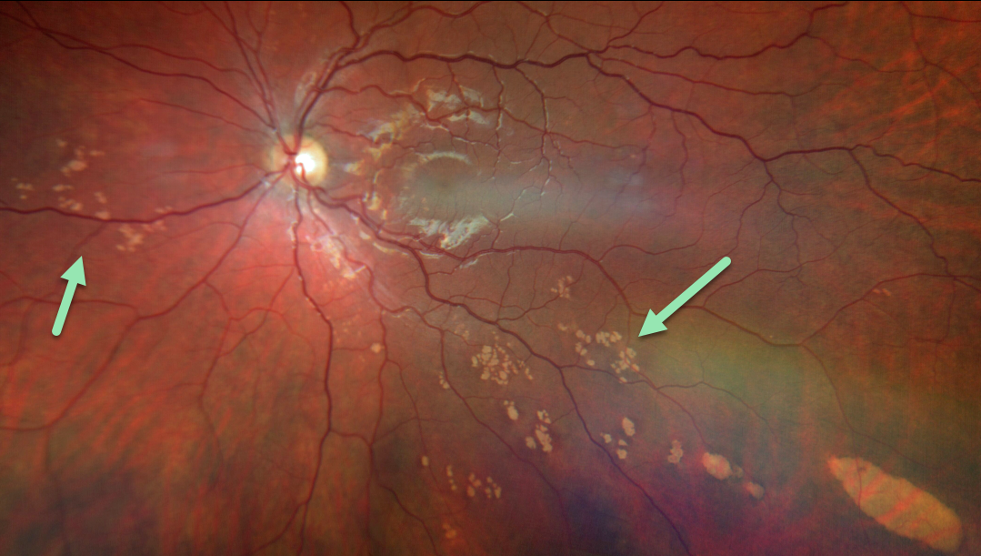
This healthy 23YO male was referred for bilateral asymptomatic retinal lesions. Scattered patches of grouped subretinal depigmented lesions resembling bear tracks are noted in each eye.
Learning Points:
These congenital albinotic spots of the retinal pigment epithelium or “polar bear tracks” are sharply circumscribed chalky white lesions with similar size and shape. They appear to be at the level of the RPE with a “dimpled” appearance due to increased white pigmentation at their periphery.
The composition of this white pigmentation is not known, and the lesions are not associated with any other ocular or systemic disorders. For a great reference for focal congenital RPE anomalies, see Gass, Eye 1989;3:1-18.
BEAR TRACKS
Originally posted on @retina.rocks February 16, 2023
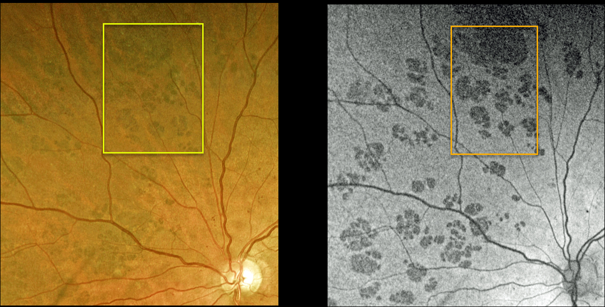
This 55YO female has a history of type 2 diabetes and moderate nonproliferative diabetic retinopathy. An incidental finding shows bear-track lesions (congenital grouped hypertrophy of the RPE) superonasally in her left eye. These lesions hypo-autofluoresce.
Learning Points:
Group-type congenital pigmented nevi of the RPE (bear tracks) is a relatively rare congenital condition characterized by well-demarcated, flat, hyperpigmented, variably sized RPE lesions resembling bear tracks.
Histopathologically, these lesions are similar to congenital hypertrophy of the RPE (CHRPE), with increased numbers of pigment granules in normal-sized RPE cells. These lesions are benign and cause no visual symptoms.
The major fluorophore with fundus autofluorescence (FAF) is lipofuscin, which is intraliposomal RPE material generated as byproducts of outer segment metabolism.
The outer retina overlying these bear track lesions shows varying degrees of atrophy. This leads to less lipofuscin production with hypo-FAF.
BEAR TRACKS
Originally posted on @retina.rocks November 16, 2022
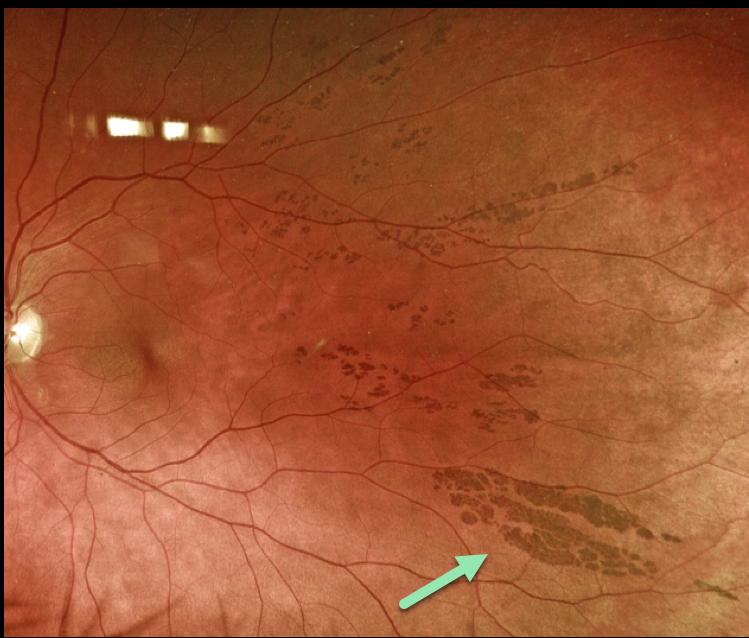
This 66YO male presented with these asymptomatic unilateral pigmented lesions in his left eye.
Learning Points:
Group-type congenital pigmented nevi of the RPE (bear tracks) is a relatively rare congenital condition characterized by well-demarcated, hyperpigmented, flat, variably-sized RPE lesions, resembling bear footprints.
Histopathologically, these lesions are similar to congenital hypertrophy of the RPE (CHRPE), with increased numbers of pigment granules in normal-sized RPE cells. These lesions are benign and cause no visual symptoms.
Bear tracks should not be mistaken for the comet-shaped lesions in familial adenomatous polyposis (FAP, or Gardner’s syndrome). FAP is an autosomal dominant syndrome associated with colonic polyps and colorectal cancer.
Although typical CHRPE lesions (large, round, unilateral, with depigmented lacunae with age) are not associated with systemic abnormalities, the multifocal, often bilateral, and small comet-shaped lesions with FAP are associated with colonic polyps and cancer (see Romania et al Ophthalmology 1992;99:911-913).
CHRPE + RETINAL BREAK
Originally posted on @retina.rocks November 15, 2022
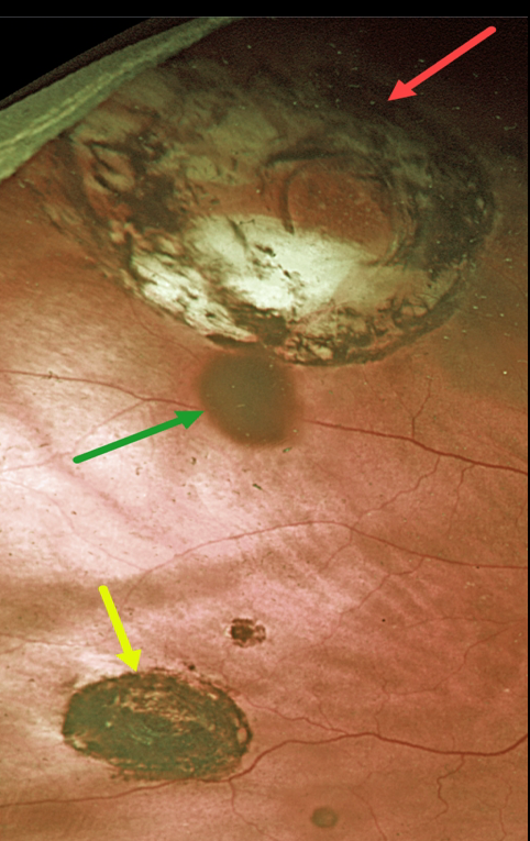
This 87YOM received thermal laser around a retinal break 23 years earlier. He presented to us without symptoms and 20/30 vision.
Optos imaging shows a small inferior congenital hypertrophy of the RPE (CHRPE) with a few tiny additional CHRPE lesions.
Confluent variably pigmented laser scarring around a superotemporal retinal break simulates an atrophic CHRPE lesion.
The operculum casts a shadow on the underlying retina, somewhat giving the impression of yet another pigmented lesion.
CRVO + CHRPE + MYELINATED NERVE FIBER LAYER
Will Gibson
Originally posted on @retina.rocks October 27, 2022
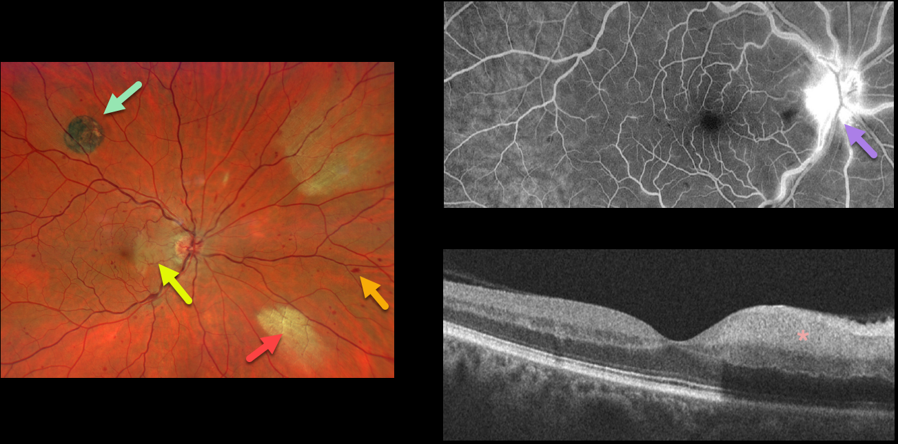
This 52YO male presented with acute visual symptoms in his right eye. Vision was 20/20.
Optos imaging shows a non-ischemic central retinal vein occlusion (CRVO) with mild scattered retinal hemorrhages in all quadrants. An acute nasal macular branch retinal artery occlusion (BRAO) is noted, along with a small round patch of congenital hypertrophy of the RPE (CHRPE) superotemporally, and two patches of myelinated nerve fiber layer (NFL) nasally.
Fluorescein angiography shows a well-perfused central retinal vein occlusion (CRVO) with late optic nerve leakage. OCT scanning shows hyperreflectivity of the inner nasal retina.
FAMILIAL ADENOMATOUS POLYPOSIS (FAP)
Originally posted on @retina.rocks August 12, 2022
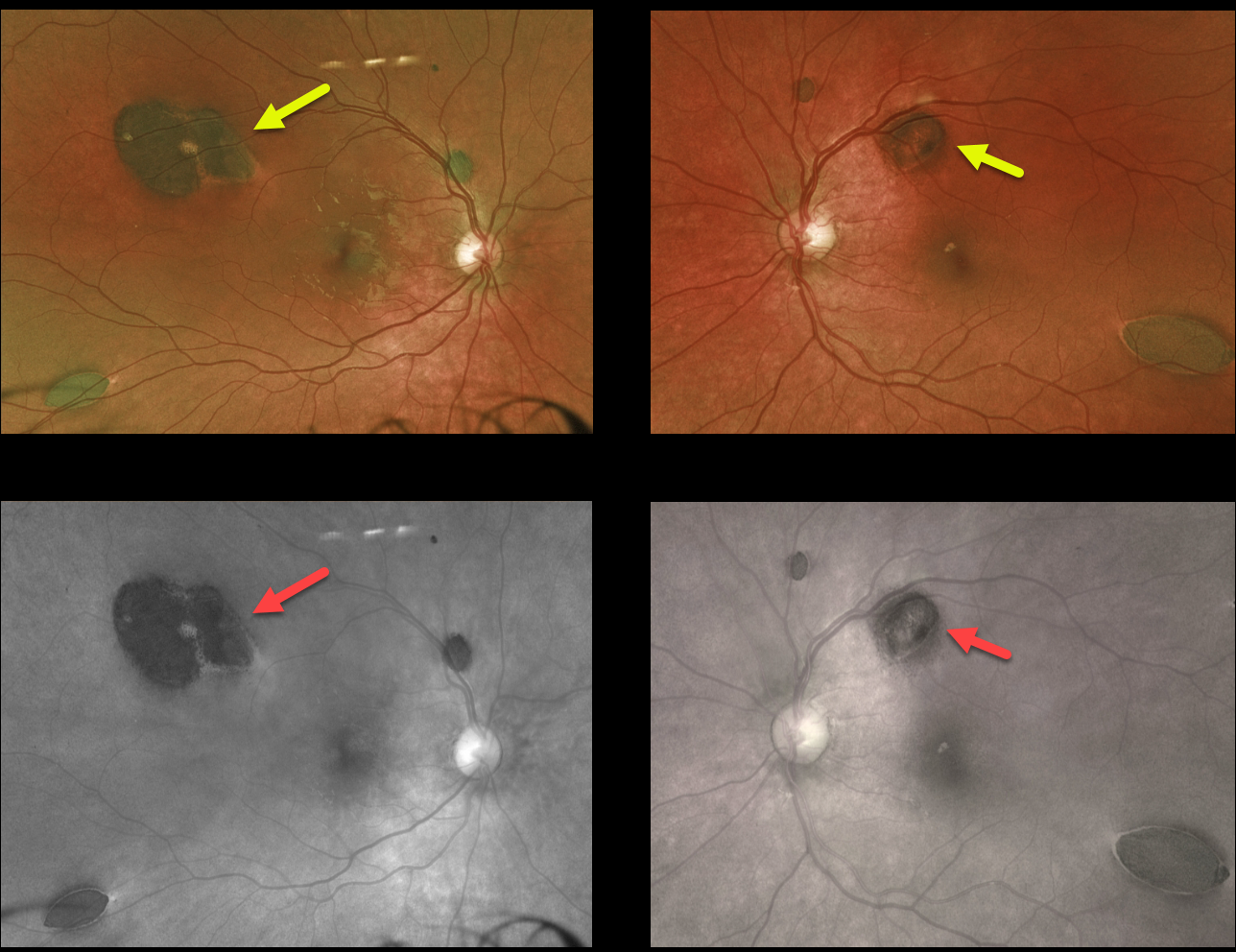
This healthy 4YO boy’s father and paternal grandfather have histories for colon cancer. He was referred to us for asymptomatic retinal findings.
Optos color and red-channel imaging shows multiple, bilateral, variably comet-shaped, jet-black subretinal lesions.
Learning Points:
Familial adenomatous polyposis (FAP) is an autosomal dominant syndrome associated with colonic polyps and colorectal cancer.
Although typical CHRPE lesions (large, round, unilateral, with depigmented lacunae with age) are not associated with systemic abnormalities, the FAP multifocal, often bilateral and smaller comet-shaped lesions, as seen in our patient, are associated with colonic polyps and cancer (see Romania et al Ophthalmology 1992;99:911-913).
The likely diagnosis of FAP was discussed with our patient’s family, and he was referred for genetic testing.
CHRPE + CHOROIDAL NEVUS
Originally posted on @retina.rocks July 19, 2022
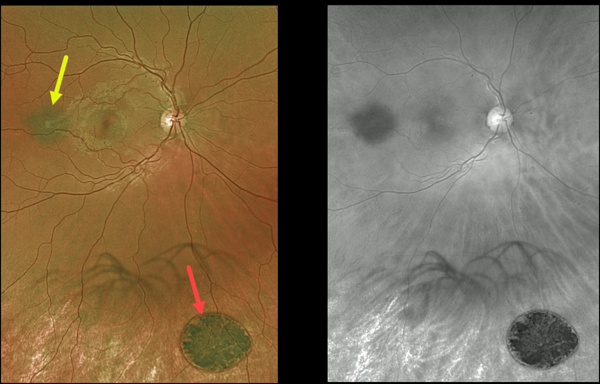
This 27YO male was referred for two distinct and asymptomatic pigmented fundus lesions.
Optos color imaging shows a small, flat choroidal nevus and a larger, flat patch of congenital hypertrophy of the RPE. The color image best shows the color difference between these lesions, with the CHRPE lesion appearing jet-black compared with the nevus.
The red channel best highlights the typical, somewhat indistinct margins of the nevus compared with the very sharp, round margins typical for the CHRPE. The patient is being followed yearly.
Learning Points:
Clinical characteristics of choroidal nevi at risk for progressing to a uveal malignant melanoma were initially described by Shields et al (Ophthalmology 1995;102:1351-1361).
The most recent version of this famous acronym (see Retina 2019;39:1840-1851) is To Find Small Ocular Melanoma Doing Imaging (TFSOM-DIM), which stands for:
To (Thickness >2mm)
Find (subretinal Fluid)
Small (Symptoms, vision <= 20/50)
Ocular (Orange pigment)
Melanoma (Melanoma hollow on ultrasonography)
DIM (DIaMeter > 5mm)
Although CHRPE lesions can enlarge with time (see Shields et al, Ophthalmology 2003;110:1968-1973), they carry virtually no malignant potential.
Histologic RPE hypertrophy presents in several ways. The typical isolated CHPRE lesions and bear tracks are not associated with intestinal polyps or cancer, and thus require no systemic evaluation. Only the multiple, small comet-shaped lesions are part of the familial polyposis spectrum.
DARK WITHOUT PRESSURE + CHRPE
Originally posted on @retina.rocks April 7, 2022
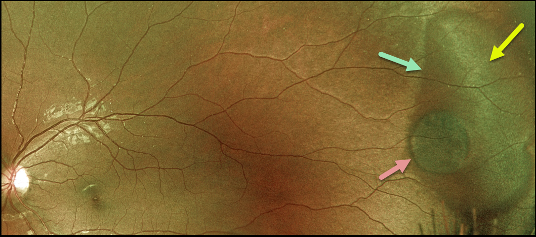
This 22YO female presented with a large temporal patch of dark without pressure (DWP), white without pressure (WWP), and a small round patch of congenital hypertrophy of the RPE (CHRPE). None of these findings is of concern, but we thought it was cool that all of them are associated with each other at this single location.
Learning Points:
WWP and DWP are relatively common benign peripheral findings most commonly found in darkly pigmented individuals. Typically, there are bilateral peripheral geographic areas of whitish (WWP) or dark (DWP) retinal discoloration.
WWP gets its name because the area is white without indentation, such as during scleral depression (white with pressure). It can sometimes be confused with retinal breaks or detachment.
The exact cause of this phenomenon is unknown. Historically, it was thought to be due to a benign vitreoretinal interface change, but more recent OCT findings show increased (WWP) or decreased (DWP) reflectivity in the outer retina (see Diaz et al., Retina 2014;34:1020-1021).
Although CHRPE lesions can enlarge with time (see Shields et al, Ophthalmology 2003;110:1968-1973), they carry virtually no malignant potential. Histologic RPE hypertrophy presents in several ways.
The typical isolated CHPRE lesions (this lesion) and bear tracks are not associated with intestinal polyps or cancer, and thus require no systemic evaluation. Only the multiple, small comet-shaped lesions are part of the familial polyposis spectrum.
BEAR TRACKS
Originally posted on @retina.rocks March 28, 2022
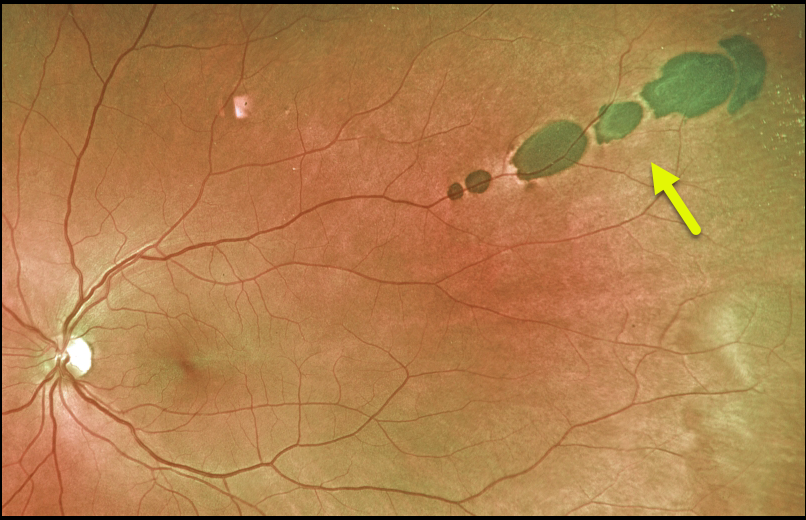
This 44YO presented with these asymptomatic pigmented lesions in his left eye. He is healthy and has no family history of colon cancer.
These lesions most likely represent an atypical case of group-type congenital pigmented nevi of the RPE (bear tracks). This is a relatively rare congenital condition characterized by well-demarcated, hyperpigmented, flat, variably sized RPE lesions resembling bear footprints.
Histopathologically, these lesions are similar to congenital hypertrophy of the RPE (CHRPE), with increased numbers of pigment granules in normal-sized RPE cells. These lesions are benign and cause no visual symptoms.
Learning Points:
Bear tracks should not be mistaken for the comet-shaped lesions in familial adenomatous polyposis (FAP, or Gardner’s syndrome). FAP is an autosomal dominant syndrome associated with colonic polyps and colorectal cancer.
Although typical CHRPE lesions (large, round, unilateral, with depigmented lacunae with age) are not associated with systemic abnormalities, the multifocal, often bilateral, and small comet-shaped lesions with FAP are associated with colonic polyps and cancer (see Romania et al Ophthalmology 1992;99:911-913).
CONGENITAL HYPERTROPHY OF THE RPE (CHRPE)
Originally posted on @retina.rocks December 2, 2021
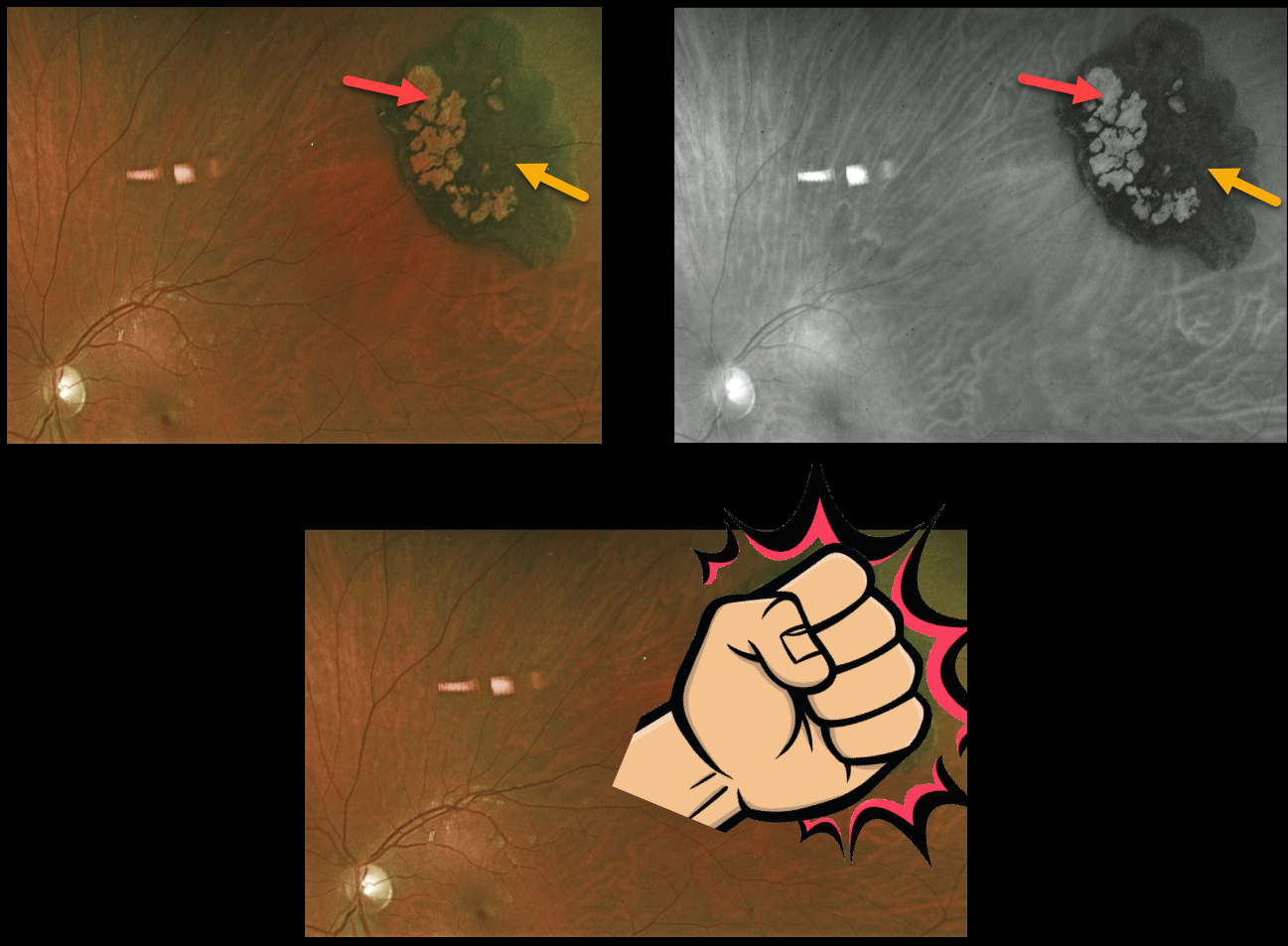
This 25YO female presented with a classic asymptomatic solitary congenital hypertrophy of the RPE (CHRPE). It almost resembles a fist!
The borders are well demarcated, with variable coal-black coloration and lacunae of depigmentation.
Learning Points:
Although CHRPE lesions can enlarge with time (see Shields et al, Ophthalmology 2003;110:1968-1973), they carry virtually no malignant potential. Histologic RPE hypertrophy has several clinical presentations.
The typical isolated CHPRE lesions (today’s post) and bear tracks are not associated with intestinal polyps or cancer, and thus require no systemic evaluation.
Only the multiple, small comet-shaped lesions are part of the familial polyposis spectrum.
CONGENITAL HYPERTROPHY OF THE RPE (CHRPE)
Leslie Cecil
Originally posted on @retina.rocks October 5, 2021
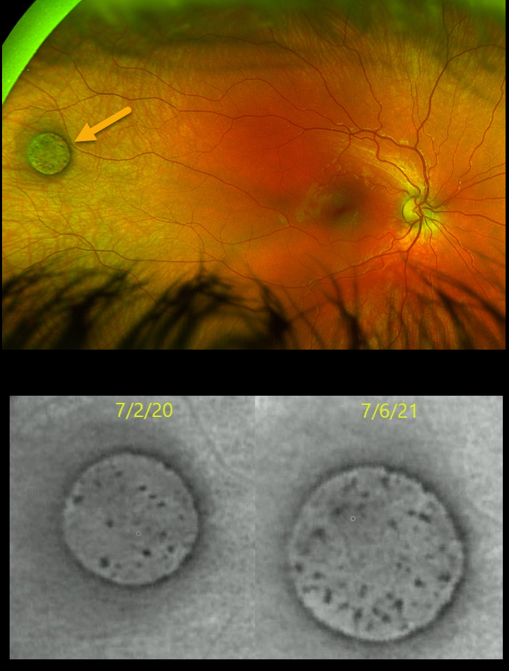
This healthy 4YO girl presented for a one-year exam to check on a benign congenital hypertrophy of the RPE (CHRPE) lesion that was noticed one year earlier. Amazingly, an Optos was obtained at that time when she was 3YO.
Optos imaging at the current visit showed subtle yet definite concentric enlargement, as well as redistribution of the lesion’s central pigmentation.
Continued observation was recommended since these lesions have virtually no malignant potential.
Learning Points:
Although these lesions were historically thought not to grow, subtle growth occurs in the vast majority of eyes (see Shields et al, Ophthalmology 2003;110;1968-1973).
CONGENTIAL HYPERTROPHY OF THE RPE (CHRPE)
Originally posted on @retina.rocks May 6, 2021
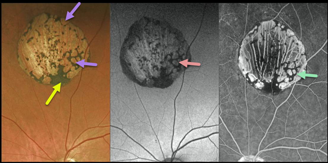
This is a gorgeous multimodal Optos image of a classic solitary congenital hypertrophy of the RPE (CHRPE). The borders are well-demarcated with a variable coal-black coloration.
With age, they usually develop multiple hypopigmented lacunae that tend to enlarge over time. These lacunae demonstrate hyperautofluorescence on fundus autofluorescence (FAF), whereas on fluorescein angiography (FA), they appear as hyperfluorescent window defects.
The CHRPE lesions can also enlarge with time, but carry virtually no malignant potential.
Learning Points:
Histologic RPE hypertrophy presents in several ways. Typical isolated CHRPE lesions and bear tracks are benign ocular findings and require no systemic evaluation. Only the multiple, small comet-shaped lesions are part of the familial polyposis spectrum.
CONGENTIAL HYPERTROPHY OF THE RPE (CHRPE)
Originally posted on @retina.rocks August 13, 2020
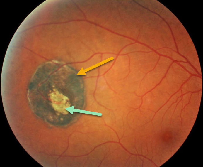
This is a picture of a classic solitary congenital hypertrophy of the RPE (CHRPE). The borders are well-demarcated with a variable coal-black coloration.
With age, they often develop multiple hypopigmented lacunae that tend to enlarge over time. The CHRPE lesions can also enlarge with time, but carry virtually no malignant potential.
Learning Points:
Histologic RPE hypertrophy presents in several ways. The typical isolated CHPRE lesions and bear tracks are not associated with intestinal polyps or cancer, and thus require no systemic evaluation. Only the multiple, small comet-shaped lesions are part of the familial polyposis spectrum.
BEAR TRACKS
Originally posted on @retina.rocks August 3, 2020
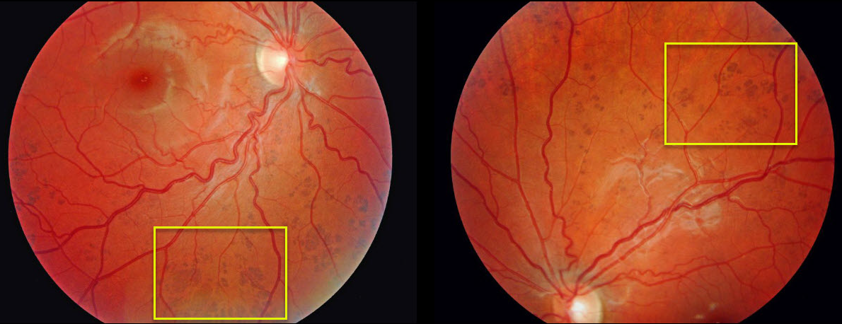
Grouped-type congenital pigmented nevi of the retinal pigment epithelium (RPE) or bear tracks is a rare congenital condition characterized by well-demarcated, hyperpigmented, flat, variably-sized RPE lesions, resembling bear footprints.
Histopathologically, these lesions are similar to congenital hypertrophy of the RPE (CHRPE), with an increased number of pigment granules in normal-sized RPE cells.
These lesions are benign and cause no visual symptoms.
Learning Points:
Bear tracks should not be mistaken for the comet-shaped lesions in familial adenomatous polyposis (Gardner’s syndrome), which are associated with intestinal polyps and cancer.
CONGENTIAL HYPERTROPHY OF THE RPE (CHRPE) FROM FAMILIAL ADENOMATOUS POLYPOSIS
Originally posted on @retina.rocks July 14, 2020
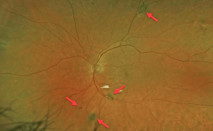
This 76yo male was asymptomatic when he presented with multiple, irregular congenital hypertrophy of the RPE (CHRPE) lesions in both eyes (only the left eye pictured).
He had a known family history of familial adenomatous polyposis (FAP) or Gardner’s Syndrome. Annual screening colonoscopies have been normal.
Learning Points:
FAP is an autosomal dominant syndrome associated with colonic polyps and colorectal cancer. Although typical CHRPE lesions (large, round, unilateral, with depigmented lacunae with age) are not associated with systemic abnormalities, multifocal, often bilateral, small comet-shaped lesions in FAP are associated with colonic polyps and cancer.


