

Rubella
<< Back to Cases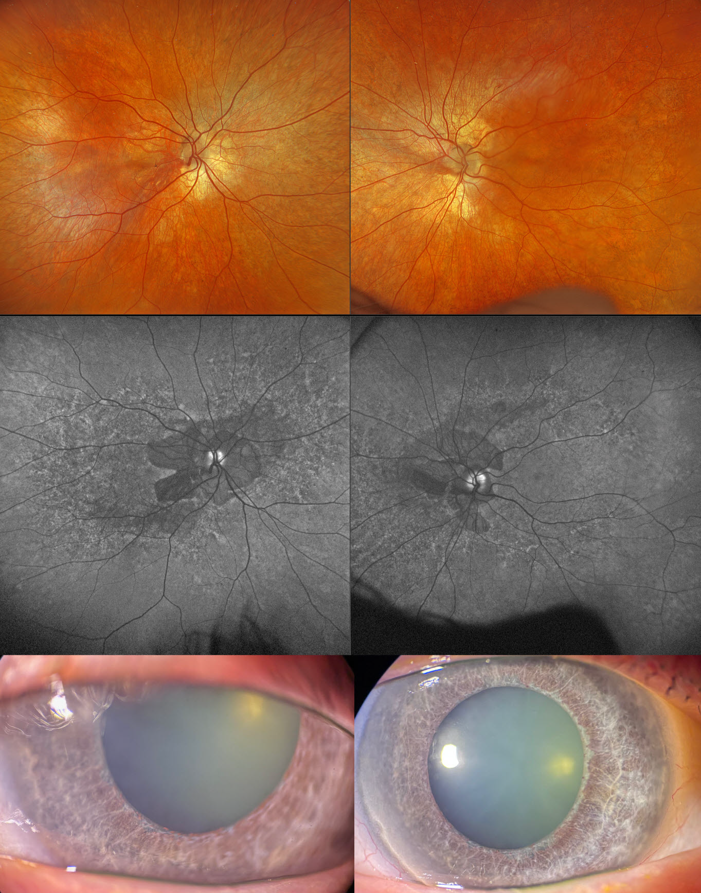
CONGENITAL RUBELLA
Fraser McKay
Originally posted on @retina.rocks December 3, 2025
This 59YO male with type 2 diabetes was examined for his yearly screening. He has a known history of congenital rubella with deafness. Vision was 20/30 OD and 20/25 OS.
Optos color RGB imaging shows a diffuse salt-and-pepper appearance in each fundus, with peripapillary atrophy. On fundus autofluorescence (FAF), the peripapillary atrophy is hypo-FAF with a speckled hyper- and hypo-FAF appearance to each posterior pole. Slit lamp photographs show diffuse iris atrophy with a relative lack of pigmentation. Triton swept-source OCT shows variable outer retinal thinning with ellipsoid zone disruption.
Learning Points:
The classic ocular findings in congenital rubella include cataracts, pigmentary retinopathy (“salt-and-pepper” fundus), congenital glaucoma, and microphthalmia. Nuclear cataracts are the most common ocular abnormality, although our patient had just trace nuclear sclerosis. Pigmentary retinopathy is characterized by diffuse mottling of the retinal pigment epithelium, which is best visualized with FAF. Systemic associations include sensorineural hearing loss and congenital heart defects.
OCULAR ALBINISM
Originally posted on @retina.rocks March 9, 2020
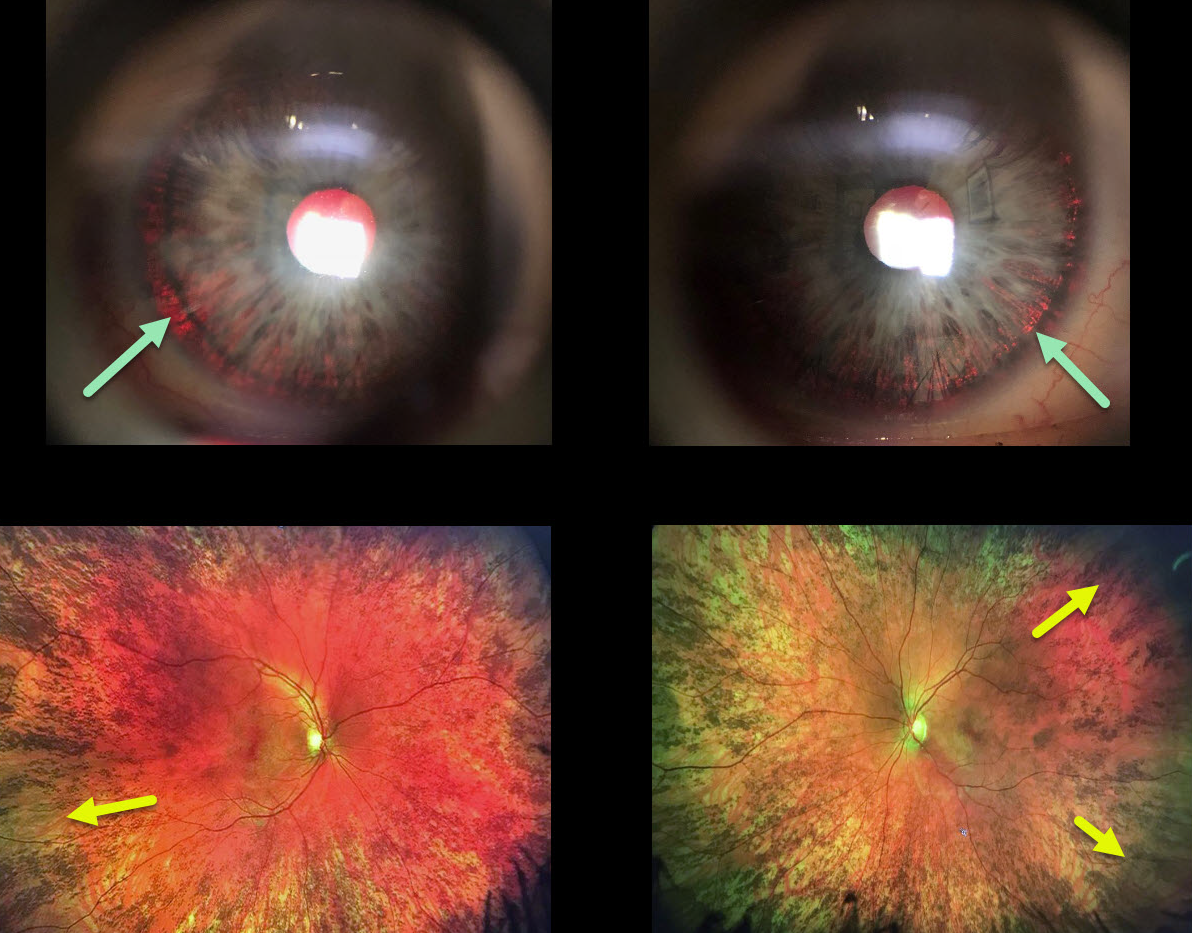
This woman has a history for both a congenital rubella infection and a positive family history for albinism in both an uncle and cousin.
We think that she is likely a carrier for OA1 (ocular albinism type 1) given the presence of iris transillumination defects and pigmentary changes of the peripheral retina, although the fundus changes may also be due to congenital rubella.
Learning Points:
Ocular albinism is a X-linked recessive disorder that affects only the eyes and is caused by a mutation in the OA1 gene. Findings seen in ocular albinism include strabismus, nystagmus iris transillumination defects, blonde fundus, and absent or blunted foveal reflex (fovea plana). There is often a higher number of crossed nerve fibers at the optic chiasm.
OA1 carriers will often have partial iris transillumination defects and up to 90% of OAT1 female carriers exhibit a salt and pepper (or mud-splattered) fundus due to the patches of amelanotic RPE.
This salt and pepper appearance can also be seen in congenital rubella, Leber’s congenital amaurosis, congenital syphilis, and phenothiazine toxicity.
Oculocutanoeus albinism involves both eye and skin findings and is caused by mutations in the tyrosine gene. There is a complete tyrosinase-negative form in which no melanin is produced and a tyrosinase-positive form in which only partial amounts of melanin are produced,


