

Sarcoidosis
<< Back to Cases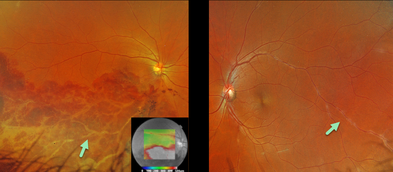
BRVO AND PERIPHLEBITIS FROM LIKELY SARCOIDOSIS
Originally posted on @retina.rocks October 8, 2024
This healthy 36YO male presented with 1 week of unilateral vision loss. Vision was 20/400 OD and 20/20 OS.
Optos color RGB imaging OD shows a major inferotemporal branch retinal vein occlusion (BRVO) with severe confluent retinal hemorrhages and a frosted appearance to the major vessels. The occlusion does not seem to arise from a more proximal arteriovenous (AV) crossing. A small area of peripheral superotemporal periphlebitis is noted OD, with more extensive periphlebitis OS. Triton swept-source OCT shows inferior macular fluid.
BRVOs nearly always arise at an AV crossing, where a thickened arterial vessel compresses the underlying vein. Sarcoidosis is the classic exception where a BRVO is not caused by an AV crossing, but by an inflammatory narrowing of the involved vein. In addition, our patients’ segmental periphlebitis (‘candle wax drippings’) is virtually pathognomonic for sarcoidosis as well.
A sarcoidosis workup was recommended, although the patient failed to show for his testing and was immediately lost to follow-up.
SARCOIDOSIS
Mattie Adams
Originally posted on @retina.rocks August 21, 2024
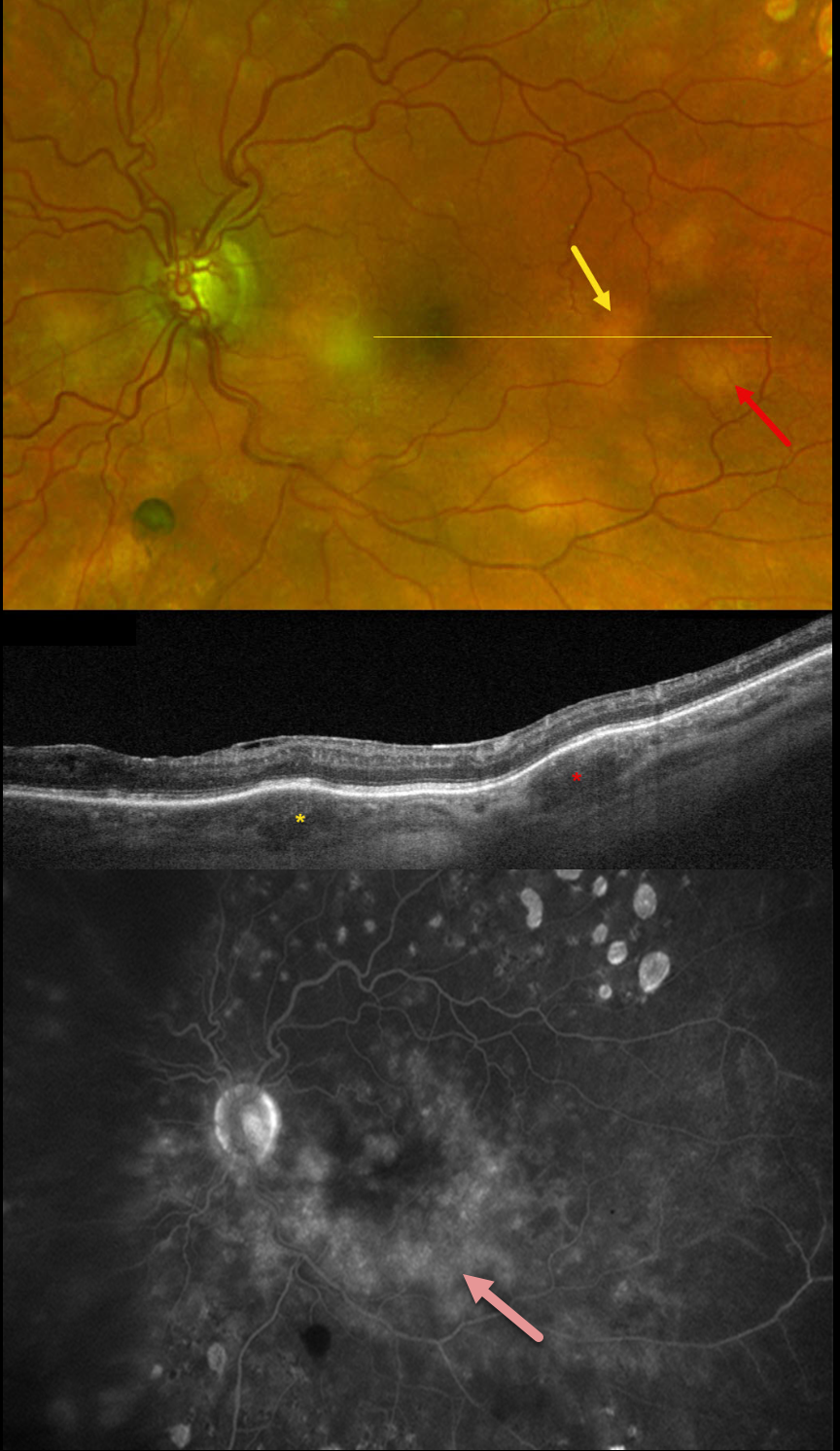
This 51YO male has a history of bilateral central retinal vein occlusions for which he has been receiving intravitreal injections. On examination on 12/21/23, vision was stable at 20/70 OD and 20/200 OS, with new elevated multifocal choroidal lesions noted bilaterally. Looking back through his prior imaging, these lesions were faintly evident 3 months earlier, but had rapidly grown since.
Optos color RG imaging of his left eye shows multifocal, elevated, creamy white temporal macula choroidal lesions. Triton swept-source OCT reveals these lesions to be hyporeflective. They show variable ill-defined staining vs leakage on fluorescein angiography.
The differential for these amelanotic choroidal lesions initially included choroidal metastasis, lymphoma, and granulomas. Extensive bloodwork, including ACE and lysozyme, was negative. Metastatic workup revealed mild splenomegaly and axial and mediastinal lymphadenopathy with a 5mm pulmonary nodule. Pulmonary biopsy showed noncaseating granulomas. Oral prednisone 60mg/day was started. When last examined on 4/12/24, vision remained at counting fingers, and his findings were relatively unchanged.
Learning Points:
Sarcoidosis, like syphilis, has a myriad of ocular presentations, including anterior, intermediate, and posterior uveitis, vasculitis, papillitis, and choroidal granulomas. Sarcoid granulomas are usually found as unilateral lesions without associated uveitis and appear as yellow choroidal lesions distinguished by an irregular, vermiform margin (Bas et al, Retina 2023;43:1842-1851). Granulomas are hyporeflective on enhanced-depth OCT imaging (Invernizzi et al, Retina 2015;35:525-531).
Establishing the diagnosis of ocular sarcoid in the absence of a known systemic diagnosis is often difficult. In our experience, chest CT is often positive despite negative bloodwork.
Given the negative metastatic workup and biopsy results, we assume the choroidal lesions represent sarcoid granulomas. These lesions have not significantly responded to prednisone. Pulmonary consultation agreed with our sarcoidosis diagnosis but suggested referring to rheumatology for help with getting the patient on a systemic steroid-sparing agent.
SARCOIDOSIS
Mattie Adams
Originally posted on @retina.rocks May 27, 2024
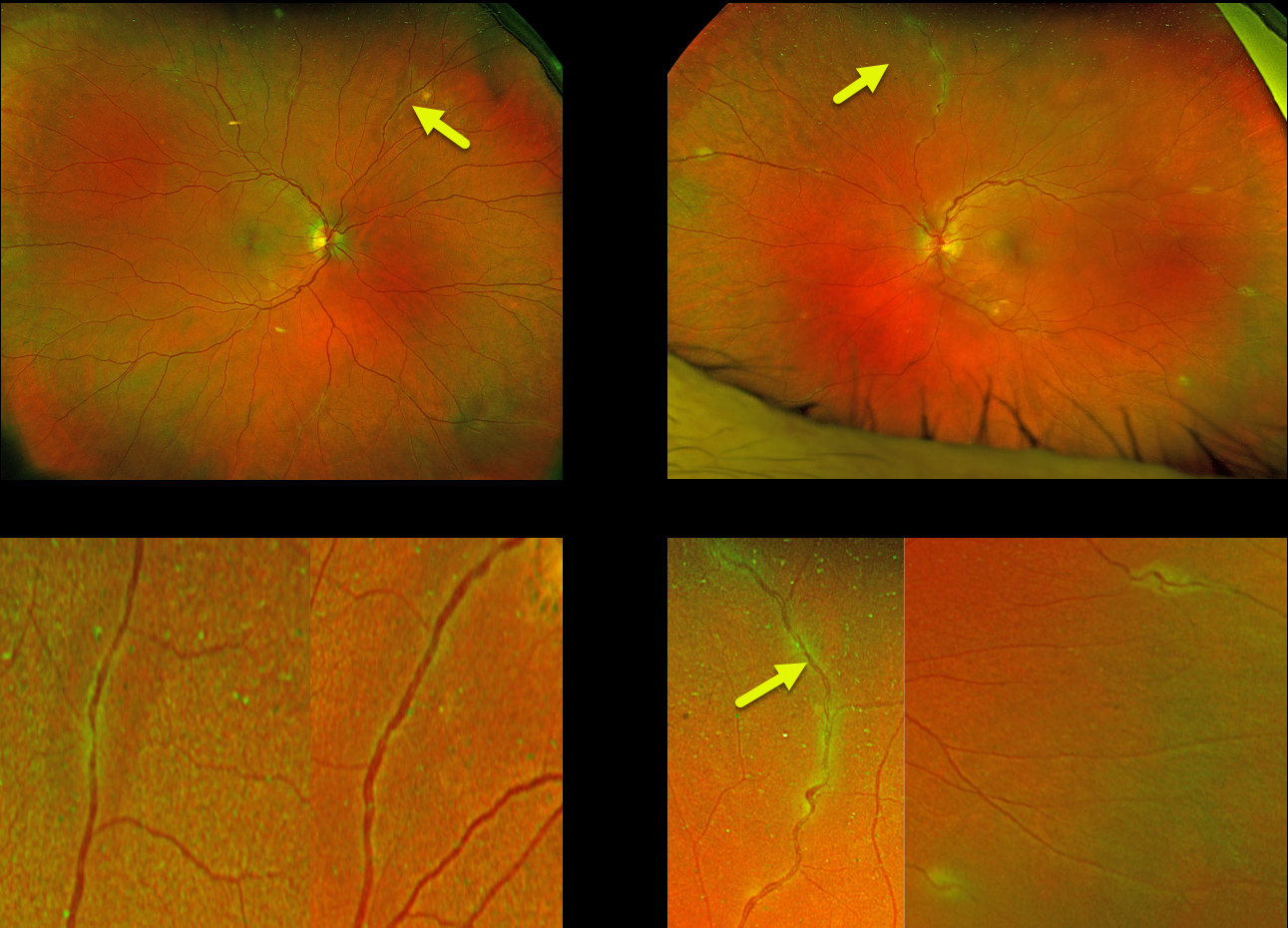
This 58YO female was referred for asymptomatic fundus changes. She gave a history of hypertension and COPD. Vision was 20/40 OU. Anterior segments were normal.
Optos color RG imaging shows bilateral multifocal areas of periphlebitis. Although sarcoid-specific bloodwork was negative (angiotensin converting enzyme, serum lysozyme, and soluble interleukin-2 receptor), chest CT showed multifocal, bilateral calcified and noncalcified pulmonary nodules throughout the lung parenchyma in addition to calcified mediastinal and hilar nodes. She was referred to a pulmonologist who agreed with our diagnosis of sarcoidosis. However, since there were no functional pulmonary deficits, he felt local treatment for the eye findings would be best. The patient will therefore be initially treated with a subtenon triamcinolone injection.
Learning Points:
Sarcoidosis, like syphilis, has a myriad of ocular presentations, including anterior, intermediate, and posterior uveitis, vasculitis, papillitis, and choroidal granuloma. Our patient’s segmental periphlebitis (‘candle wax drippings’) is virtually pathognomonic for sarcoidosis.
In our personal experience, as in our patient, there doesn’t appear to be a strong association between active systemic disease and ocular inflammation. Establishing the diagnosis of ocular sarcoid in the absence of a known systemic diagnosis is often difficult.
Sensitivity and specificity are highest for chest CT, and combined with elevated serum ACE and lymphopenia, are strongly suggestive of systemic sarcoidosis (Nieder and Sims, AJO 1019;206:149-153). Another recent publication found bilateral hilar lymphadenopathy on chest CT and sIL-2R as the most sensitive biomarkers for diagnosing systemic sarcoidosis (Japanese J Ophthalmology 2021;65:191-198).
SARCOIDOSIS
Originally posted on @retina.rocks December 30, 2021
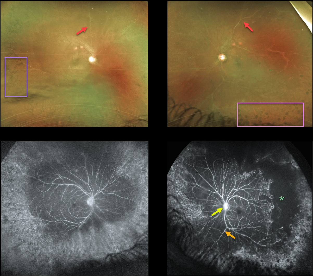
This 43YO male has a long history of panuveitis from sarcoidosis, and was previously seen 4 years earlier before being lost to follow-up.
He presented recently with a few weeks of symptomatic panuveitis. Vision was 20/100 OD and 20/200 OS with active granulomatous anterior uveitis.
Optos imaging shows a hazy view of each fundus, with diffusely white retinal vessels. Inferior vitreous puff balls were noted clinically but not imaged on Optos.
Fluorescein angiography shows focal areas of vascular staining/leakage, severe peripheral ischemia, and late optic nerve staining/leakage (especially OS). There is encircling peripheral intraretinal pigment migration OD with apparent peripheral scatter laser scarring OS.
He is being treated with topical steroids since the majority of active inflammation is anterior.
Further scatter laser to the untreated ischemic retina will be recommended if posterior segment neovascularization develops.
FROSTED BRANCH ANGIITIS FROM SARCOIDOSIS
David Kilpatrick
Originally posted on @retina.rocks October 11, 2021
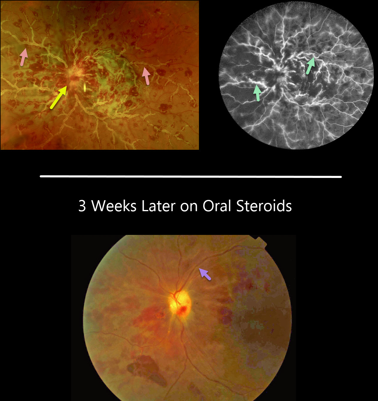
This 34YO African American female presented with several days of painless vision loss in her left eye. Vision was 20/30 OD and HM OS with an afferent defect OS. Anterior segments were normal.
There was mild disc edema OD (not shown) and florid disc edema OS with retinal vasculitis predominantly involving the veins.
On fluorescein angiography, the retinal veins in the left eye were diffusely stained.
On further review of systems, she had a new skin rash on her right forearm, but was otherwise asymptomatic. Testing revealed elevated angiotensin-converting enzyme and lysozyme levels, and a chest X-ray showed bilateral perihilar fullness.
With a working diagnosis of sarcoidosis, she is currently being managed with oral steroids and has been evaluated by both rheumatology and pulmonology.
While her retinal vasculitis has responded well to treatment, her vision remains HM three weeks later.
Learning Points:
Frosted branch angiitis is a retinal vasculitis that gets its name due to the involved vessels resembling the branches of a tree that are “frosted” with snow. Although these findings may be isolated, they are often considered a phenotype found in a multitude of inflammatory conditions, most commonly CMV retinitis.
The inflammation is usually highly responsive to steroids, and the visual prognosis is generally good, although some may develop secondary neovascularization in the anterior and posterior segments due to widespread ischemia.
SARCOIDOSIS
Originally posted on @retina.rocks July 9, 2021
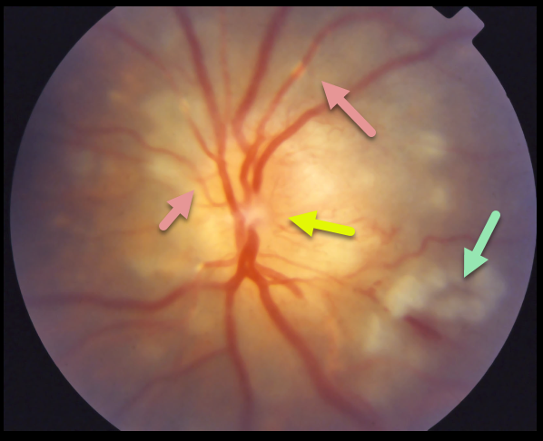
This patient with a known history of sarcoidosis presented with a swollen optic nerve, vasculitis, and patches of inner retinal opacification.
Learning Points:
Sarcoidosis, similar to syphilis, has a myriad of ocular presentations, including anterior, intermediate, and posterior uveitis, vasculitis, papillitis, and choroidal granuloma.
In our experience, there appears to be little association between active systemic disease and ocular inflammation. Establishing the diagnosis of ocular sarcoid in the absence of a known systemic diagnosis is often difficult.
Sensitivity and specificity are highest for chest CT and, when combined with elevated serum ACE and lymphopenia, are strongly suggestive of systemic sarcoidosis (see Nieder and Sims, AJO 1019;206:149-153).
Another recent publication identified soluble interleukin-2 receptor (sIL-2R) and chest CT evidence of bilateral hilar lymphadenopathy as the most sensitive biomarkers for diagnosing systemic sarcoidosis (Japanese J Ophthalmology 2021;65:191-198).


