

Scleral Buckle
<< Back to Cases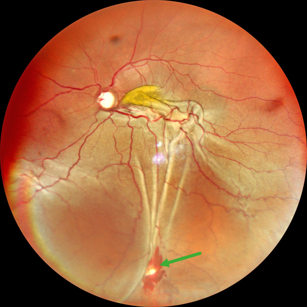
RHEGMATOGENOUS RD WITH RETINAL INCARCERATION FOLLOWING FAILED SCLERAL BUCKLING
Malvika Singh and Manish Nagpal
Originally posted on @retina.rocks November 4, 2025
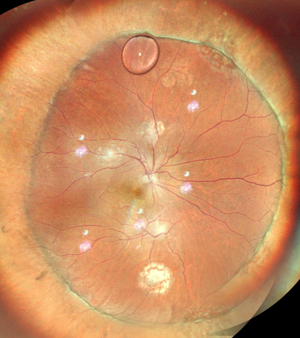
SILICONE OIL DROPLET
Ankit Jain and Manish Nagpal
Originally posted on @retina.rocks September 17, 2025
This 56YO male underwent successful retinal reattachment with vitrectomy, scleral buckling, and silicone oil. One month following silicone oil removal, he presented complaining of an inferior floater. Vision was 20/40.
Color SLO imaging shows an attached retina with an encircling buckle. A residual silicone oil droplet is noted superiorly along the buckle edge. Observation was recommended.
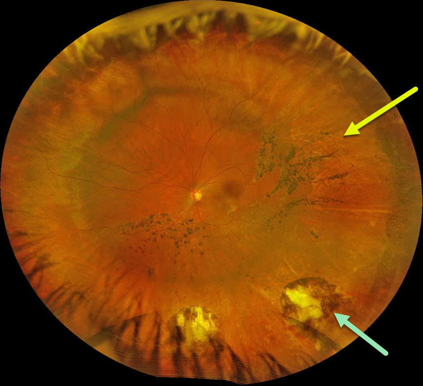
SCLERAL BUCKLE
Originally posted on @retina.rocks October 28, 2024
This 31YO female underwent successful scleral buckling surgery about 7 years earlier. Optos color RGB imaging shows a broad encircling buckle. Cryotherapy scarring is noted inferior to the buckle. Multiple demarcation lines with some intraretinal pigment migration are noted within the region of the prior detachment.
Learning Points:
The normal neurosensory retina remains attached to the RPE due to numerous physiologic mechanisms, including the inner (retinal vascular endothelium) and outer (tight junctions at the apex of the RPE cells) blood retinal barriers, hyperosmotic choroid passively drawing fluid out of the subretinal space, RPE pump, interphotoreceptor matrix, and friction between the RPE microvilli and the photoreceptor outer segments. Rhegmatogenous retinal detachment (RRD) occurs when liquid vitreous, entering the subretinal space through a retinal break, overwhelms these forces, favoring retinal detachment.
Repairing RRD involves finding and closing all breaks, essentially denying the liquid vitreous access to the subretinal space. The break(s) can be closed internally with a gas bubble with pneumatic retinopexy or vitrectomy surgery, or externally by sewing a piece of silicone material to the eye wall (scleral buckle). This indents, or buckles, the RPE and sclera up against the retinal break and formed vitreous.
The choice of procedure to reattach the retina is complex, depending on each patient’s unique anatomy and the surgeon’s preference. Historically, scleral buckling was the only method until vitrectomy surgery appeared in the 1970’s. There are advantages and disadvantages to all techniques. With one or more surgeries, there is a high anatomic success rate regardless of the method of retinal reattachment.
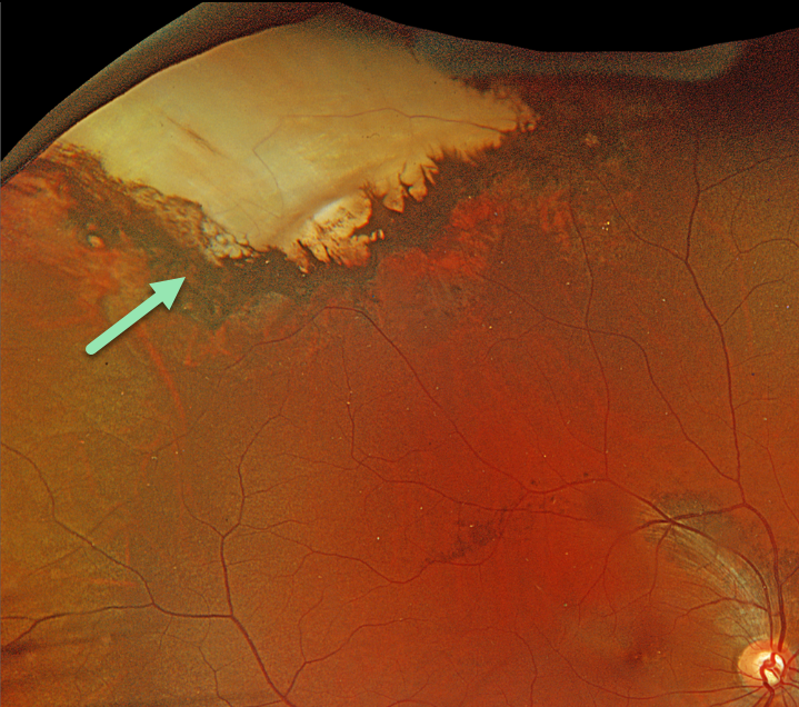
SCLERAL BUCKLE
Originally posted on @retina.rocks March 21, 2024
This 31YO male gave a history of scleral buckling surgery in Cuba 14 years earlier. There were no visual complaints, and vision was 20/70.
Optos color RGB imaging shows an intruding scleral buckle in the superotemporal periphery. The buckle is covered by bare neurosensory retina with some surrounding hyperpigmented chorioretinal scarring. Externally, the eye was quiet, and the conjunctiva completely covered the buckle.
Learning Points:
Intrusion of a scleral buckle is now a rare event with modern surgical techniques and buckling materials. This was most common following MIRAgel explants, which have since been removed from the market (Davuluri et al, JAMA Ophthalmology 2023;141:1081-1082).
An intruding buckle is usually asymptomatic but can cause vitreous hemorrhage (Zarei et al, Int J Retina Vitreous 2020;6;7). Observation for our patient was recommended.
MACULAR HOLE
Veronika Matello and Barbara Parolini
Originally posted on @retina.rocks October 25, 2022
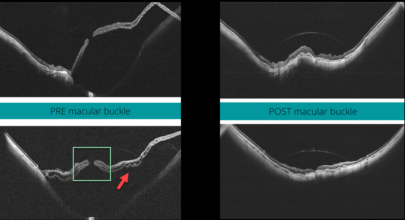
This 58YO highly myopic female presented with 20/400 vision in her left eye. The axial length was 32.45 mm, and her refractive error prior to LASIK surgery was 13D.
Widefield OCT scanning (Canon Xephilio SQ) shows a large macular retinal detachment with (or associated to) a full-thickness macular hole, defined as stage 4c according to the Myopic Traction Maculopathy Staging System (Parolini et al, Eur J Ophthalmol 2021;31:1299-1312).
Pars plana vitrectomy with an ILM flap and macular buckling was performed. One month postoperatively, the retina was flat, and the macular hole closed. Vision was 20/80.
Learning Points:
In our experience, these detachments are totally tractional, with the macular hole developing before or after the retina detaches. The macular buckle treats both the macular schisis and detachment, whereas the vitrectomy with ILM peeling closes the macular hole.
SCLERAL BUCKLE
Barbara Parolini and Veronika Matello
Originally posted on @retina.rocks September 30, 2021
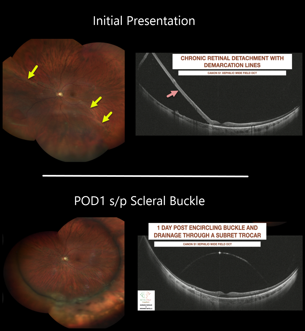
This 36YO male presented with 20/20 vision despite a chronic rhegmatogenous retinal detachment with a demarcation line of his left eye. The wide-field OCT shows the chronic retinal detachment.
The patient underwent scleral buckling with drainage of subretinal fluid through a trocar. The retina is completely attached on post-operative day one.
Learning Points:
The normal neurosensory retina remains attached to the RPE due to numerous physiologic mechanisms, including the inner and outer blood-retinal barriers, the hyperosmotic choroid passively drawing fluid into the subretinal space, the RPE pump, the interphotoreceptor matrix, and friction between the RPE microvilli and the photoreceptor outer segments.
Rhegmatogenous retinal detachment (RRD) occurs when liquid vitreous, entering the subretinal space through a retinal break, overwhelms these forces, favoring retinal detachment.
Repairing RRD involves finding and closing all breaks, essentially denying the liquid vitreous access to the subretinal space. The break(s) can be closed internally with a gas bubble via pneumatic retinopexy or vitrectomy surgery, or externally by securing a piece of silicone material to the sclera (scleral buckle). This indents, or buckles, the RPE against the retinal break.
SCLERAL BUCKLE
Originally posted on @retina.rocks July 23, 2021
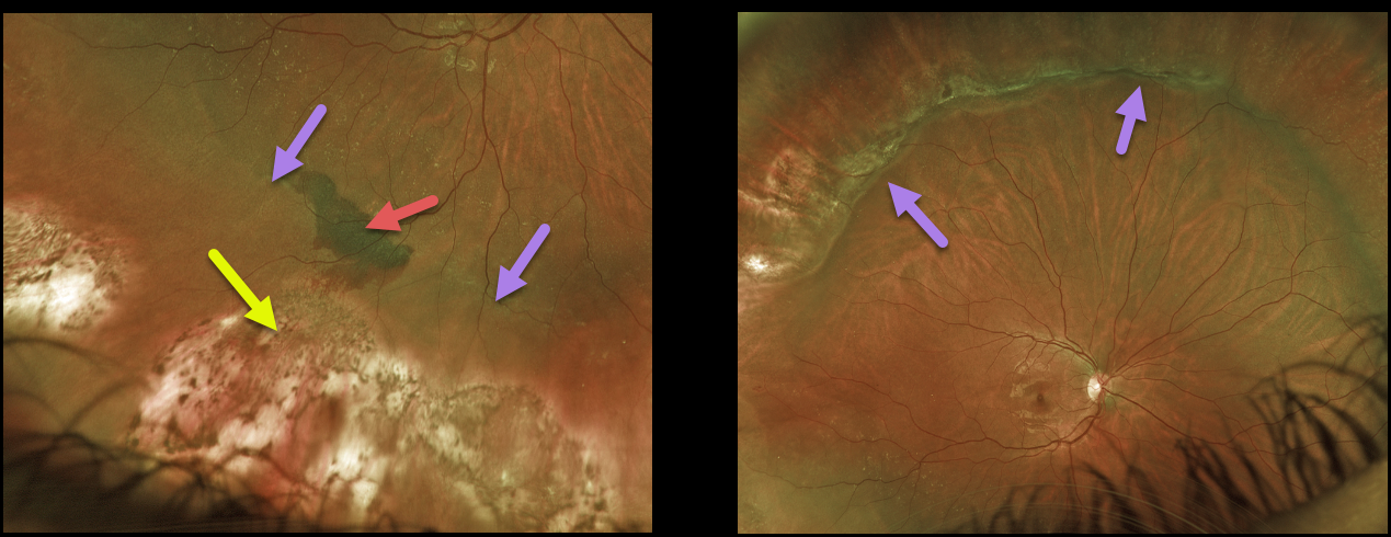
This 23YO female underwent successful retinal reattachment surgery with drainage of subretinal fluid and an encircling buckle. The drainage was uncomplicated, but at the completion of surgery, indirect ophthalmoscopy revealed a localized subretinal hemorrhage posterior to the drainage site.
One week later, Optos imaging shows resolving subretinal blood. Cryotherapy scarring of the causative lattice and breaks is noted with a depigmented drainage site. A shallow buckling effect is seen.
Learning Points:
Drainage of subretinal fluid at the time of scleral buckling is usually performed if the break cannot be opposed to the RPE with buckling alone.
Subretinal bleeding sometimes follows puncture of the choroid with either a needle or a laser. This is usually self-limited and resolves postoperatively without treatment.
SCLERAL BUCKLE
Originally posted on @retina.rocks May 12, 2021
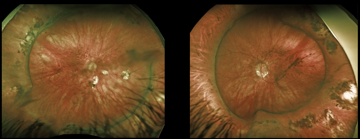
This is a beautiful Optos image of bilateral scleral buckles in a 51YO female myopic patient who underwent successful retinal reattachment surgery years ago.
Learning Points:
The normal neurosensory retina remains attached to the RPE due to numerous physiologic mechanisms, including the inner and outer blood-retinal barriers, the hyperosmotic choroid passively drawing fluid into the subretinal space, the RPE pump, the interphotoreceptor matrix, and friction between the RPE microvilli and the photoreceptor outer segments.
Rhegmatogenous retinal detachment (RRD) occurs when liquid vitreous, entering the subretinal space through a retinal break, overwhelms these forces, favoring retinal detachment.
Repairing RRD involves finding and closing all breaks, essentially denying the liquid vitreous access to the subretinal space. The break(s) can be closed internally with a gas bubble via pneumatic retinopexy or vitrectomy surgery, or externally by securing a piece of silicone material to the sclera (scleral buckle). This indents, or buckles, the RPE against the retinal break.
VASOPROLIFERATIVE TUMOR
Originally posted on @retina.rocks June 29, 2020
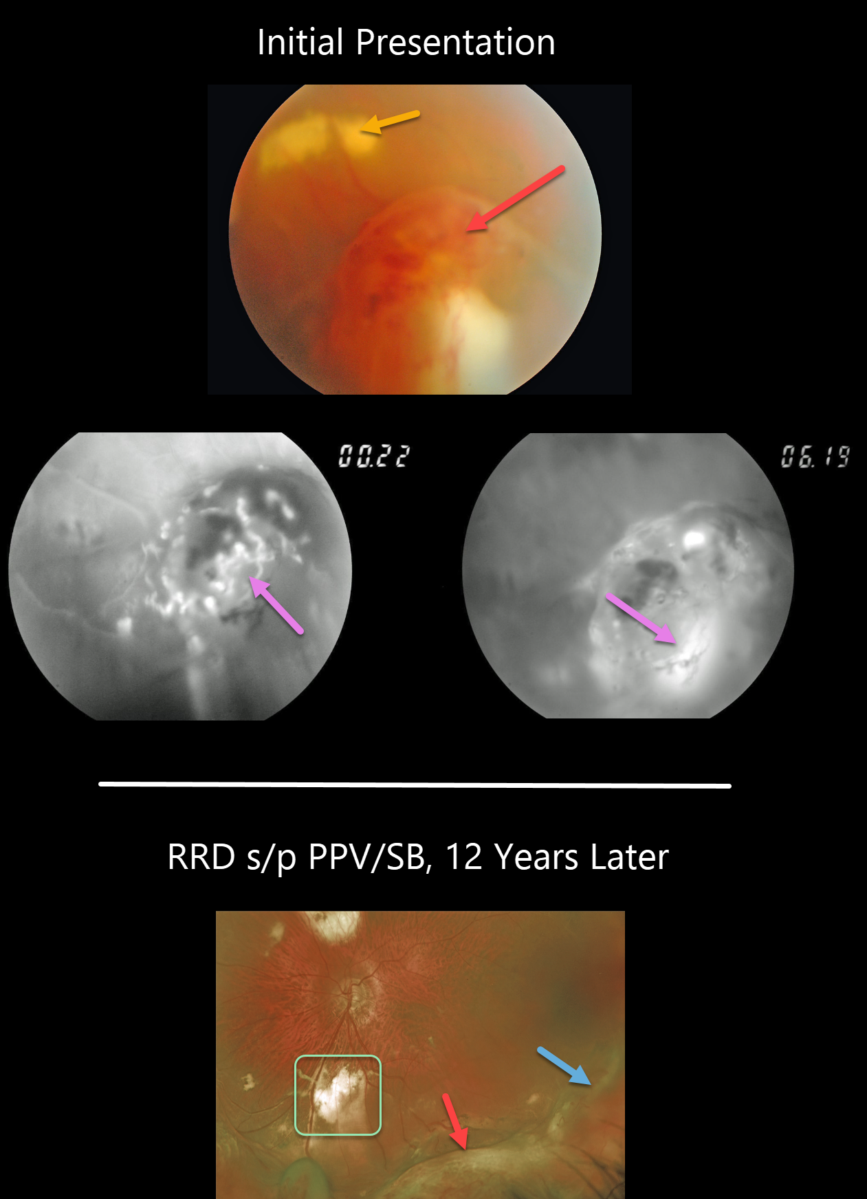
This 46YO presented with an asymptomatic vasoproliferative tumor (VPT). The VPT initially appeared as a tongue-like, reddish, elevated, peripheral mass with more posterior subretinal lipid. Fluorescein angiography (FA) revealed the vascular nature of the lesion.
He then developed a rhegmatogenous retinal detachment that was repaired with a combined vitrectomy and scleral buckle. The indentation from the buckle is visible more temporally and distinct from the complex enlarged VPT more inferiorly. The subretinal lipid resolved with more extensive subretinal scarring.
Learning Points:
Vasoproliferative tumors are benign globular, dome-shaped lesions arising in the peripheral retina. They can be yellow or red and cause subretinal exudation and detachment, anterior or posterior segment neovascularization, and vitreous hemorrhage.
They can be primary idiopathic lesions or a vascular response to various entities, including intermediate uveitis, retinitis pigmentosa, Coats disease, and prior retinal detachment.
For lesions causing symptomatic exudation or neovascularization, treatment with thermal laser, cryotherapy, and/or anti-VEGF injections is indicated.
SCLERAL BUCKLE
Originally posted on @retina.rocks June 1, 2020
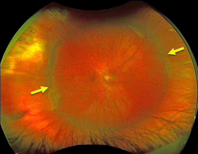
This patient has a broad encircling scleral buckle that’s beautifully imaged in this Optos photograph.
Learning Points:
The normal neurosensory retina remains attached to the RPE due to numerous physiologic mechanisms, including the inner (retinal vascular endothelium) and outer (tight junctions at the apex of the RPE cells) blood retinal barriers, hyperosmotic choroid passively drawing fluid into the subretinal space, RPE pump, interphotoreceptor matrix, and friction between the RPE microvilli and the photoreceptor outer segments.
Rhegmatogenous retinal detachment (RRD) occurs when liquid vitreous, entering the subretinal space through a retinal break, overwhelms these forces, favoring retinal detachment.
Repairing RRD involves finding and closing all breaks, essentially denying the liquid vitreous access to the subretinal space. The break(s) can be closed internally with a gas bubble via pneumatic retinopexy or vitrectomy surgery, or externally by securing a piece of silicone material to the sclera (scleral buckle). This indents, or buckles, the sclera against the retinal break.
The choice for what procedure to use to reattach the retina is complex, depending on each patient’s unique anatomy and on the surgeon’s preference. Historically, scleral buckling was the only method before vitrectomy surgery was introduced by Machemer in the early 1970’s.
There are advantages and disadvantages to all techniques. With one or more surgeries, there is a high anatomic success rate regardless of how the retina is reattached.


