

Scleritis
<< Back to Cases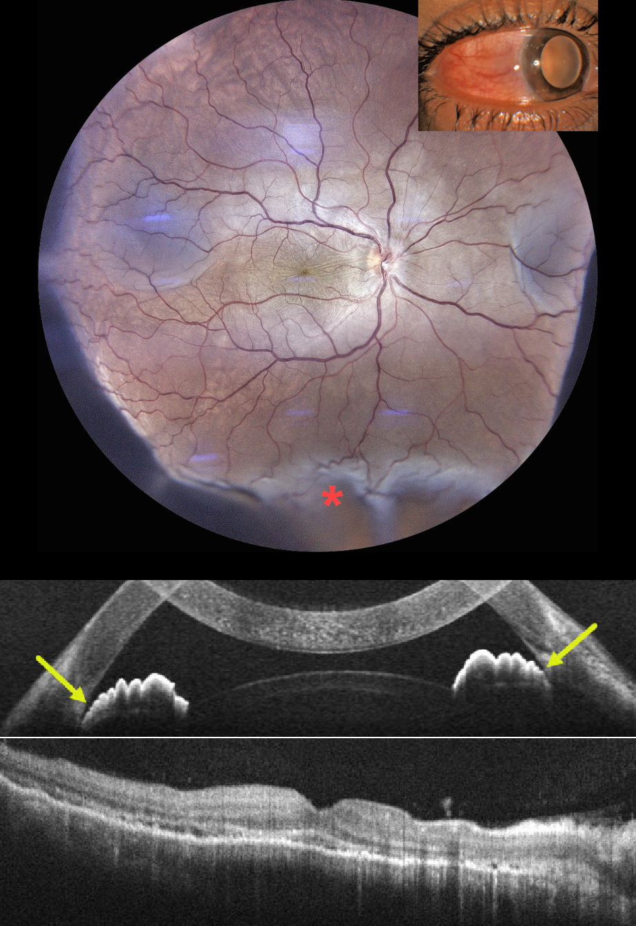
TUBERCULOUS POSTERIOR SCLERITIS
Aniruddh Soni
Originally posted on @retina.rocks July 31, 2026
This previously healthy 35YO male presented with 1 week of pain, redness, and decreased vision in his right eye. He had been treated by an outside doctor for conjunctivitis. Vision 20/200 OD and 20/20 in his normal OS. Slit lamp examination showed temporal scleral injection. Intraocular pressure was 23 mm Hg.
Color photography shows radiating chorioretinal folds throughout the macula with an inferior exudative retinal detachment. OCT scanning of the macula shows chorioretinal folds, with anterior chamber OCT showing a narrow-to-closed angle. B-scan ultrasonography showed subtenon’s fluid (not shown).
Following a 3-week course of tapering oral steroids and glaucoma drops, vision improved to 20/40 with reduced chorioretinal folds and complete resolution of the exudative detachment. Rheumatologic workup was negative, but Mantoux skin testing and QuantiFERON-TB GOLD were positive, with a subsequent diagnosis of latent tuberculosis (TB). Antituberculous therapy was recommended, but the patient refused treatment.
Learning Points:
Tuberculous posterior scleritis is a rare manifestation of ocular TB (Agarwal and Majumder, Indian J Ophthalmol 2019;67:1362-1365), which itself occurs in approximately 1.5% of patients with confirmed systemic TB. Ocular findings include optic disc edema, choroiditis, and sclerochoroidal thickening with widening of the sub-Tenon space. It may occur via direct hematogenous dissemination of Mycobacterium tuberculosis or through a hypersensitivity-mediated immune response to the organism and can develop even in the absence of clinically active pulmonary disease.
Diagnosis, as in our patient, is largely presumptive, requiring a high index of suspicion supported by bloodwork, chest imaging to assess pulmonary involvement, and exclusion of mimics such as sarcoidosis, syphilis, and metastatic disease.
POSTERIOR SCLERITIS
Will Gibson
Originally posted on @retina.rocks September 26, 2023
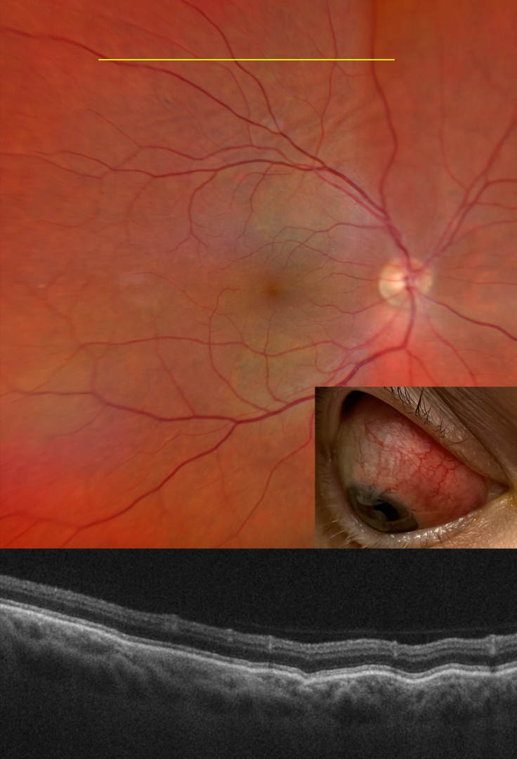
This 48YO female with a history of psoriatic arthritis presented on 10/25/22 with painful anterior scleritis. Vision was 20/20. The patient was started on 50mg of prednisone and slowly tapered off by rheumatology.
She returned on 7/19/23 with pain behind her right eye. Vision was 20/20. Color imaging shows subtle chorioretinal folds in the superior midperiphery.
OCT B-scan through this region confirmed the chorioretinal folds. B-scan ultrasonography revealed bilateral T signs.
Learning Points:
Posterior scleritis can present either as a diffuse thickening of the entire posterior sclera or as a nodular type with localized scleral thickening (see Agrawal et al, Retina 2016;36:392-401).
Nodular inflammation appears as a posterior pole subretinal yellowish mass lesion, often with overlying serous fluid and chorioretinal folds. B-scan ultrasonography may show fluid in the Tenon space (“T” sign).
Unlike anterior scleritis, the eyes are usually quiet and pain-free. Initial treatment is usually with oral non-steroidal anti-inflammatories or corticosteroids, although patients may also require immunosuppressive therapy. For a large case series of posterior scleritis, see McCluskey et al, Ophthalmology 1999;106:2380-2386.
Oral prednisone was restarted and slowly tapered. Further labs and chest X-ray were performed to rule out other possible causes of scleritis, and she was started on Humira.
POSTERIOR SCLERITIS
Originally posted on @retina.rocks June 17, 2021
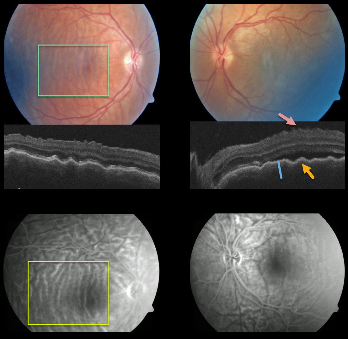
This patient presented with bilateral posterior pole chorioretinal folds, confirmed on OCT as inner retinal folds, outer retinal/RPE folds, and thickened choroid, and on fluorescein angiography.
Our patient most likely had the diffuse type of posterior scleritis. Unfortunately, we don’t have any further images or clinical information.
Learning Points:
Posterior scleritis can present either as a diffuse thickening of the entire posterior sclera or as a nodular type with localized scleral thickening (see Agrawal et al, Retina 2016;36:392-401).
Nodular inflammation appears as a posterior pole subretinal yellowish mass lesion, often with overlying serous fluid and chorioretinal folds.
B-scan ultrasonography may show fluid in the Tenon space (“T” sign). Unlike anterior scleritis, the eyes are usually quiet and pain-free.
Initial treatment is usually with oral non-steroidal anti-inflammatories or corticosteroids, although patients may also require immunosuppressive therapy.
For a large case series of posterior scleritis, see McCluskey et al, Ophthalmology 1999;106:2380-2386.


