

Sickle Cell
<< Back to Cases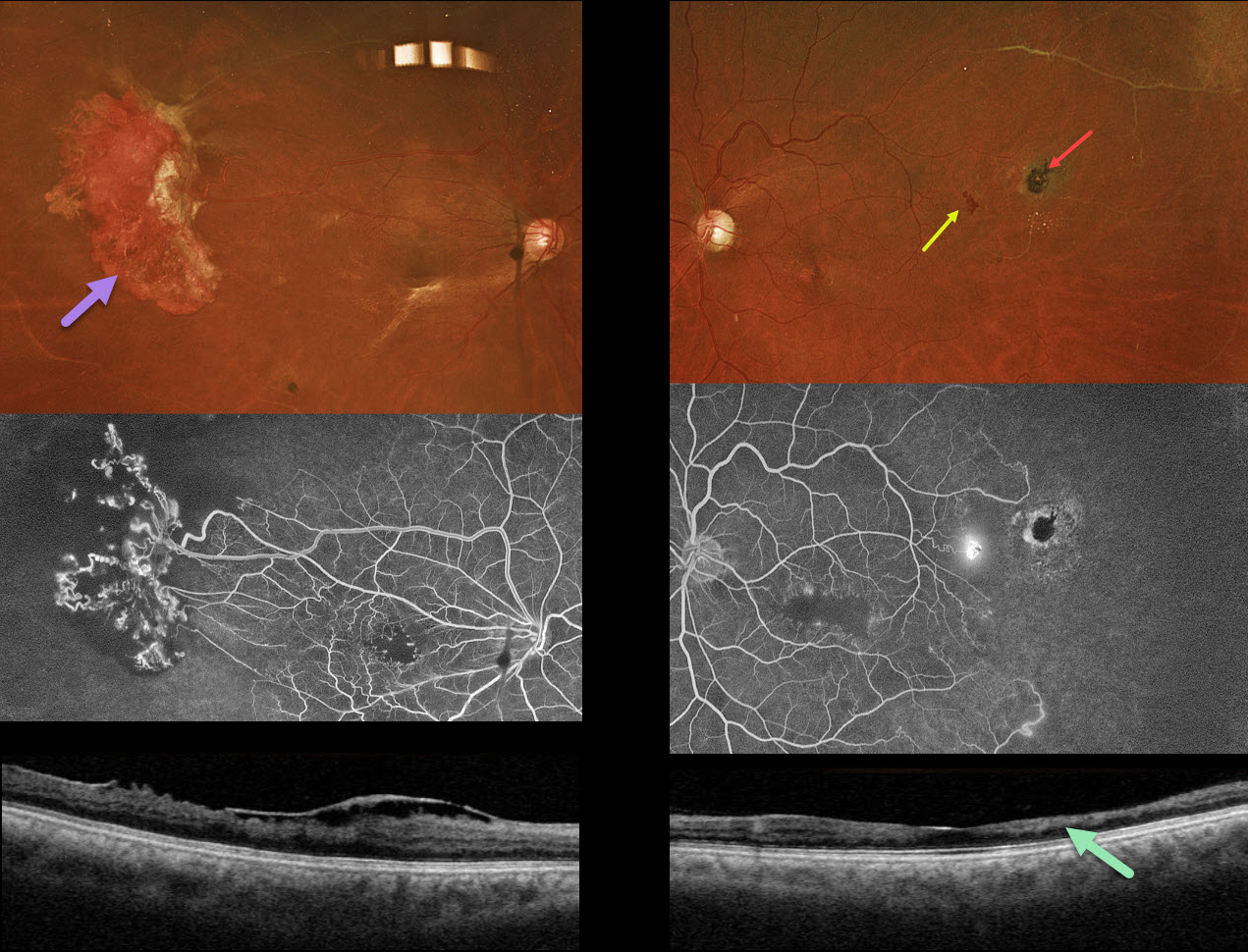
SICKLE CELL RETINOPATHY
Originally posted on @retina.rocks May 5, 2026
This 44YO male with a known history of sickle cell disease was referred for a possible detached retina. Vision has decreased over the past few years. Vision was 20/200 OU.
Optos color RGB imaging shows a large area of active neovascularization in the right temporal midperiphery associated with extensive angiographic retinal ischemia. Similar ischemia is noted OS with a small patch of leaking neovascularization. There is a small, more distal black sunburst lesion. The foveal avascular zones are enlarged with surrounding telangiectasia. This is reflected in bilateral inner retinal thinning with disorganization of inner retinal layers on OCT scanning (DRIL). An epimacular membrane is also noted OD.
Learning Points:
Patients with sickle cell disease develop symptoms throughout their bodies as stiffened, sickled red blood cells block capillary beds. In the eye, this can cause proliferative sickle cell retinopathy (PSR) with sea-fan-shaped areas of peripheral retinal neovascularization developing at the junction of perfused and ischemic retina. Increasing macular ischemia correlates with peripheral angiographic nonperfusion, as seen in our patient (Han et al., Ophthalmology Retina 2019;2:599-605). Complications include vitreous hemorrhage and tractional retinal detachment. Although patients with SS disease tend to have more systemic complications, SC disease is usually associated with more severe ocular findings.
Treatment with sectoral (Farber et al Arch Ophthalmology 1991;109:363-367) or circumferential (Kimmel et al Ophthalmology 1986;93:1429-1434) scatter laser photocoagulation is somewhat controversial since, unlike more typical proliferative retinopathies (ie from diabetes and branch retinal vein occlusions), these new vessels tend to auto-infarct with a good natural history for untreated disease (Downes et al Ophthalmology 2005;112:1869-1875). Based on the active appearance of the large temporal neovascularization OD, we elected for prophylactic scatter laser throughout the ischemic retina.
SICKLE CELL RETINOPATHY
Originally posted on @retina.rocks January 1, 2026
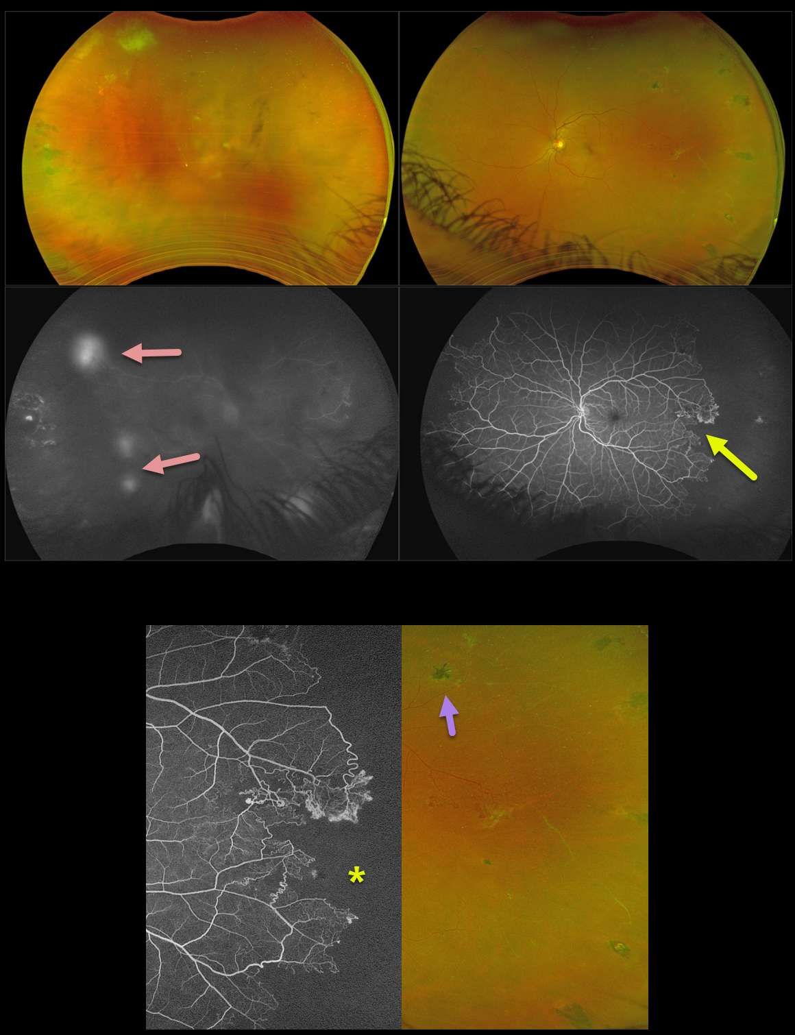
This 45YO male with a known history of sickle cell SC disease presented with new floaters and blurred vision in his right eye. Vision was 20/70 OD and 20/30 OS.
Optos color RG imaging shows a fairly dense vitreous hemorrhage OD, with variably fibrosed peripheral retinal neovascularization superotemporally. The left fundus shows temporal retinal ischemia with white vessels and mostly active retinal neovascularization along its posterior extent. Scattered black sunburst lesions are noted temporally as well. Fluorescein angiography OS shows an irregular, abrupt margin of peripheral complete capillary nonperfusion, especially temporally. The neovascularization in his OD shows late leakage.
Avastin was injected OD. Given the active vessels in his OS, along with the recent vitreous hemorrhage OD, we decided to prophylactically treat the left ischemic retina with scatter laser.
Learning Points:
Patients with sickle cell disease develop symptoms throughout their bodies as stiffened, sickled red blood cells block capillary beds. In the eye, this can cause proliferative sickle cell retinopathy (PSR) with sea-fan-shaped areas of peripheral retinal neovascularization developing at the junction of perfused and ischemic retina. The black sunburst lesions are thought to occur from intraretinal RPE migration in response to photoreceptor damage from intraretinal and subretinal hemorrhage (Asdourian et al, BJO 1975;59:710-716).
Complications include vitreous hemorrhage and traction retinal detachment. Although patients with SS disease tend to have more systemic complications, SC disease is usually associated with more severe ocular findings. Treatment with peripheral scatter laser photocoagulation is somewhat controversial since, unlike more typical proliferative retinopathies (ie from diabetes and branch retinal vein occlusions), these new vessels tend to auto-infarct.
For a great review article on sickle cell disease and the eye, see Elagouz et al, Surv Ophthalmology 2010;55;359-377.
SICKLE CELL RETINOPATHY
Originally posted on @retina.rocks July 10, 2025
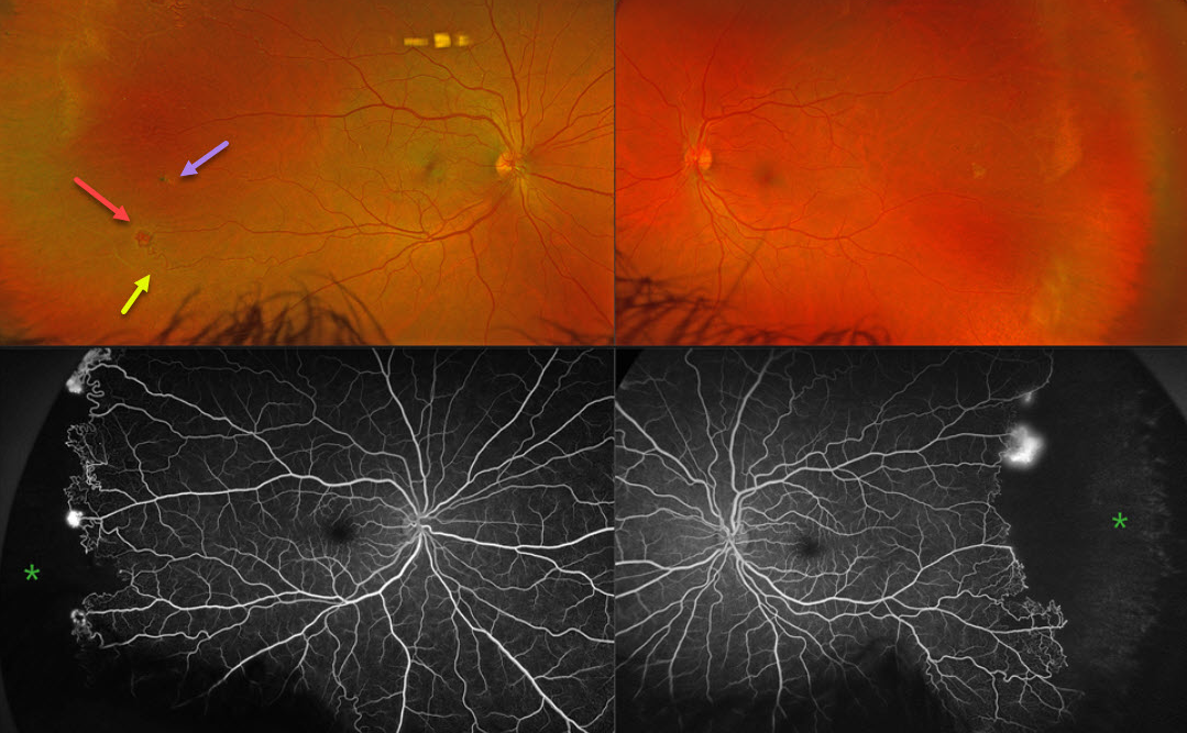
This 43YO male was referred for asymptomatic retinal findings. He has a known history of sickle cell disease. Vision was 20/20 OU.
Optos color RG imaging shows bilateral peripheral temporal and featureless retinas with white vessels. There is a small temporal pigmented chorioretinal scar OD, indicating a possible black sunburst lesion. The retinal vascular bed is remodeled along the border of perfused and ischemic retina with retinal arteriovenous shunting and neovascularization. The complete temporal nonperfusion with bilateral neovascularization is confirmed on fluorescein angiography.
Learning Points:
Patients with sickle cell disease develop symptoms throughout their bodies as stiffened, sickled red blood cells block capillary beds. In the eye, this can cause proliferative sickle cell retinopathy (PSR) with sea-fan-shaped areas of peripheral retinal neovascularization developing at the junction of perfused and ischemic retina. Complications include vitreous hemorrhage and tractional retinal detachment. Although patients with SS disease tend to have more systemic complications, SC disease is usually associated with more severe ocular findings.
Treatment with sectoral (Farber et al Arch Ophthalmology 1991;109:363-367) or circumferential (Kimmel et al Ophthalmology 1986;93:1429-1434) scatter laser photocoagulation is somewhat controversial since, unlike more typical proliferative retinopathies (ie from diabetes and branch retinal vein occlusions), these new vessels tend to auto-infarct with a good natural history for untreated disease (Downes et al Ophthalmology 2005;112:1869-1875). We elected to closely follow him for now with observation.
SICKLE CELL RETINOPATHY
Will Gibson
Originally posted on @retina.rocks April 24, 2023
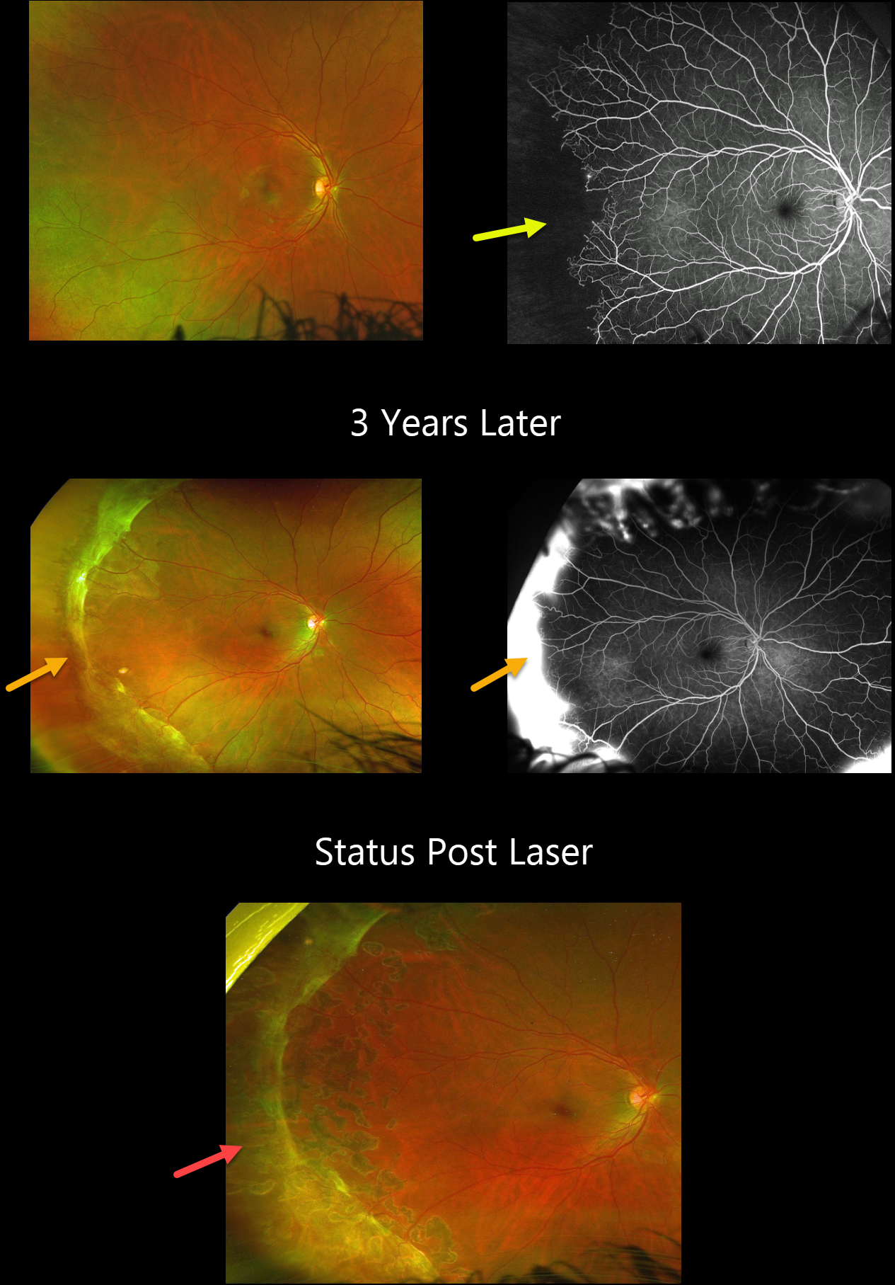
This 19YO male has a known history of sickle disease (Hb SC). On 7/30/20, vision was 20/25 OD.
Optos color imaging shows temporal vascular whitening, and angiography shows marked temporal ischemia.
He was lost to follow-up until 1/10/23 when he presented with hand motions vision from a vitreous hemorrhage OS (not shown). Although vision was still 20/20 OD, a broad band of fibrosed neovascularization was noted in the periphery. A peripheral scatter laser was applied to the right eye at the time of OS vitrectomy, and 6 weeks later, all neovascularization had regressed.
Learning Points:
Patients with sickle cell disease develop symptoms throughout their bodies as stiff, sickled red blood cells block capillary beds. In the eye, this can cause proliferative sickle cell retinopathy with sea-fan shaped areas of peripheral retinal neovascularization developing at the junction of perfused and ischemic retina. Complications include vitreous hemorrhage and traction retinal detachment. Although patients with SS disease tend to have more systemic complications, SC disease is usually associated with more severe ocular findings.
SICKLE CELL RETINOPATHY
Lucas Zago, Julia Jiquilin Carvalho and Mauricio Maia
Originally posted on @retina.rocks April 3, 2023
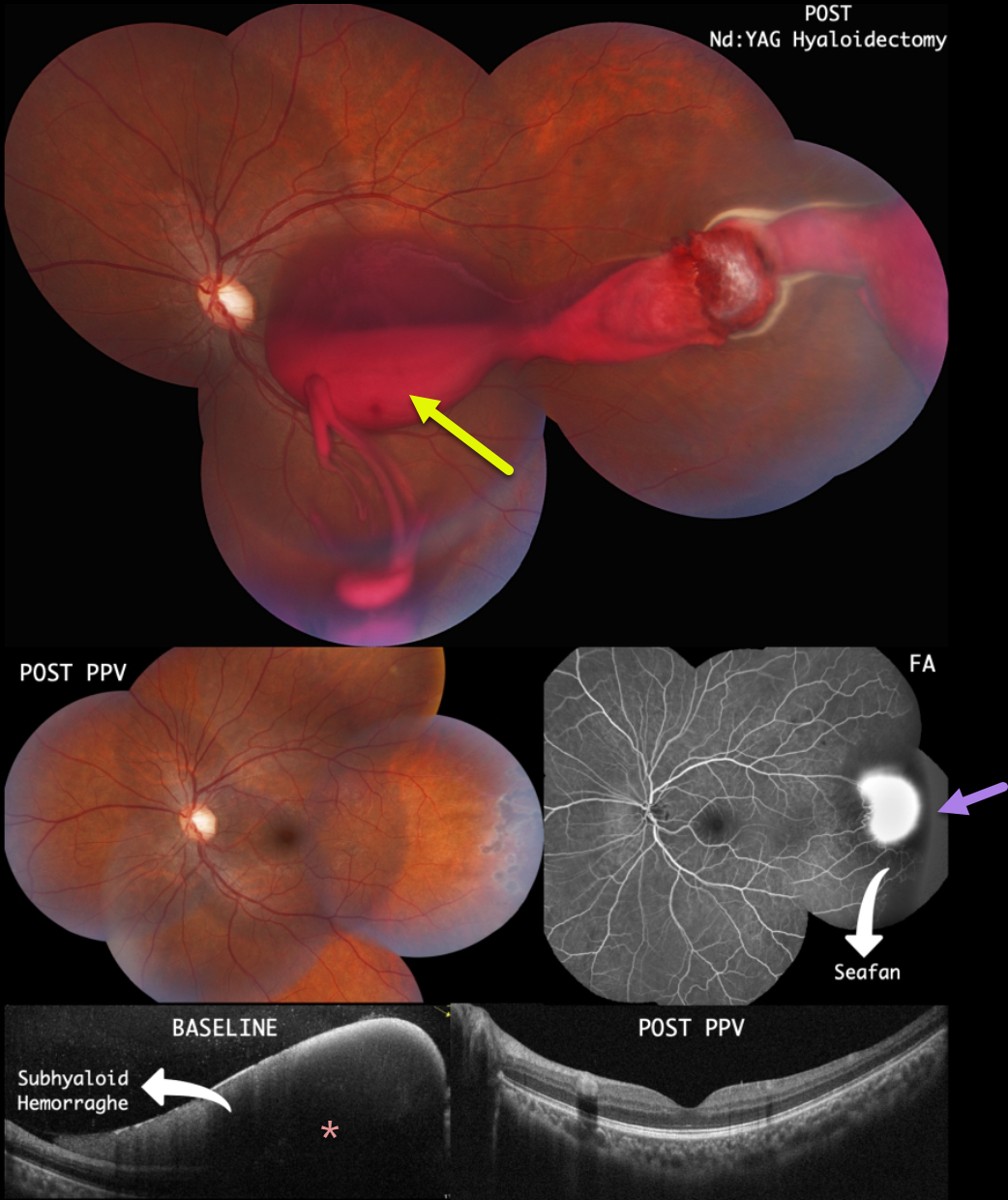
This 22YO male presented with 3 days of vision loss in his left eye. There was a past medical history of Hemoglobin SC Disease. He denied any Valsalva maneuvers. He remembered seeing a retinal doctor several months earlier with the diagnosis of proliferative sickle retinopathy. Vision was counting fingers OS.
Color imaging shows a large subhyaloid/vitreous hemorrhage extending from the temporal midperiphery into the macula. The blood seemed to originate from a probable area of neovascularization (sea fan). OCT scanning confirmed the subhyaloid location of the blood.
Immediately following Nd:YAG laser hyaloidotomy, the blood began to drain into the inferior vitreous. Peripheral scatter laser into the temporal ischemic retina was also performed.
However, he subsequently developed a dense vitreous hemorrhage requiring vitrectomy with endolaser. One month after surgery, the vitreous was clear, the neovascularization had regressed, and the macular appearance had normalized. Vision was 20/20.
SICKLE CELL RETINOPATHY
Paulo Eduardo Stanga, Javier Valentin Bravo, Sebastian Stanga, and Ursula Reinstein
Originally posted on @retina.rocks March 13, 2023
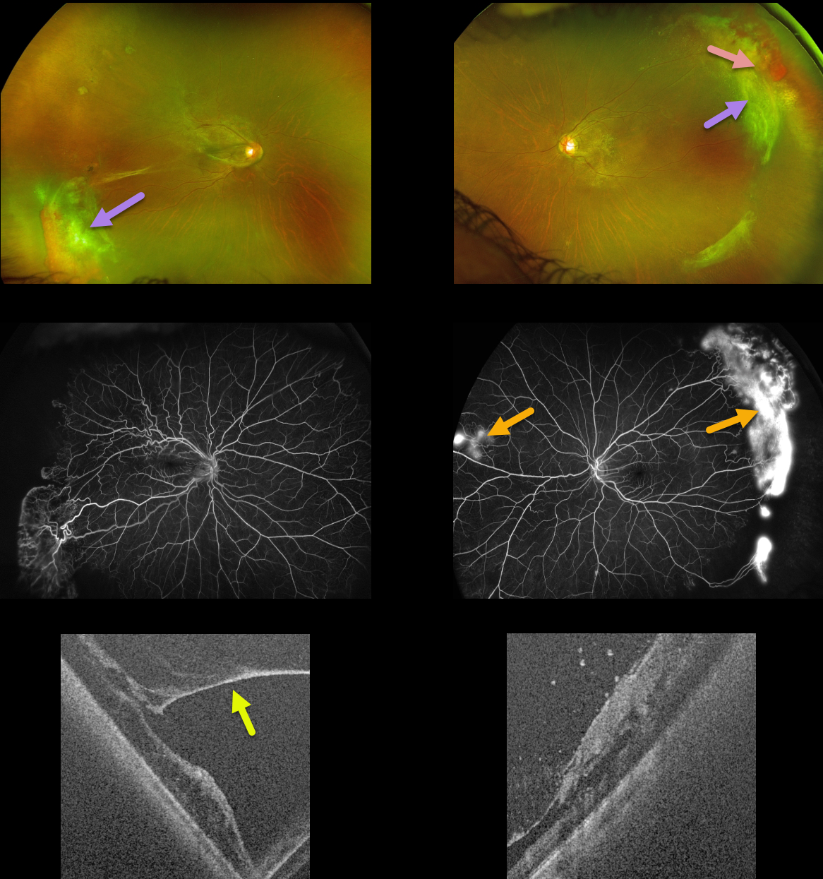
This 45YO female with a known history of sickle SC disease presented with intermittent bilateral floaters following a COVID infection four months earlier. Vision was 20/40 OD and 20/16 OS.
Optos color imaging shows bilateral temporal fibrosed retinal neovascularization (sea fans) associated with some localized vitreous blood. The right eye had a significant epimacular membrane with traction, with a milder adherent membrane in her left eye.
Fluorescein angiography shows bilateral far temporal ischemia with leaking neovascularization. Some nasal neovascularization is also noted in her left eye. Ultrawidefield OCT shows vitreous traction adherent to the neovascularization.
We elected to closely follow her for now with observation.
Learning Points:
Patients with sickle cell disease develop symptoms throughout their bodies as stiff, sickled red blood cells block capillary beds. In the eye, this can cause proliferative sickle cell retinopathy (PSR) with sea-fan-shaped areas of peripheral retinal neovascularization developing at the junction of perfused and ischemic retina.
Complications include vitreous hemorrhage and traction retinal detachment. Although patients with SS disease tend to have more systemic complications, SC disease is usually associated with more severe ocular findings.
Treatment with sectoral (Farber et al Arch Ophthalmology 1991;109:363-367) or circumferential (Kimmel et al Ophthalmology 1986;93:1429-1434) scatter laser photocoagulation is somewhat controversial since, unlike more typical proliferative retinopathies (i.e., from diabetes and branch retinal vein occlusions), these new vessels tend to auto-infarct with a good natural history for untreated disease (Downes et al Ophthalmology 2005;112:1869-1875).
SICKLE CELL RETINOPATHY
Originally posted on @retina.rocks October 21, 2022
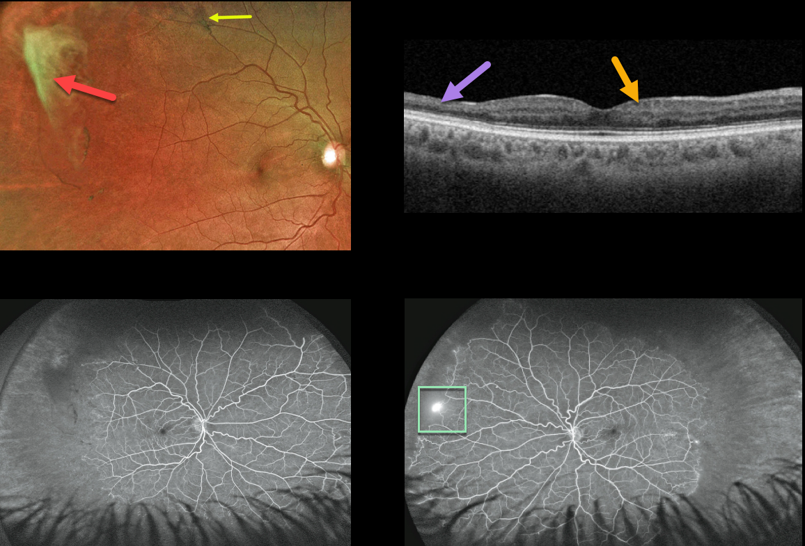
This 39YO African American male presented with asymptomatic proliferative sickle cell retinopathy with spontaneously avulsed bilateral superotemporal peripheral fibrosed retinal neovascularization (sea fans). A small midperipheral black sunburst lesion is seen superotemporally in his right eye.
Swept-source OCT shows variable inner retinal thinning and disorganization of the retinal inner layers (DRIL).
Fluorescein angiography shows a patch of retinal neovascularization OS at the junction of perfused and non-perfused retina.
Since these lesions often auto-infarct, as they did in our patient, we elected to observe the small area of neovascularization in his right eye. He is also at low risk for a vitreous hemorrhage or traction detachment since the vitreous is already detached in this area.
Learning Points:
Patients with sickle cell disease develop symptoms throughout their bodies as stiff, sickled red blood cells block capillary beds. In the eye, this can cause proliferative sickle cell retinopathy (PSR) with sea-fan-shaped areas of peripheral retinal neovascularization developing at the junction of perfused and ischemic retina.
The variable macular thinning noted on OCT is caused by microinfarcts to the retinal microcirculation. Enlargement of the foveal avascular zone is a common OCT angiographic finding (Fares, AJO 2021;224:7-17).
The black sunburst lesions are thought to occur from intraretinal RPE migration in response to photoreceptor damage from intraretinal and subretinal hemorrhage (see Ausdourian et al, BJO 1975;59:710-716).
SICKLE CELL RETINOPATHY
Originally posted on @retina.rocks August 1, 2022
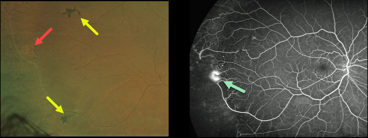
This 56YO African American male presented with floaters in his right eye and a history of sickle cell anemia. Vision was 20/30 OU.
A mild vitreous hemorrhage was noted inferiorly, along with an area of suspected peripheral retinal neovascularization. There were several areas of midperipheral black sunburst lesions. Fluorescein angiography confirmed a patch of retinal neovascularization at the junction of perfused and non-perfused retina. Due to the symptomatic vitreous hemorrhage, scatter laser to the peripheral ischemic retina was recommended.
Learning Points:
Patients with sickle cell disease develop symptoms throughout their bodies as stiff, sickled red blood cells block capillary beds. In the eye, this can cause proliferative sickle cell retinopathy (PSR) with sea-fan-shaped areas of peripheral retinal neovascularization developing at the junction of perfused and ischemic retina.
The black sunburst lesions are thought to occur from intraretinal RPE migration in response to photoreceptor damage from intraretinal and subretinal hemorrhage (see Ausdourian et al, BJO 1975;59:710-716). Complications include vitreous hemorrhage and traction retinal detachment. Although patients with SS disease tend to have more systemic complications, SC disease is usually associated with more severe ocular findings.
Treatment with peripheral scatter laser photocoagulation is somewhat controversial because, unlike in more typical proliferative retinopathies (e.g., those from diabetes and branch retinal vein occlusions), these new vessels tend to auto-infarct.
For a great review article on sickle cell disease and the eye, see Elagouz et al, Surv Ophthalmology 2010;55:359-377.
SICKLE CELL RETINOPATHY
Originally posted on @retina.rocks November 12, 2021
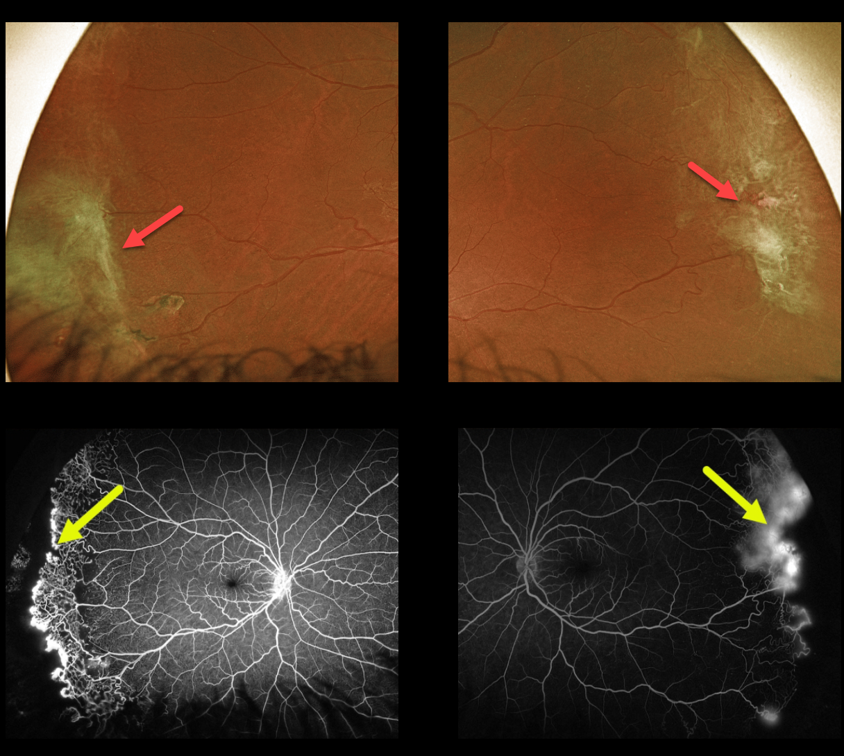
This 43YO female with a known history of SC disease presented with these asymptomatic fundus changes captured with Optos ultra-widefield imaging. Fibrosed areas of peripheral neovascularization are noted in each temporal periphery.
Fluorescein angiography shows a broad, well-demarcated zone of temporal peripheral retinal neovascularization at the junction of perfused and nonperfused retina.
Prophylactic scatter laser to the areas of capillary loss was recommended.
Learning Points:
Patients with sickle cell disease develop symptoms throughout their bodies as stiff, sickled red blood cells block capillary beds.
In the eye, this can cause proliferative sickle cell retinopathy (PSR), with sea-fan-shaped areas of peripheral retinal neovascularization developing at the junction of perfused and ischemic retina. Complications include vitreous hemorrhage and traction retinal detachment.
Although patients with SS disease tend to have more systemic complications, SC disease is usually associated with more severe ocular findings.
Treatment with peripheral scatter laser photocoagulation is somewhat controversial because, unlike more typical proliferative retinopathies (i.e., those from diabetes and branch retinal vein occlusions), these new vessels tend to auto-infarct.
For a great review article on sickle cell disease and the eye, see Elagouz et al, Surv Ophthalmology 2010;55:359-377.
SICKLE CELL RETINOPATHY
Originally posted on @retina.rocks April 2, 2020
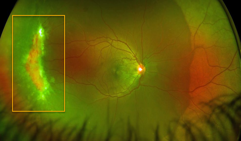
This patient, courtesy of Southern College of Optometry, presented with a large temporal peripheral area of active neovascularization from proliferative sickle cell retinopathy in the right eye. The left fundus was normal.
Learning Points:
Patients with sickle cell disease develop symptoms throughout their body from stiffened sickled red blood cells blocking capillary beds.
In the eye, this manifests as proliferative sickle cell retinopathy (PSR) with sea-fan shaped areas of peripheral retinal neovascularization developing at the junction of perfused and ischemic retina.
Complications include vitreous hemorrhage and traction retinal detachment.
Treatment with peripheral scatter laser is somewhat controversial since, unlike more typical proliferative retinopathies (ie from diabetes and branch retinal vein occlusions), these new vessels tend to auto-infarct.


