

Silicone Oil + Perfluoron
<< Back to Cases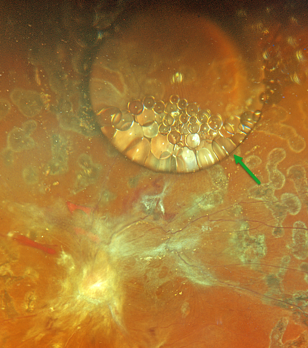
SILICONE FISH EGGS
Originally posted on @retina.rocks July 21, 2026
This 43YO male underwent a vitrectomy elsewhere for a diabetic traction retinal detachment about 5 months earlier. Vision was 20/400.
Optos color RGB imaging shows a large, round silicone oil droplet in the superior midperiphery. This bubble acts like a convex lens, magnifying the details of the underlying retina. Suspended within and along the edges of this droplet are numerous fish egg droplets. Residual fibrotic membranes extend from the nerve into the macula and along the superotemporal arcade. Repeat vitrectomy was scheduled.
EMULSIFIED SILICONE OIL WITH REVERSE PSEUDOHYPOPYON
Originally posted on @retina.rocks February 3, 2026
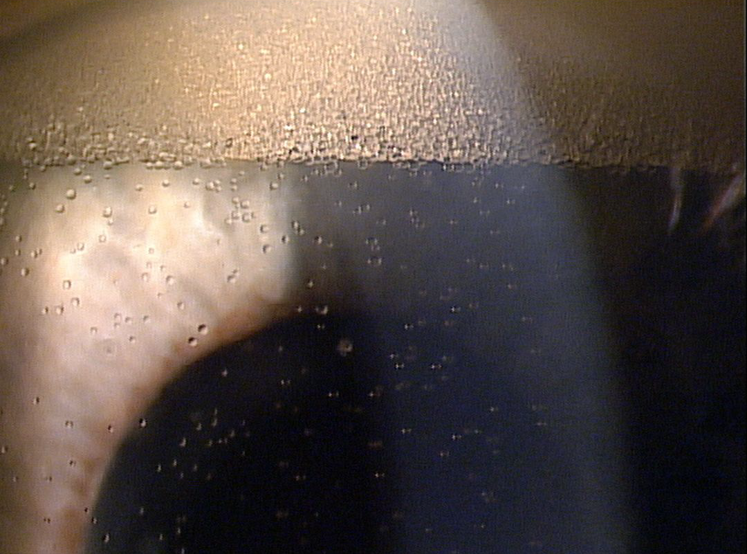
This patient underwent prior successful retinal reattachment surgery with silicone oil and presented with these asymptomatic findings.
Slit lamp photography shows a reverse pseudohypopyon of emulsified silicone oil droplets floating in the anterior chamber. Suspended droplets of oil are noted throughout the aqueous more inferiorly.
Learning Points:
Silicone oil as an adjunct for repairing complex retinal detachments was introduced by Paul Cibis in the early 1960s (Cibis et al, Arch Ophthalmol 1962;68:590-599). It is used primarily for complex detachments at high risk for failure, including severe proliferative vitreoretinopathy and viral retinitis. Although silicone oil is generally well tolerated as a long-term tamponade, it can rarely emulsify, resulting in numerous tiny, opaque droplets. The development of emulsification is multifactorial, including the properties of the oil, the surgical procedure, and postoperative factors.
When severe, the emulsified droplets can cause significant vision loss, requiring their removal. The droplets can also migrate into the anterior chamber, causing a reverse ‘hypopyon’ with secondary elevated intraocular pressure or corneal decompensation.
SILICONE OIL DROPLET
Ankit Jain and Manish Nagpal
Originally posted on @retina.rocks September 17, 2025
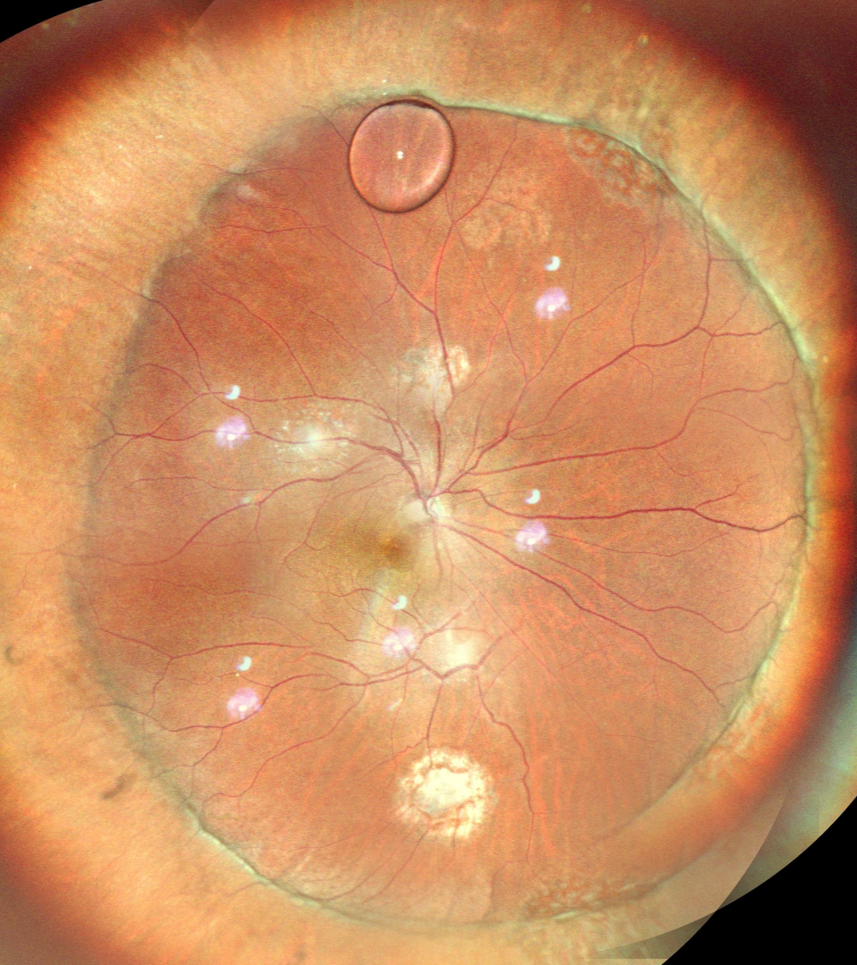
This 56YO male underwent successful retinal reattachment with vitrectomy, scleral buckling, and silicone oil. One month following silicone oil removal, he presented complaining of an inferior floater. Vision was 20/40.
Color SLO imaging shows an attached retina with an encircling buckle. A residual silicone oil droplet is noted superiorly along the buckle edge. Observation was recommended.
SUBRETINAL PERFLUORON
Originally posted on @retina.rocks March 10, 2025
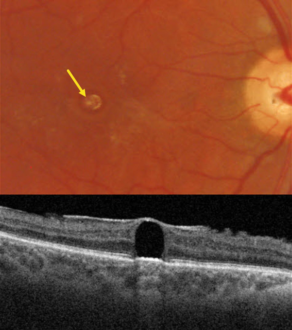
This 33YO male presented with stable vision following prior vitrectomy for a rhegmatogenous retinal detachment (RRD) in his right eye. Vision was 20/400.
Triton color imaging shows a round yellowish subfoveal lesion. Swept-source OCT shows a hyporeflective subretinal lesion compressing the overlying neurosensory retina into a thin inner retinal hyperreflective line. The underlying RPE band is slightly anteriorly displaced, with hyperreflective choroidal shadowing. A mild epiretinal membrane is also noted.
Learning Points:
Perfluorooctane (PFO) is a liquid heavier than water used intraoperatively for complex retinal detachments, including proliferative vitreoretinopathy and giant retinal tears. The PFO is removed during the fluid-air exchange. Rarely, PFO droplets can develop intraoperatively and migrate subretinally. These were much more common from intraocular turbulence prior to the introduction of modern valved cannulas.
Subretinal PFO bubbles have a characteristic appearance on OCT, including an acute angle between the bubble and the RPE, thinned, ill-defined overlying retinal layers, elevation of the underlying RPE band, and hyperreflective choroidal shadowing (Figueroa and Contreras, Retina 2012;32:2177-2178). The subretinal PFO can be removed using a variety of surgical maneuvers, although we recommend observation given its chronicity and absence of new visual complaints.
SILICONE OIL ON CAPSULE
Sharat Hegde
Originally posted on @retina.rocks December 30, 2024
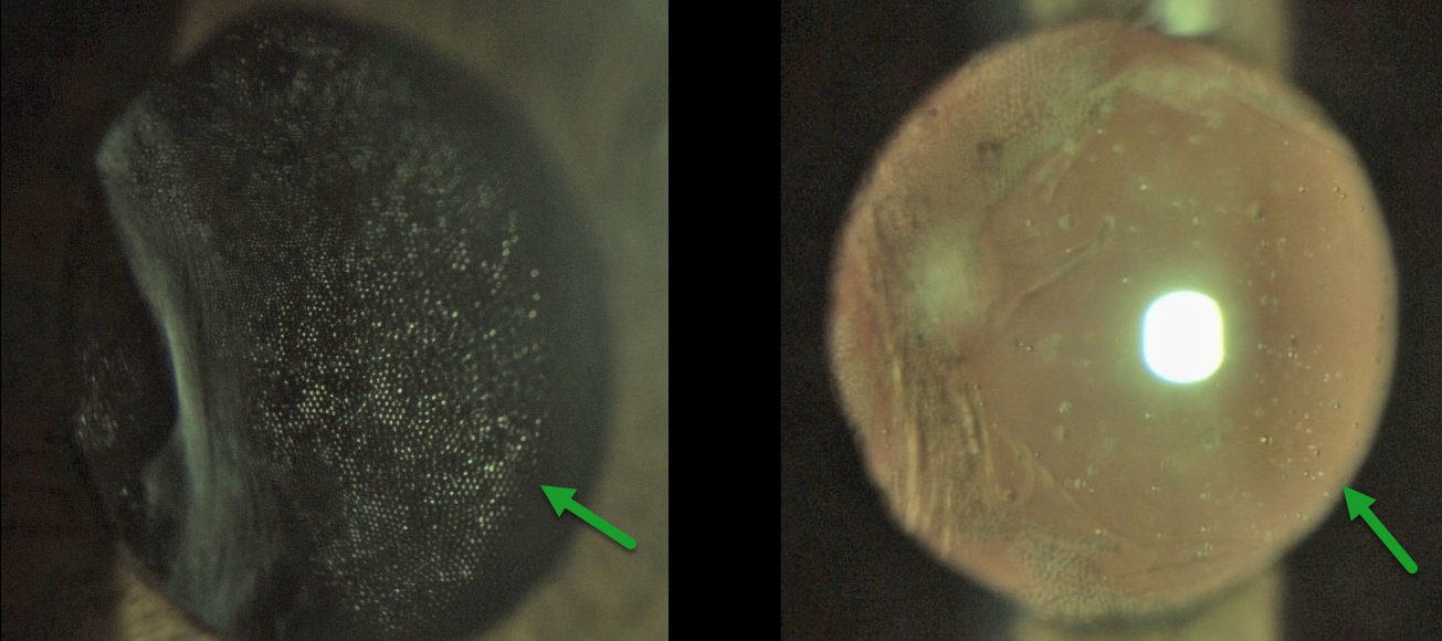
This 70 YO male presented with 2 months of blurred vision in his right eye. Vision was counting fingers OD and 20/20 in his normal OS. There was a history of a prior retinal detachment that was repaired with vitrectomy and silicone oil. Removal of the silicone oil, along with combined cataract surgery with a hydrophilic intraocular lens (IOL), was performed 6 months earlier.
Slit lamp photography shows a honeycomb pattern of adherent silicone oil bubbles along the intact posterior capsule.
Learning Points:
Intravitreal silicone oil droplets can irreversibly adhere to the surface of IOLs, particularly silicone lenses, where they can cause significant glare and vision loss (Apple et al, Ophthalmology 1996;103:1555-1562). Silicone oil can also rarely cause IOL opacification (Oner et al, Ophthalmic Lasers Imaging Retina 2021;52:37-43). The adherent oil cannot be removed, and these lenses often require explantation.
Our case is somewhat unique in that the oil droplets adhered to the posterior capsule rather than the IOL. YAG capsulotomy displaced the oil bubbles, and vision improved to 20/60.
EMULSIFIED SILICONE OIL
Originally posted on @retina.rocks September 25, 2023
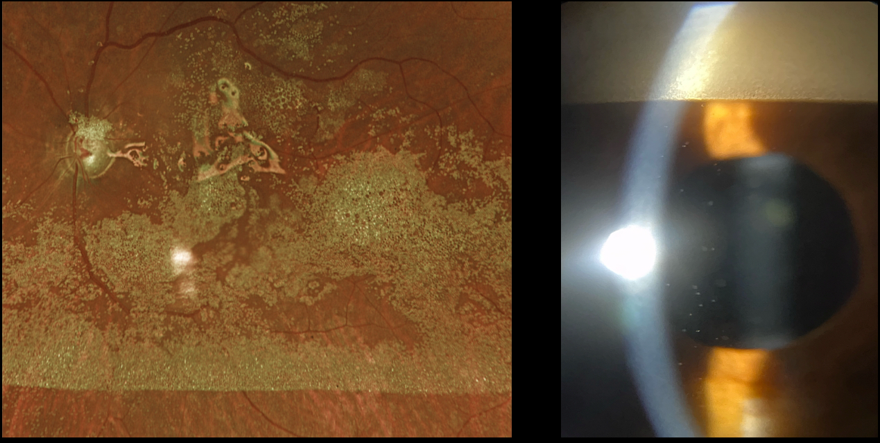
This 65YO male underwent vitrectomy surgery with silicone oil 5 years earlier for a complex rhegmatogenous retinal detachment with proliferative vitreoretinopathy. Vision has remained at counting fingers since the surgical repair, and he presented for his annual visit without new complaints.
Optos RG imaging shows innumerable tiny droplets of emulsified silicone oil coating the posterior pole. Slit lamp photography shows a reverse ‘hypopyon’ of emulsified oil superiorly. Suspended oil droplets are also noted throughout the anterior chamber.
Learning Points:
Silicone oil as an adjunct for repairing complex retinal detachments was introduced by Paul Cibis in the early 1960s (Cibis et al, Arch Ophthalmol 1962;68:590-599). It is used primarily for complex detachments at high risk for failure, including severe proliferative vitreoretinopathy and viral retinitis.
Although silicone oil is generally well-tolerated as a long-term tamponade, it can occasionally emulsify, resulting in numerous tiny, opaque droplets. Emulsification is multifactorial, involving the properties of the oil, the surgical procedure, and postoperative factors.
When severe, emulsified droplets can cause significant vision loss and require removal. The droplets can also migrate into the anterior chamber, causing a reverse ‘hypopyon’ with secondary elevated intraocular pressure or corneal decompensation.
Our patient’s increased intraocular pressure of 32 mmHg, which was elevated for the first time, was attributed to emulsified oil clogging the trabecular meshwork. Topical glaucoma drops were started. If the pressure cannot be adequately controlled medically, then vitrectomy with removal of the emulsified oil and likely exchange for new silicone oil will be recommended.
EMULSIFIED SILICONE OIL
Originally posted on @retina.rocks May 12, 2022
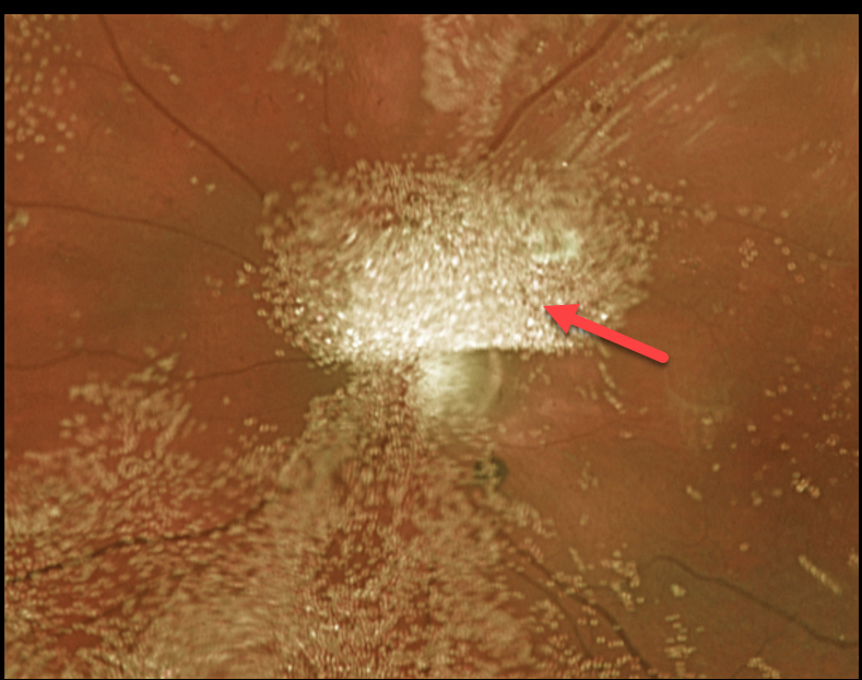
Eight years earlier, this 42YO female underwent successful retinal reattachment surgery elsewhere with scleral buckling, vitrectomy, and silicone oil. Vision was light perception.
Optos imaging shows extensive droplets of emulsified silicone oil.
We decided to observe our patient due to the long history of profound vision loss in a pain-free eye.
Learning Points:
Silicone oil as an adjunct for repairing complex retinal detachments was introduced by Paul Cibis in the early 1960s (Cibis et al, Arch Ophthalmol 1962;68:590-599). It is used primarily for complex detachments at high risk of failure, including severe proliferative vitreoretinopathy and viral retinitis.
Although silicone oil is generally well-tolerated as a long-term tamponade, it can rarely emulsify, resulting in numerous tiny opaque droplets.
The development of emulsification is multifactorial, including the properties of the oil, the surgical procedure, and postoperative factors.
When severe, the emulsified droplets can cause significant vision loss and may require removal. The droplets can also migrate into the anterior chamber, causing a ‘reverse hypopyon’ with secondary elevated intraocular pressure or corneal decompensation.
RHEGMATOGENOUS RETINAL DETACHMENT
Originally posted on @retina.rocks December 17, 2021
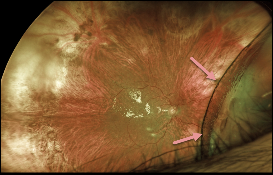
This patient presented with light perception vision following a 1-year-old total retinal detachment with proliferative vitreoretinopathy. At vitrectomy, all membranes were peeled, and the anterior loop traction was relieved (or so we thought). The retina was mobile and completely flattened with fluid-air exchange. 1000 centistoke silicone oil was infused.
At the one-day and one-week postoperative visits, vision remained light perception. The retina was completely attached except for the nasal periphery, which was locally detached with subretinal silicone oil.
Since the surgical goal was to prevent total blindness and possible phthisis, we have decided to observe for now as long as the residual detachment remains localized.
What would your management be at this point?
SILICONE OIL DROPLET
Originally posted on @retina.rocks September 20, 2021
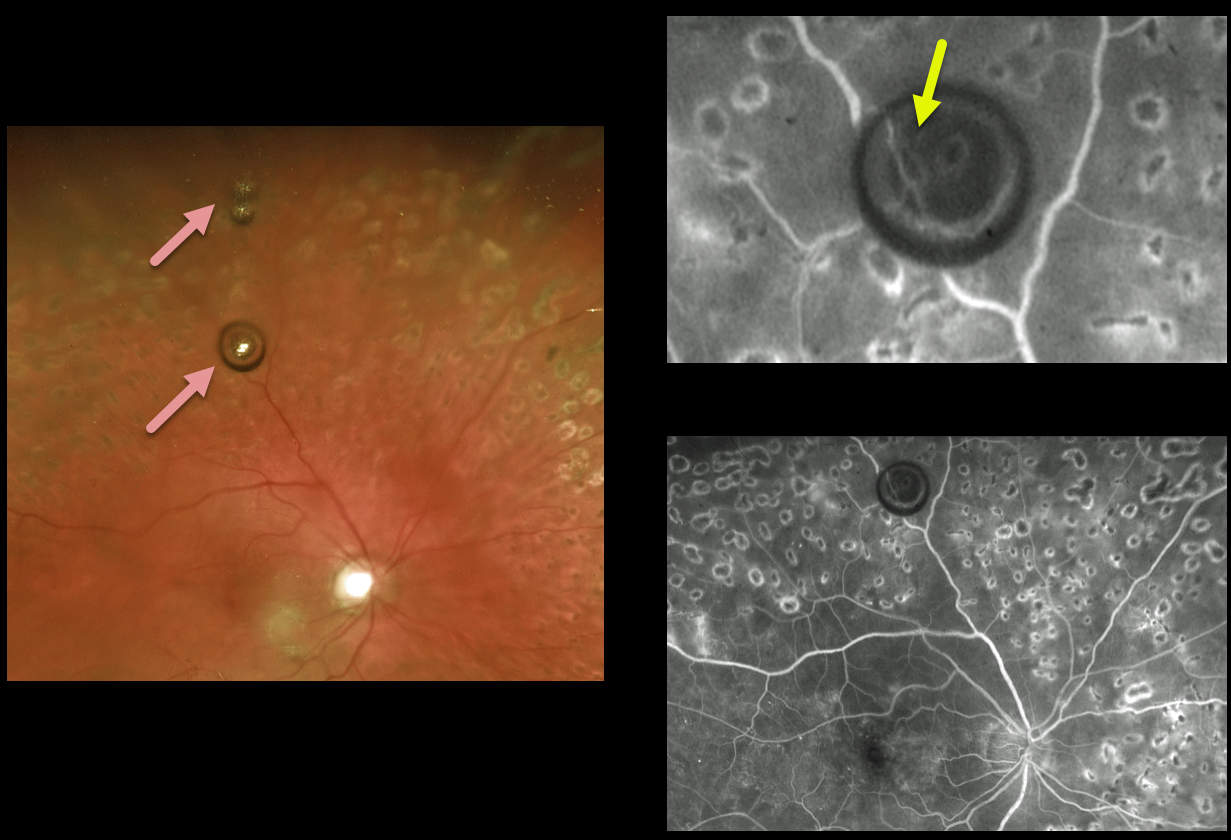
This patient with involuted proliferative diabetic retinopathy received prior intravitreal Avastin injections for diabetic macular edema.
On routine examination, a few asymptomatic silicone oil bubbles were suspended in the superior vitreous.
Fluorescein angiography best shows how these bubbles distort and minify the underlying retinal detail.
Learning Points:
The insulin syringe needles provided by compounding pharmacies for Avastin injections are lubricated with silicone.
Many eyes receiving ongoing intravitreal Avastin developed these vitreous silicone droplets, most of which were completely asymptomatic.
Once knowledge of these silicone droplets became available, use of these insulin syringes was discontinued.
RHEGMATOGENOUS RETINAL DETACHMENT
Originally posted on @retina.rocks January 25, 2021
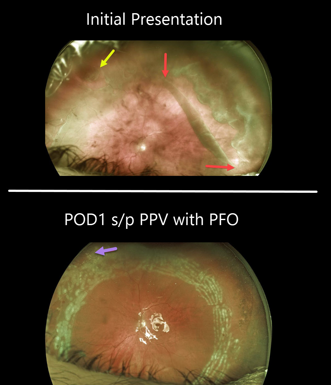
This patient presented with a symptomatic macula-on giant-tear retinal detachment (RD) in his left eye. The giant tear extends from about 12 to 4 o’clock with an additional tear noted superonasally.
One day after vitrectomy with Perfluoron (PFO, perfluoro octane) injection, the retina is completely attached, with fresh white encircling laser scarring. Some PFO bubbles can be seen superonasally.
The PFO liquid was removed two weeks later, and his retina has remained attached.
Learning Points:
By definition, giant retinal tears extend at least 3 clock hours. Surgical repair is greatly facilitated by using PFO liquids, which are heavier than water and help unroll the posterior retina and prevent posterior slippage of the detachment. The PFO can either be removed at the initial repair or left in place for a week or two before being removed with a secondary procedure.
RHEGMATOGENOUS RETINAL DETACHMENT
Originally posted on @retina.rocks June 22, 2020
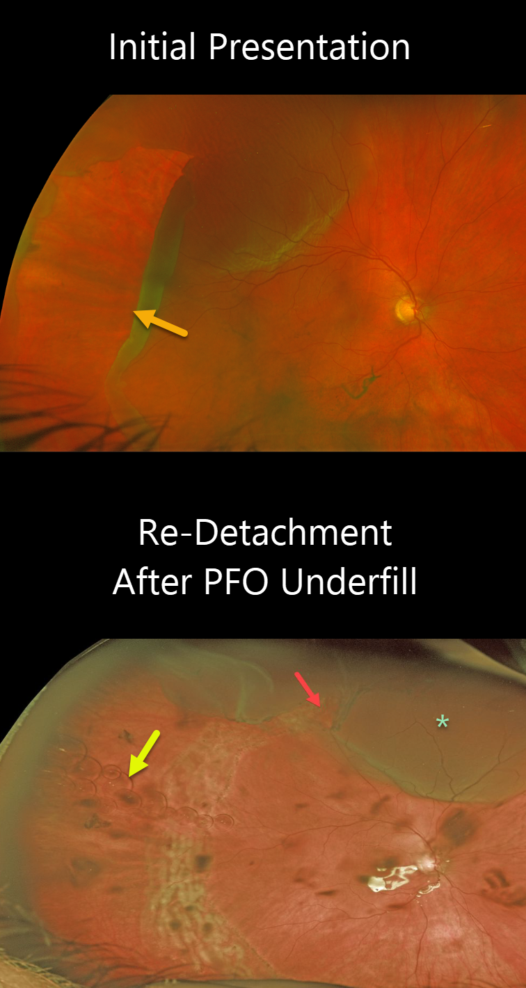
This patient presented with a retinal detachment caused by a giant retinal tear extending from 5 to 11 o’clock.
Unfortunately, the retina re-detached since the eye was inadvertently underfilled with perfluorocarbon (PFO) at the time of vitrectomy. The superior edge of the giant tear is open, resulting in a superior detachment. There are numerous small PFO bubbles floating along the interface between the PFO meniscus below and the liquid vitreous above.
The patient underwent subsequent surgery to remove the Perfluoron and successfully repair the superior detachment.
Learning Points:
PFO is a transparent, heavier-than-water liquid, often used during vitrectomy for these types of detachments. The PFO liquid is injected over the posterior pole, and as it fills the eye, it simultaneously unrolls the giant tear and flattens the retina. Traditionally, the PFO is exchanged for air, then for a long-acting gas. However, this often results in posterior retinal slippage.
Although PFO is not meant as a long-term vitreous substitute, it can be left in the eye for a week or two. Postoperatively, the patient sleeps on their back, allowing the PFO to tamponade the retina while the retinopexy begins to scar. Patients are then taken back to the operating room, where the PFO can be removed and replaced with a gas or silicone oil, without concern about the retina slipping intraoperatively.
SILICONE OIL DROPLET
Originally posted on @retina.rocks January 2, 2020
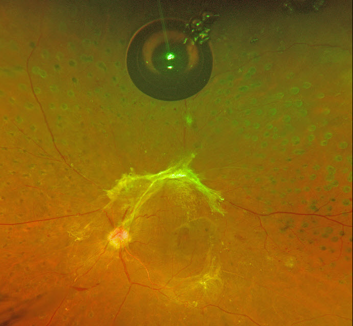
This patient with proliferative diabetic retinopathy with tractional detachment was noted to have a large asymptomatic silicone oil bubble following multiple prior Avastin injections.
Learning Points:
The older insulin syringe needles provided by compounding pharmacies for the Avastin (bevacizumab) injections were lubricated with silicone.
Many eyes receiving intravitreal Avastin developed small vitreous silicone droplets, although the droplet is unusually large in this patient. Since then, these insulin syringe needles have been discontinued.


