

Situs Inversus
<< Back to Cases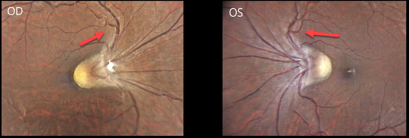
SITUS INVERSUS
Gil Calvão-Santos
Originally posted on @retina.rocks January 15, 2026
This healthy 38YO highly myopic female presented with 6 months of decreased vision in her right eye. Her current refraction was -15.00 D OD and -11.00 -2.25 × 10° OS. Vision was 20/200 OD and 20/25 OS.
Color photography shows the retinal vessels emerge from slightly tilted discs to initially course nasally and superiorly before turning temporally toward the macula. The macular center is normally located about 2-disc diameters temporal to the nerve. The right fovea cannot be visualized and likely lies just temporal to the edge of the nerve, and about one-half disc diameter temporal to the left nerve.
Learning Points:
Situs inversus occurs in 5% of normal eyes and 70% of eyes with tilted discs (Witmer et al, Surv Ophthalmology 2010;55:403-428) and presents with the retinal vessels exiting the optic disc nasally. Vision is usually normal unless other pathology is present.
Despite the abnormal macular architecture, our patient reportedly had normal vision until recently, and was found to have a cataract, which was consistent with her acuity. Cataract surgery was recommended, and vision improved to 20/25 postoperatively.
DEGENERATIVE RETINOSCHISIS + SITUS INVERSUS
Sayena Jabbehdari and Ahmed Sallam
Originally posted on @retina.rocks March 5, 2025
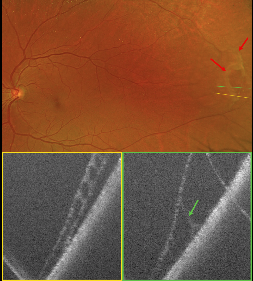
This 64YO female was referred for a possible asymptomatic rhegmatogenous retinal detachment (RRD) in her left eye. Vision was 20/20 in her normal OD and 20/20 OS.
Optos color RG imaging shows superotemporal degenerative retinoschisis with a few outer-layer holes (OLH). OCT confirms the schisis as well as the outer layer hole.
Learning Points:
Clinically, degenerative retinoschisis usually appears as a smooth, dome-shaped peripheral retinal elevation without the outer retinal corrugations classically seen in RRD (Oquendo et al, AJO 2024;268:212-221). Degenerative retinoschisis is virtually never prophylactically treated (Byer 1986;93:1127-1137). Although the schisis cavities can enlarge, prophylactic treatment, including thermal laser, does not prevent further spread and should not be performed (Ness et al, Surv Ophthalmology 2022;67:892-907).
Situs inversus of the optic disc was identified as a benign incidental finding in our patient. This occurs in 5% of normal eyes and 70% of eyes with tilted discs (Witmer et al, Surv Ophthalmology 2010;55:403-428). It can be mistaken for other congenital disc anomalies.
RETINOPATHY OF PREMATURITY
Originally posted on @retina.rocks May 13, 2021
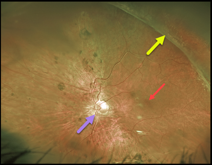
For over 20 years, we’ve been following this 39YO male with stable regressed retinopathy of prematurity in his left eye. Vision is NLP OD and 20/80 OS.
Optos imaging shows that the macular center is dragged temporally due to vitreoretinal traction from regressed neovascularization.
The macular center is usually about 2 disc diameters temporal to the edge of the optic nerve, and our patient’s is about 3 disc diameters. This dragging is usually accompanied by a straightening of the temporal arcades.
However, in our patient, this is masked by situs inversus, a congenital abnormality in which the retinal vessels exit the optic nerve with a nasal bend before coursing temporally.
Learning Points:
These outcomes can be minimized through aggressive neonatal screening and treatment with laser photocoagulation and anti-VEGF agents.
SITUS INVERSUS
Originally posted on @retina.rocks March 17, 2021
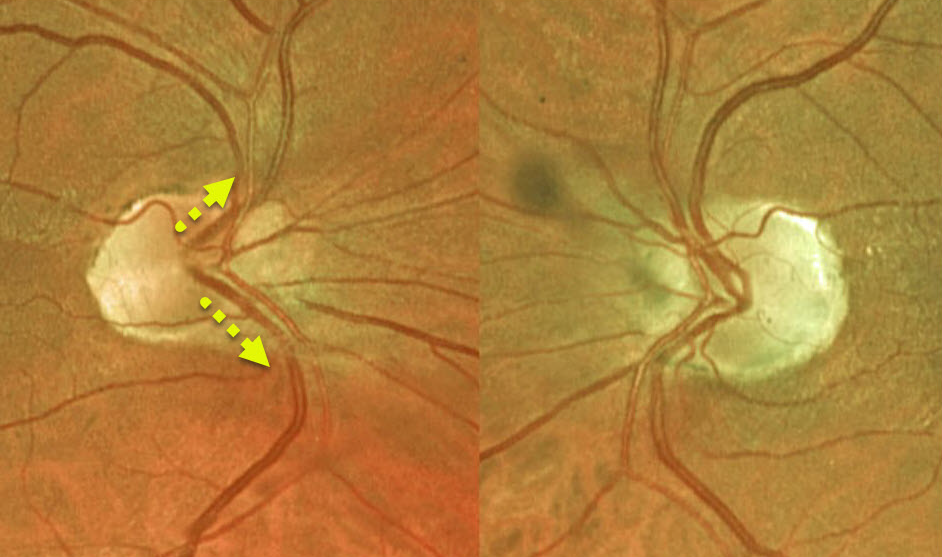
Situs inversus is a congenital abnormality where the retinal vessels exit the optic nerve with a nasal bend prior to heading temporally.
Learning Points:
Situs inversus occurs in 1 to 2% of the population and is sometimes associated with a tilted optic disc.


