

Solar + Welder's Retinopathy
<< Back to Cases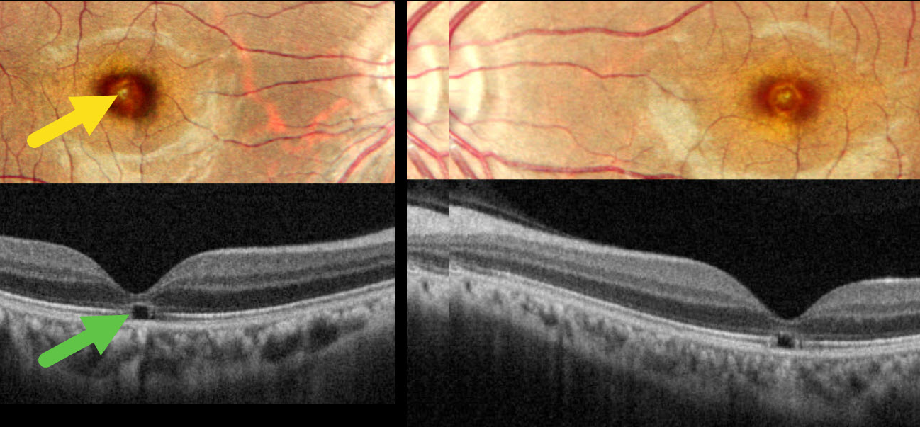
WELDING ARC MACULOPATHY
Malvika Singh and Manish Nagpa
Originally posted on @retina.rocks December 24, 2025
This 31YO male presented with 2 years of bilateral vision loss. He works as a welder and admits to rarely wearing his safety glasses. Vision was 20/30 OU.
Pseudocolor SLO imaging shows symmetrical, tiny yellow foveal scars. OCT shows outer foveal microdefects characterized by hyporeflectivity due to loss of all bands deep to the external limiting membrane.
Learning Points:
The welding process emits optical radiation across various wavelengths and intensities, including infrared, visible, and ultraviolet (UV) light. Exposure to this UV light without proper eye protection can result in keratoconjunctivitis (the most common clinical presentation) and foveal lesions similar to solar retinopathy. Findings include a small yellowish foveal spot or scar with an outer foveal microdefect on OCT (Singh et al, Surv Ophthalmology 2023;68:655-668). Vision is usually good despite the persistent clinical and OCT findings.
Our patient was lucky to have good vision despite his poor work habits. He was advised to use protective eyewear during welding.
WELDING ARC MACULOPATHY
Rohan Jain and Manish Nagpal
Originally posted on @retina.rocks May 1, 2025
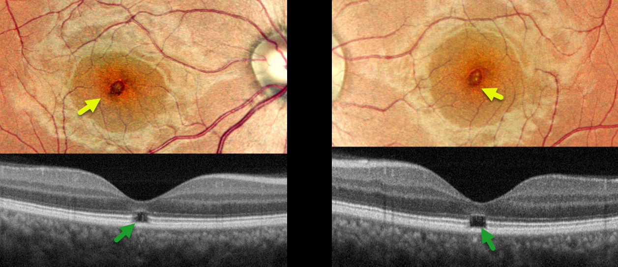
This 38YO male presented with 6-7 months of bilateral vision loss. He works as a welder and admits to rarely wearing his safety glasses. Vision was 20/25 OD and 20/20 OS.
Pseudocolor SLO imaging shows symmetrical, tiny yellow foveal scars. OCT shows outer foveal microdefects with hyporeflectivity due to loss of all bands deep to the external limiting membrane.
Learning Points:
The welding process emits optical radiation across various wavelengths and intensities, including infrared, visible, and ultraviolet (UV) light. Exposure to this UV light without proper eye protection can result in keratoconjunctivitis (the most common clinical presentation) and foveal lesions similar to those seen in solar retinopathy. Findings include a small foveal yellowish spot or scar with an outer foveal microdefect on OCT (Singh et al, Surv Ophthalmology 2023;68;655-668). Vision is usually good despite the persistent clinical and OCT findings.
Our patient was lucky to have good vision despite his poor work habits. He was advised to use protective eyewear during welding.
SOLAR RETINOPATHY
Originally posted on @retina.rocks August 28, 2023
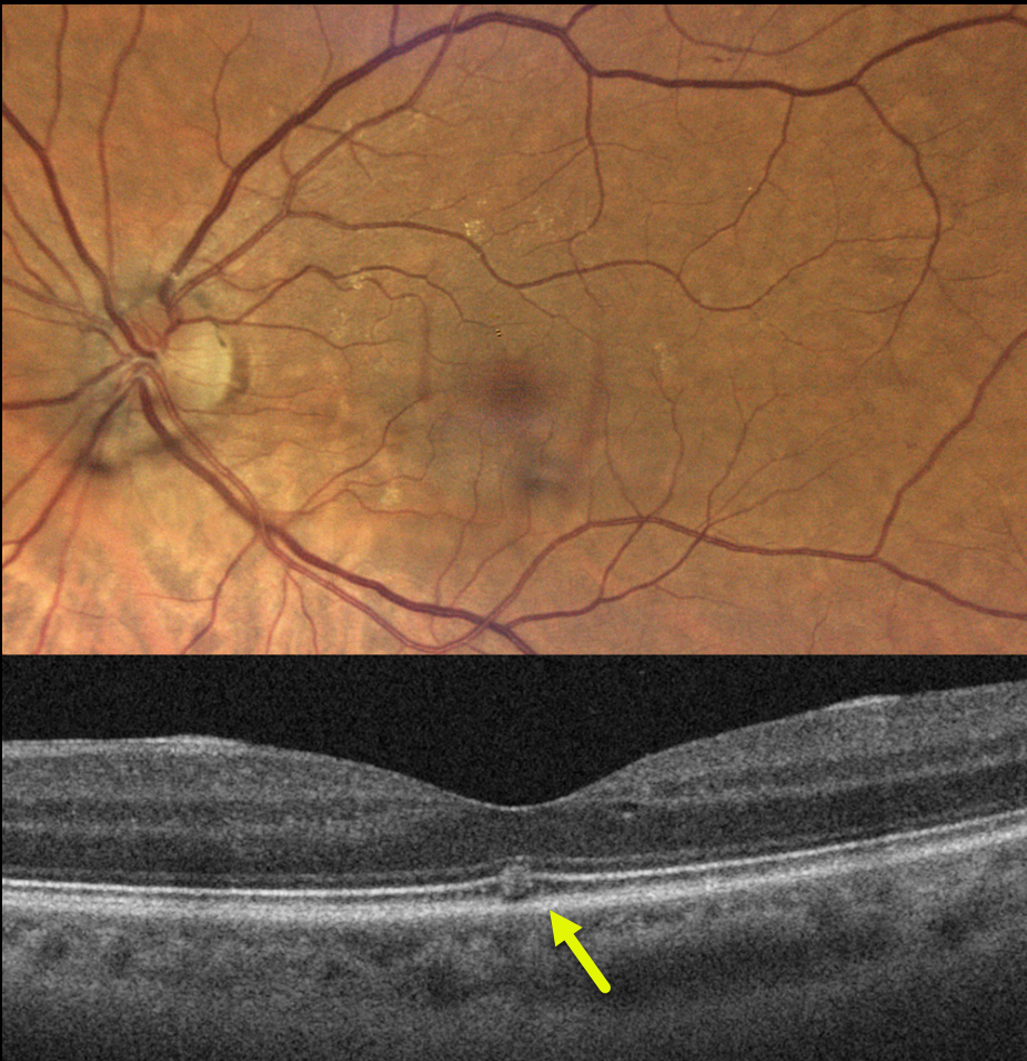
This 67YO female was referred for 3 weeks of vision loss in her left eye. Vision was 20/25 bilaterally.
The fundus photo is normal, but her OCT shows a focal area of hyperreflectivity interrupting the ellipsoid zone.
On further questioning, her symptoms developed while driving, immediately following a transformer explosion.
Learning Points:
The outer retinal damage in solar/welder’s retinopathy is thought to be caused by photochemical outer retinal and RPE damage instead of a thermal burn. Vision is usually fairly good despite the findings in the outer retina and RPE.
SOLAR RETINOPATHY
Originally posted on @retina.rocks February 27, 2023
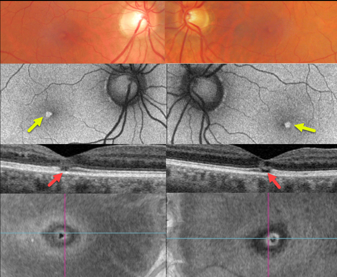
This healthy 46YO male complained of mild bilateral central vision loss for about 5 years. Vision was 20/25 OD and 20/30 OS.
Optos color imaging shows subtle central foveal pigment loss. Fundus autofluorescence (FAF) shows these lesions to be hyper-FAF.
Swept-source OCT shows small central foveal outer retinal defects, and en face OCT of the outer retina shows a unique perspective of these lesions.
On further questioning, he noted that his visual symptoms immediately followed his viewing the Great Eclipse of 2017 without proper eye protection.
Learning Points:
The outer retinal damage in solar retinopathy is thought to be caused by photochemical outer retinal and RPE damage instead of a thermal burn. Vision is usually fairly good despite the outer retinal and RPE findings.
SOLAR RETINOPATHY
Originally posted on @retina.rocks April 27, 2022
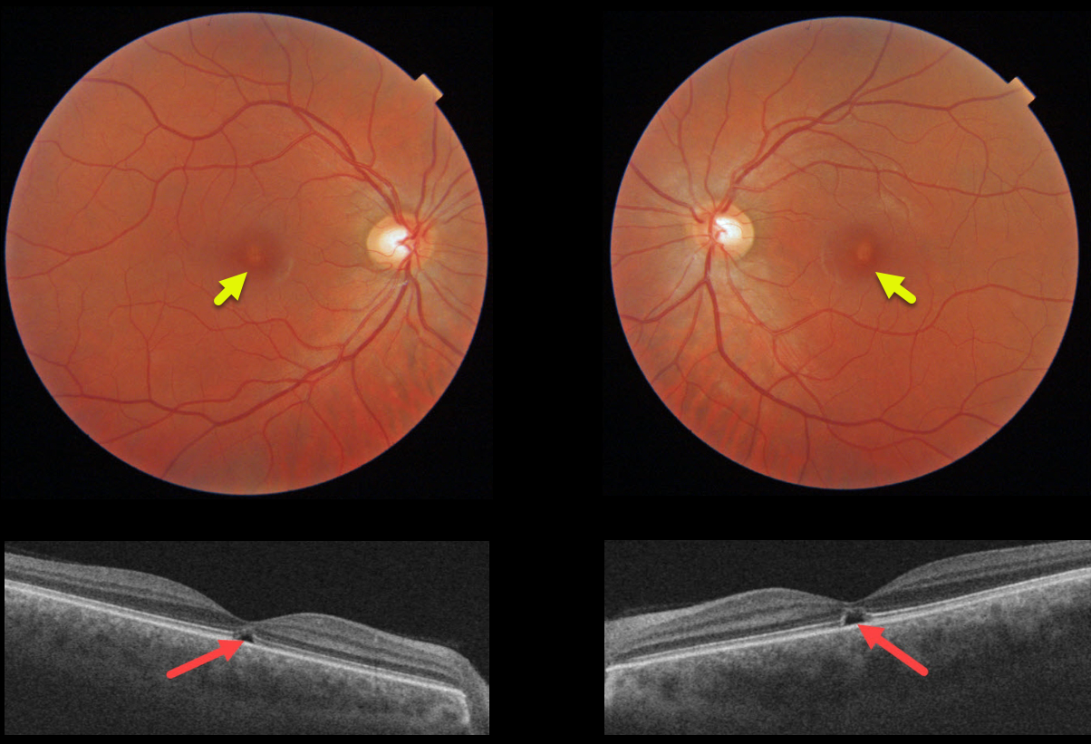
This 31YO male was referred for relatively asymptomatic central macular pigmentary changes. He gave a long history of twice-daily sun gazing, which always caused a very pleasurable sneezing episode. Vision was 20/30 OD and 20/40 OS.
Triton color imaging shows tiny foveal pigment loss. Swept-source OCT shows bilateral ellipsoid zone and outer segment defects.
We diagnosed him with the ACHOO reflex (Autosomal Dominant Compelling Helioophthalmic Outburst). And no, this isn’t an April Fools post, and we’re not talented enough to even make this stuff up if we wanted to! Patients with the ACHOO reflex sneeze when exposed to bright lights, typically sunlight.
We advised him to stop his sun gazing ritual, although we’re not sure this will be possible for him.
Learning Points:
The outer retinal damage in solar retinopathy is thought to be caused by photochemical outer retinal and RPE damage instead of a thermal burn. Vision is usually fairly good despite the outer retinal and RPE findings.
SOLAR RETINOPATHY
Originally posted on @retina.rocks February 24, 2022
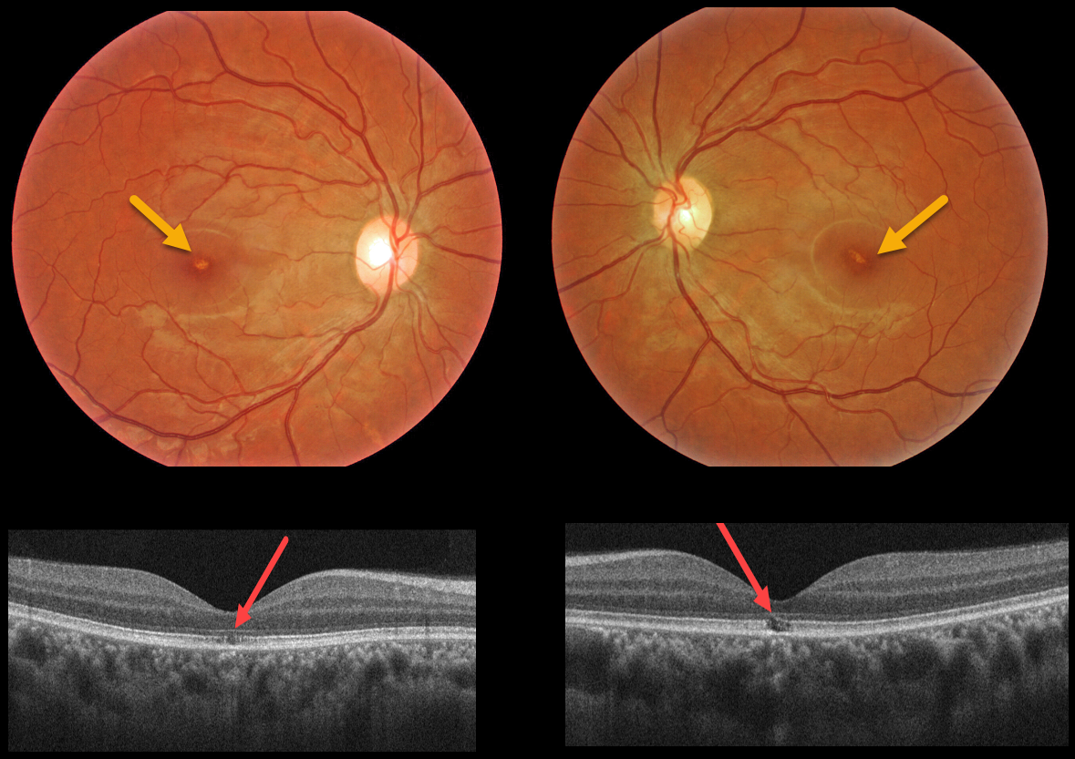
This 32YO female with schizophrenia reported regular sun gazing every morning for many years. Vision was 20/40 OU.
Triton color imaging shows tiny foveal pigment loss. Swept-source OCT shows bilateral ellipsoid zone and outer segment defects.
Learning Points:
The outer retinal damage in solar retinopathy is thought to be caused by photochemical outer retinal and RPE damage instead of a thermal burn.
Vision is usually fairly good despite the outer retinal and RPE findings.
WELDER’S FLASH RETINOPATHY
Originally posted on @retina.rocks December 16, 2021
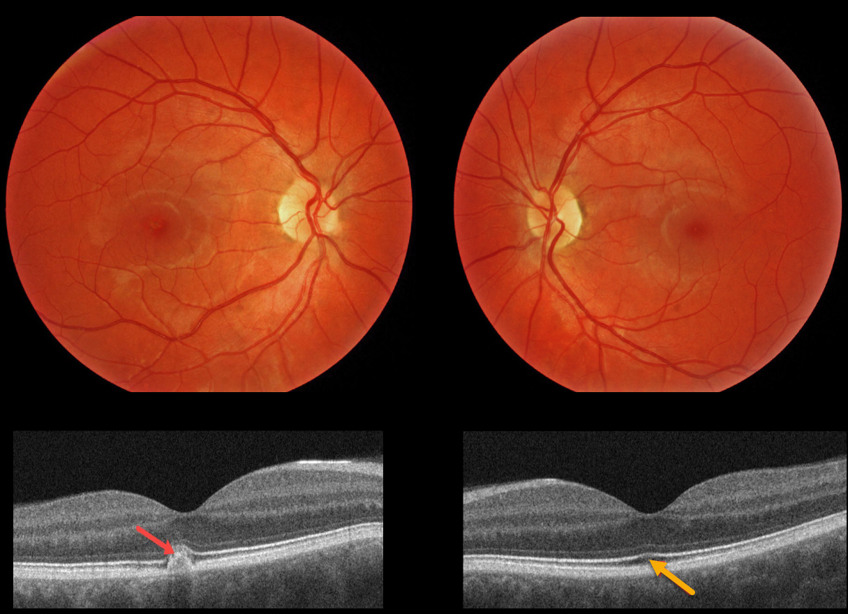
This 29YO male subjectively noted a persistent central visual disturbance in his right eye following a welder’s flash. Vision was 20/20 OU.
Triton color imaging shows subtle, tiny foveal pigmentary changes. Swept-source OCT shows an RPE and outer retinal hyperreflective mound OD, and very subtle ellipsoid zone and outer segment changes OS.
SOLAR RETINOPATHY
Originally posted on @retina.rocks July 31, 2020
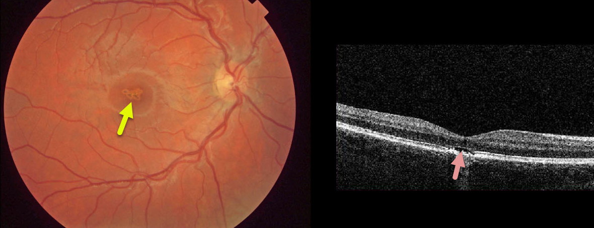
This asymptomatic 10YO boy presented with 20/40 vision OD and 20/20 vision OS.
There were multifocal small subretinal pigment clumps with surrounding pigment loss in the right central macula.
OCT shows foveal outer segment loss with ragged RPE loss. The left macula was normal.
He denied looking at a laser pointer but did admit to a history of sun-gazing. He likely closed his left eye each time he looked at the sun, which explains the unusual finding of unilateral solar retinopathy.
Learning Points:
The outer retinal damage in solar retinopathy is thought to be caused by photochemical outer retinal and RPE damage instead of a thermal burn. Vision is usually fairly good.


