

Stargardt Disease + Fundus Flavimaculatus + ABCA4
<< Back to Cases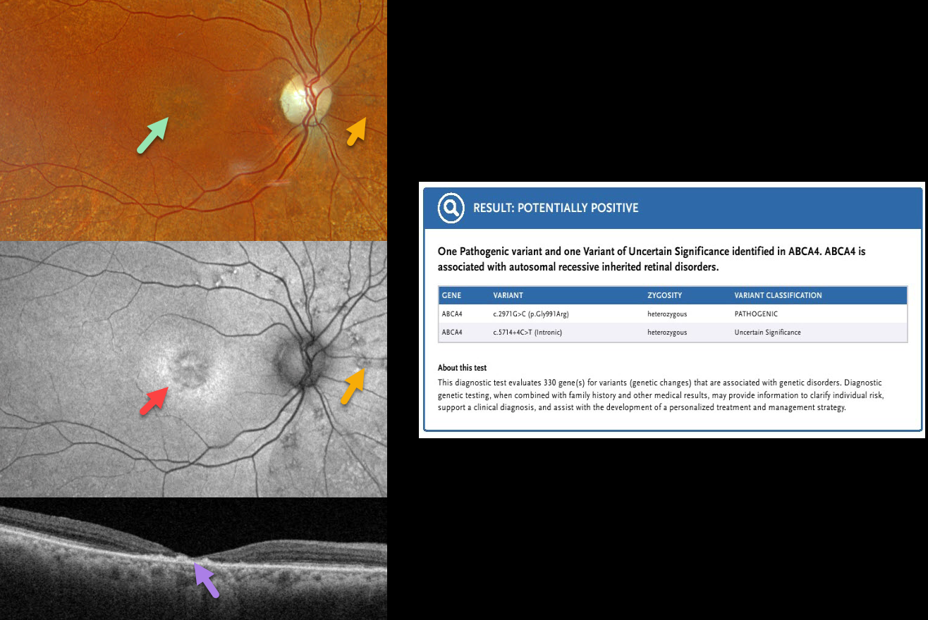
ABCA4 RETINOPATHY
Originally posted on @retina.rocks May 19, 2026
This 88YO female was diagnosed with age-related macular degeneration elsewhere. Vision was 20/400 OD and 20/50 OS.
Optos color RGB imaging OD shows a round area of subfoveal pigmentary changes with extensive drusen-like lesions extending outwards from the peripheral macula. Fundus autofluorescence (FAF) shows central hypo-FAF with a ring of hyper-FAF around this foveal lesion. Scattered lesions consisting of small dots of hyper-FAF with surrounding hypo-FAF are scattered around the nerve. Swept-source OCT shows severe central neurosensory atrophy with a macular pseudohole. Some clumps of hyperreflective material are noted within the area of atrophy, along with variable ellipsoid zone loss elsewhere. Similar findings were present in her left eye (not shown). Genetic testing revealed heterozygous ABCA4 mutations, including a pathogenic variant (c.2971G>C, p.Gly991Arg) and a variant of uncertain significance (c.5714.+4C-T, intronic).
Learning Points:
The ABCA4 protein is located in the photoreceptor outer segments and is involved in the recycling of 11-cis-retinal. The mutation of this gene causes the accumulation of lipofuscin in the RPE, which eventually leads to photoreceptor and RPE degeneration. ABCA4 disorders include Stargardt disease, fundus flavimaculatus, cone-rod dystrophy, retinitis pigmentosa, and age-related macular degeneration.
STARGARDT DISEASE
Shraddha Raj Shrivastava and Manish Nagpal
Originally posted on @retina.rocks December 31, 2025
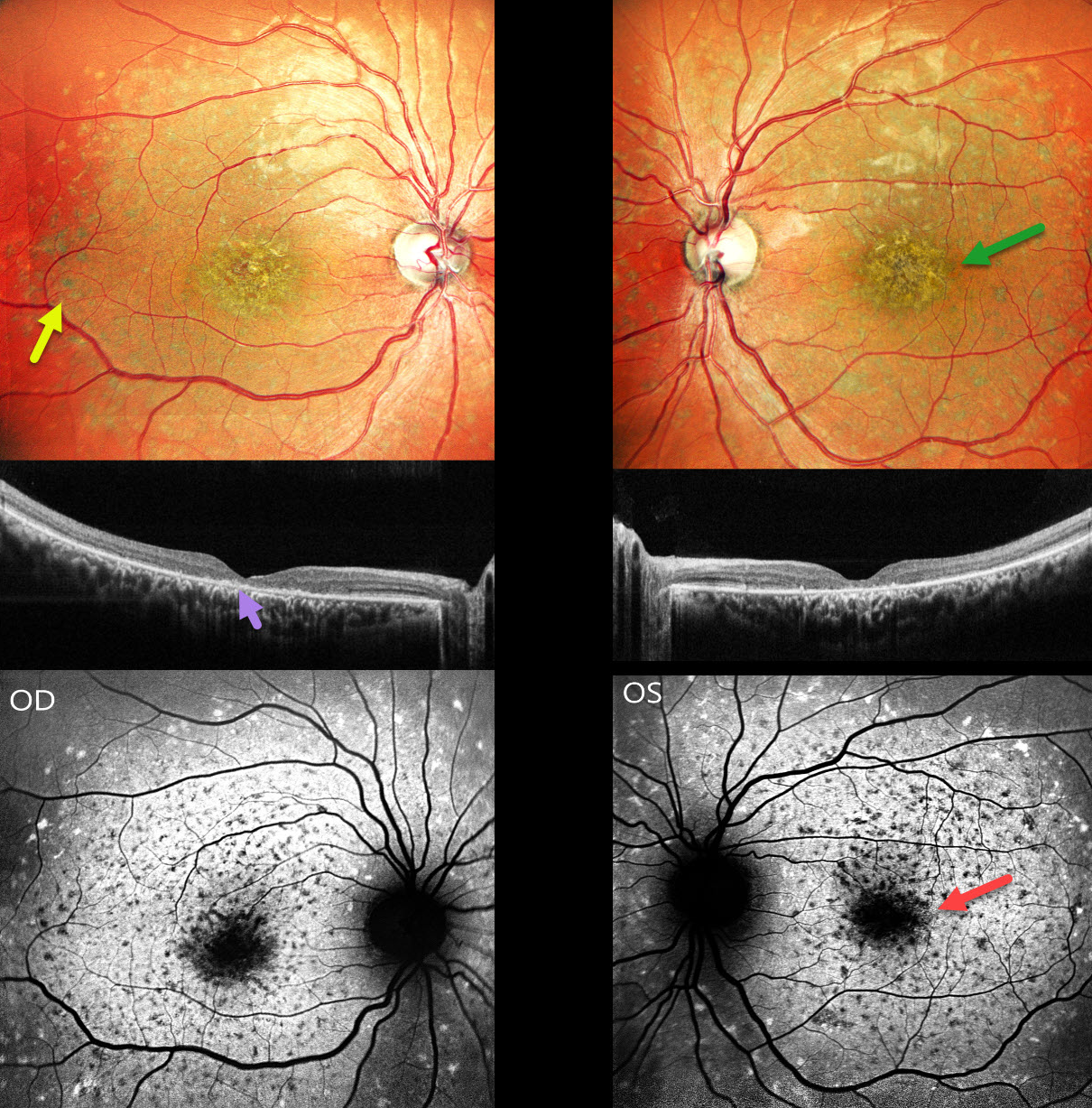
This 26YO male presented with 3 years of bilateral decreased vision along with night vision difficulties since childhood. Family history was negative. Vision was counting fingers at 3 meters OU.
Pseudocolor SLO imaging shows a symmetrical ‘beaten-bronze’ sheen to each central macula with surrounding yellowish pigmented flecks. OCT shows central loss of the photoreceptor layers with hyperreflective deposits above the RPE. Green fundus autofluorescence (FAF) shows foveal hypo-FAF. The clinical flecks are hypo-FAF centrally and hyper-FAF more peripherally. There is relative peripapillary sparing.
Learning Points:
Stargardt disease is an autosomal recessive disorder caused by a mutation in the ABCA4 gene located on chromosome 1. The ABCA4 transmembrane protein localizes to the photoreceptor outer segments and is involved in the recycling of 11-cis-retinal. Mutations of this gene cause the accumulation of lipofuscin in the RPE, which eventually leads to photoreceptor and RPE degeneration. Other ABCA4 disorders include fundus flavimaculatus, cone-rod dystrophy, retinitis pigmentosa, and age-related macular degeneration.
Historically, Stargardt disease was used to describe patients with macular involvement only, and fundus flavimaculatus for those with flecks extending more peripherally. Both are now considered different retinal phenotypes within the ABCA4 spectrum.
BULLSEYE MACULOPATHY PROBABLY FROM ABCA4 MUTATION
Moazzam Parvez and Krishnendu Nandi
Originally posted on @retina.rocks June 11, 2025
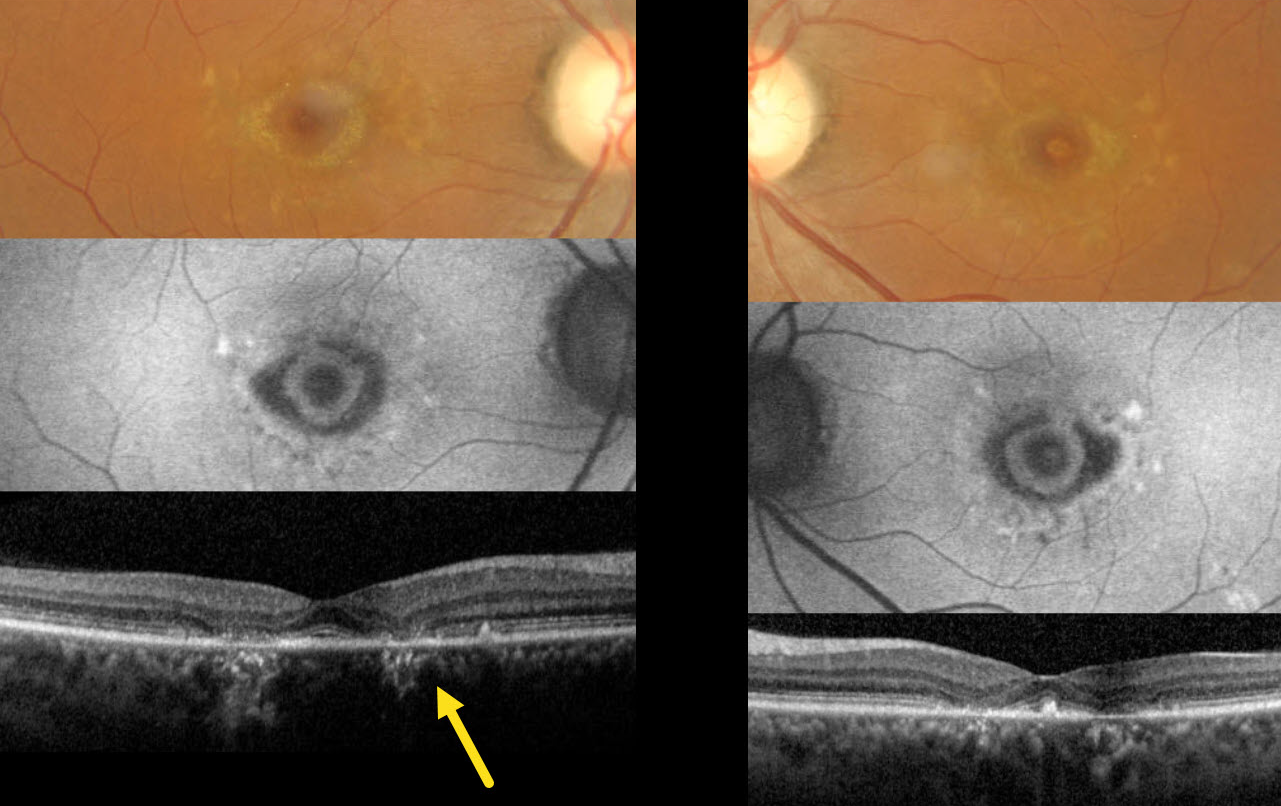
This 40YO female presented with 5 years of stable, mild, distorted vision bilaterally. There was no family history of eye disease. Vision was stable at 20/20 OU.
Color imaging shows bilaterally symmetric bullseye lesions with surrounding subretinal yellow macular flecks. On fundus autofluorescence (FAF), these lesions are hypo-FAF with an inner hyper-FAF ring. Pericentral outer retinal atrophy is noted on OCT.
The funduscopic appearance is most consistent with ABCA4 disease, including Stargardt disease and fundus flavimaculatus. The patient declined genetic testing due to cost concerns, and continued observation was recommended.
STARGARDT DISEASE
Yuenpang Cheung and Stephen Tsang
Originally posted on @retina.rocks September 4, 2024
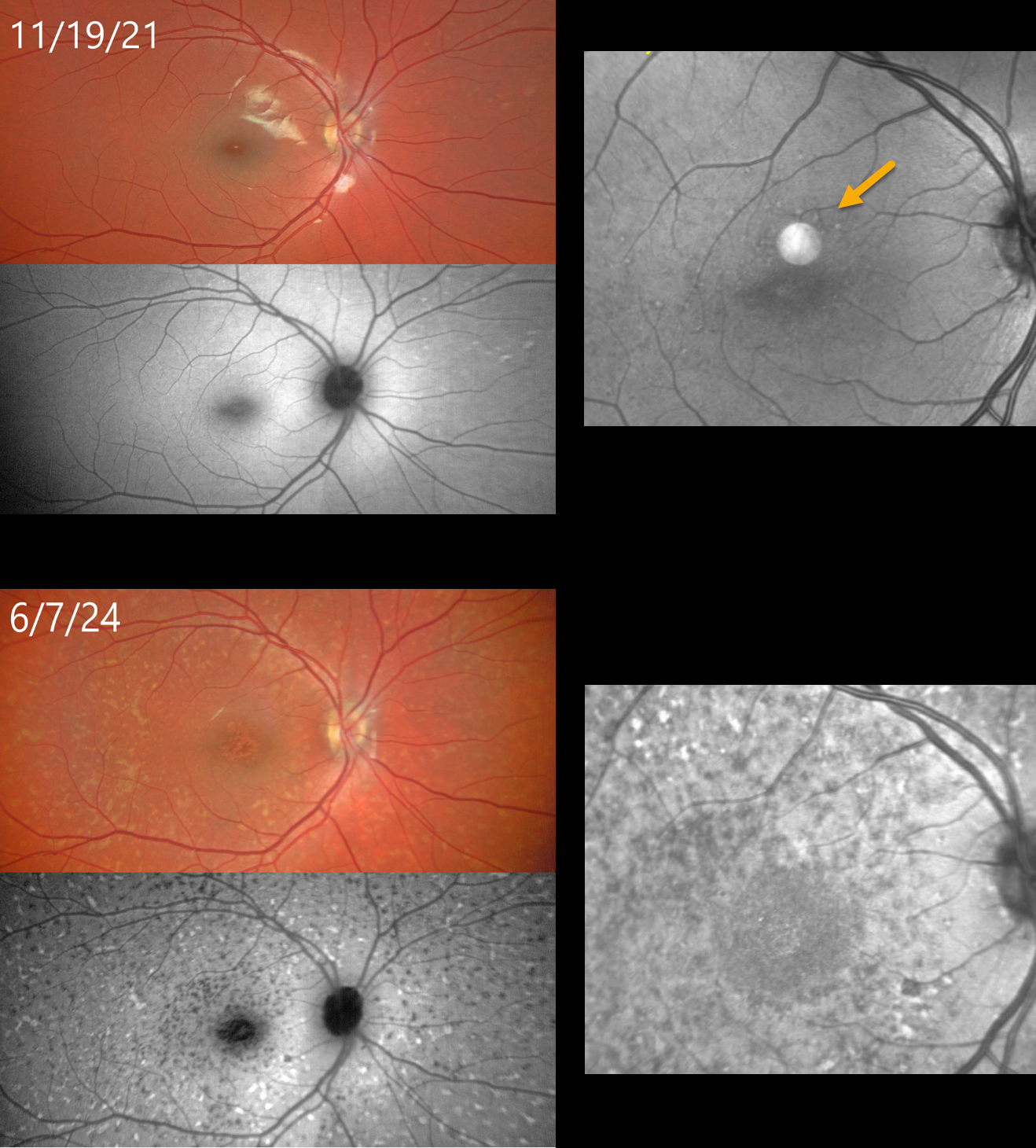
This 9YO female had a known history of Stargardt disease at the time of examination on 11/19/21. Prior genetic testing revealed two pathogenic ABCA4 mutations.
On 11/19/21, color photography, fundus autofluorescence, and OCT en face of the outer retina are fairly normal except for some faint hyper-FAF flecks outside the macula and faint hyperreflective macular lesions.
When last examined on 6/7/24, vision was 20/100 OD and 20/80 OS. Despite the relatively stable vision, there was a dramatic progression of her disease on all multimodal images.
Similar findings were noted in her left eye (not shown).
Learning Points:
Stargardt disease is an autosomal recessive disorder caused by a mutation in the ABCA4 gene. The ABCA4 protein is located in the photoreceptor outer segments and is involved in the recycling of 11-cis-retinal. The mutation in this gene causes lipofuscin accumulation in the RPE, which eventually leads to photoreceptor and RPE degeneration. Other ABCA4 disorders include fundus flavimaculatus, cone-rod dystrophy, retinitis pigmentosa, and age-related macular degeneration.
FUNDUS FLAVIMACULATUS
Originally posted on @retina.rocks March 26, 2024
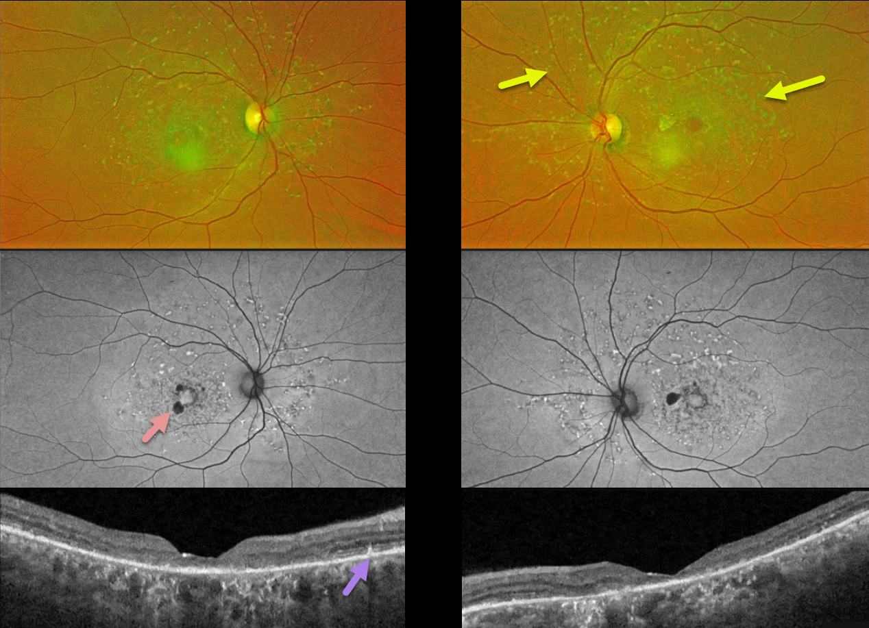
This 58YO female recently noticed difficulties with depth perception. There was no family history of eye disease. Vision was 20/50 OD and 20/80 OS. Genetic testing revealed two heterozygous pathogenic ABCA4 mutations.
Optos color RG imaging shows multiple yellow fleck-like lesions throughout each posterior pole. Fundus autofluorescence (FAF) of these flecks shows variable hyper-FAF, with patches of paracentral hypo-FAF.
Triton swept-source OCT shows variable outer retinal atrophy and disorganization. Some hyperreflective spots are noted above the RPE.
Learning Points:
Fundus flavimaculatus is a genetic disorder, usually transmitted as an autosomal recessive trait, that is caused by mutations involving the ABCA4 gene. This gene encodes one of a family of ATP-binding cassette (ABC) transmembrane proteins that mediate the active transport of various substrates across cellular membranes. The ABCA4 protein is present in the photoreceptor outer segment disc membranes, where it is involved in recycling 11-cis-retinal.
Stargardt disease is believed to be fundus flavimaculatus without peripheral findings. Vision in fundus flavimaculatus is often better than that in Stargardt due to later disease onset and less macular involvement. Both likely represent an RPE lipofuscin storage disease. The RPE cells become engorged with lipofuscin, causing a dark choroid on fluorescein angiography.
CONE DYSTROPHY
Gökşen Gökçen
Originally posted on @retina.rocks February 21, 2024
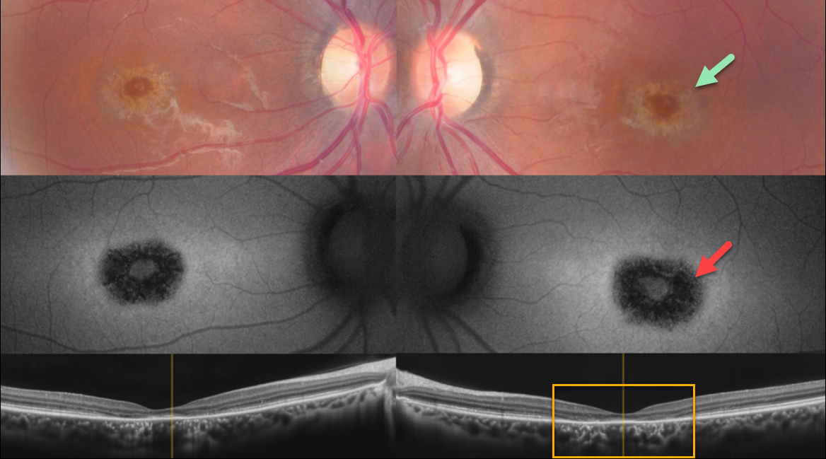
This 22YO male medical student gave a history of relatively rapid bilateral central vision loss 6 years earlier. His symptoms have been stable since, and he continues to function well visually, personally, and academically.
Color imaging shows a bullseye of pigmentary loss around each fovea. The area of pigment loss is hypo-autofluorescent. OCT scanning shows central outer retinal atrophy, particularly in the perifovea. The changes are markedly symmetrical between the eyes.
Genetic testing revealed two pathogenic ABCA4 mutations.
Learning Points:
Cone-rod dystrophy is a group of inherited retinal disorders characterized by loss of cone photoreceptors followed by rod photoreceptors. Symptoms include decreased visual acuity, central vision loss, color vision abnormalities, and photophobia.
Several genes have been implicated, including ABCA4, CRX, GUCY2D, and RPGR. ABCA4 mutations are found in autosomal recessive cone-rod dystrophy and Stargardt disease.
For an excellent review of the complex clinical and genetic spectrum of ABCA4 disorders, see Cremers et al, Progress Retinal Eye Research 2020;79;100861.
STARGARDT DISEASE
Originally posted on @retina.rocks October 10, 2023
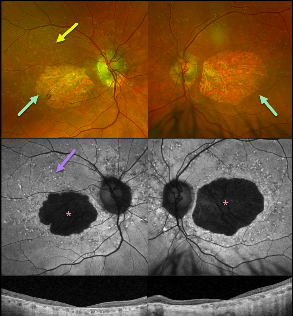
This 83YO female presented for a retinal examination without a new complaint. There was a known history of Stargardt disease from a pathogenic ABCA4 mutation. Vision was stable at 20/200 OD and 20/400 OS.
Optos color RG imaging shows bilateral macular atrophy with yellow subretinal flecks more peripherally. Fundus autofluorescence (FAF) shows hypo-FAF from the macular atrophy with variable hyper-FAF from the subretinal flecks. Diffuse outer retinal atrophy is seen on Triton swept-source OCT.
Learning Points:
Stargardt disease is an autosomal recessive disorder caused by a mutation in the ABCA4 gene. The ABCA4 protein is located in the photoreceptor outer segments and is involved in the recycling of 11-cis-retinal. The mutation in this gene causes lipofuscin accumulation in the RPE, which eventually leads to photoreceptor and RPE degeneration. Other ABCA4 disorders include fundus flavimaculatus, cone-rod dystrophy, retinitis pigmentosa, and age-related macular degeneration.
FUNDUS FLAVIMACULATUS
Originally posted on @retina.rocks February 21, 2023
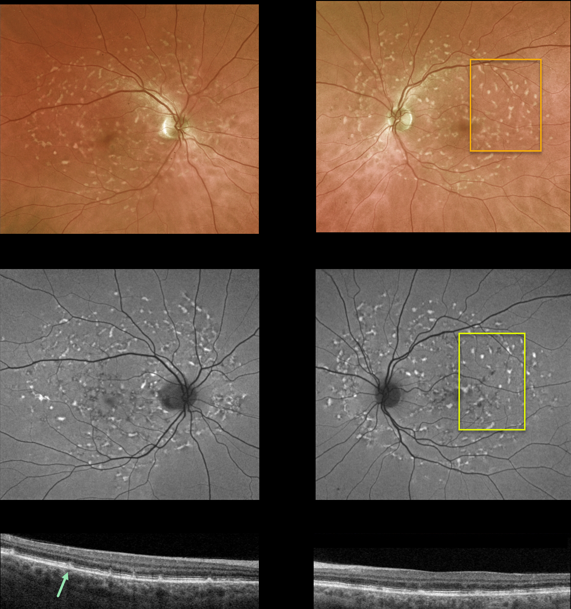
This 55YO female was referred for a possible white dot syndrome. She had no ocular symptoms, and vision was 20/30/ OU. There was no family history of ocular disease.
Optos color imaging shows multiple yellow fleck-like lesions throughout each posterior pole. Fundus autofluorescence of these lesions shows variable hyper-FAF.
OCT scanning shows scattered defects in the EZ with hyperreflective dots extending from the RPE into the EZ and outer nuclear layer.
Learning Points:
Fundus flavimaculatus is a genetic disorder, usually transmitted as an autosomal recessive trait, that is caused by mutations involving the ABCA4 gene. This gene encodes one of a family of ATP-binding cassette (ABC) transmembrane proteins, which mediate the active transport of various substrates across cellular membranes. The ABCA4 protein is present in the photoreceptor outer segment disc membranes, where it participates in the recycling of 11-cis-retinal.
Stargardt disease is believed to be fundus flavimaculatus without peripheral findings. Vision in fundus flavimaculatus is often better than that in Stargardt due to later disease onset and less macular involvement. Both likely represent an RPE lipofuscin storage disease. The RPE cells become engorged with lipofuscin, causing a dark choroid on fluorescein angiography.
See Cremers et al (Progress Retinal Eye Research 2020;79;100861) for a wonderful review of the complex spectrum of ABCA4 disorders.
STARGARDT DISEASE
Omar Mulki
Originally posted on @retina.rocks November 25, 2022
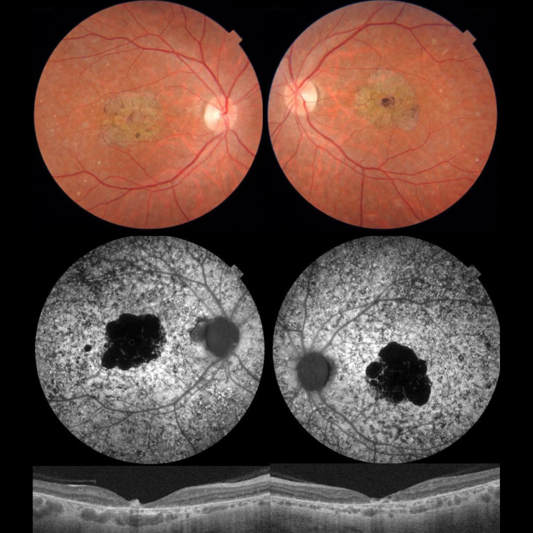
This 33YO male presented with counting fingers bilaterally from Stargardt disease. There was a positive family history for Stargardt, and genetic testing in the past was positive for a pathogenic ABCA4 mutation.
Color photographs show a bilateral beaten-bronze appearance at the macular centers, with more subtle pigmentary changes throughout the fundi.
Fundus autofluorescence (FAF) shows hypo-FAF in the macular lesions and, more dramatically, diffuse pigmentary changes elsewhere.
OCT scanning shows marked central foveal retinal thinning, with variable more peripheral outer retinal atrophy.
Learning Points:
Stargardt disease is an autosomal recessive disorder caused by a mutation in the ABCA4 gene. The ABCA4 protein is located in the photoreceptor outer segments and is involved in the recycling of 11-cis-retinal.
The mutation of this gene causes the accumulation of lipofuscin in the RPE, which eventually leads to photoreceptor and RPE degeneration. Other ABCA4 disorders include fundus flavimaculatus and cone-rod dystrophy.
STARGARDT DISEASE
Originally posted on @retina.rocks April 26, 2022
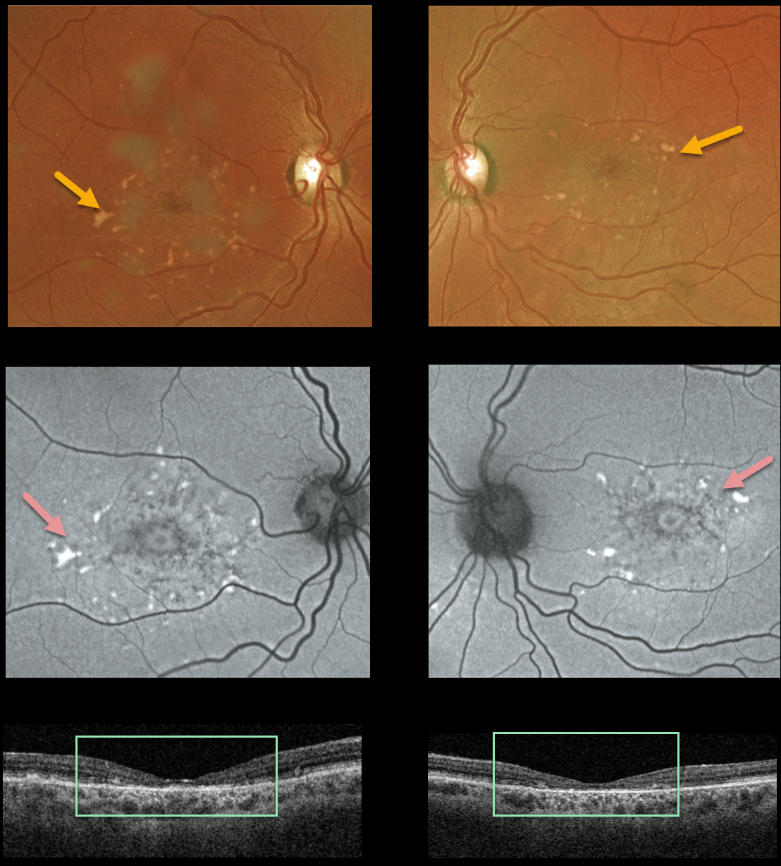
This 37YO female presented with vision of 20/80 OU and classic findings for Stargardt disease, captured with Optos multimodal imaging and Triton swept-source OCT, including bilateral perifoveal yellow subretinal flecks that are variably hyperautofluorescent and variable outer retinal and RPE atrophy.
Learning Points:
Stargardt disease is an autosomal recessive disorder caused by a mutation in the ABCA4 gene. The ABCA4 protein is located in the photoreceptor outer segments and is involved in the recycling of 11-cis-retinal.
Mutation of this gene causes the accumulation of lipofuscin in the RPE, which eventually leads to photoreceptor and RPE degeneration. Other ABCA4 disorders include fundus flavimaculatus and cone-rod dystrophy.
FUNDUS FLAVIMACULATUS
Originally posted on @retina.rocks November 18, 2021
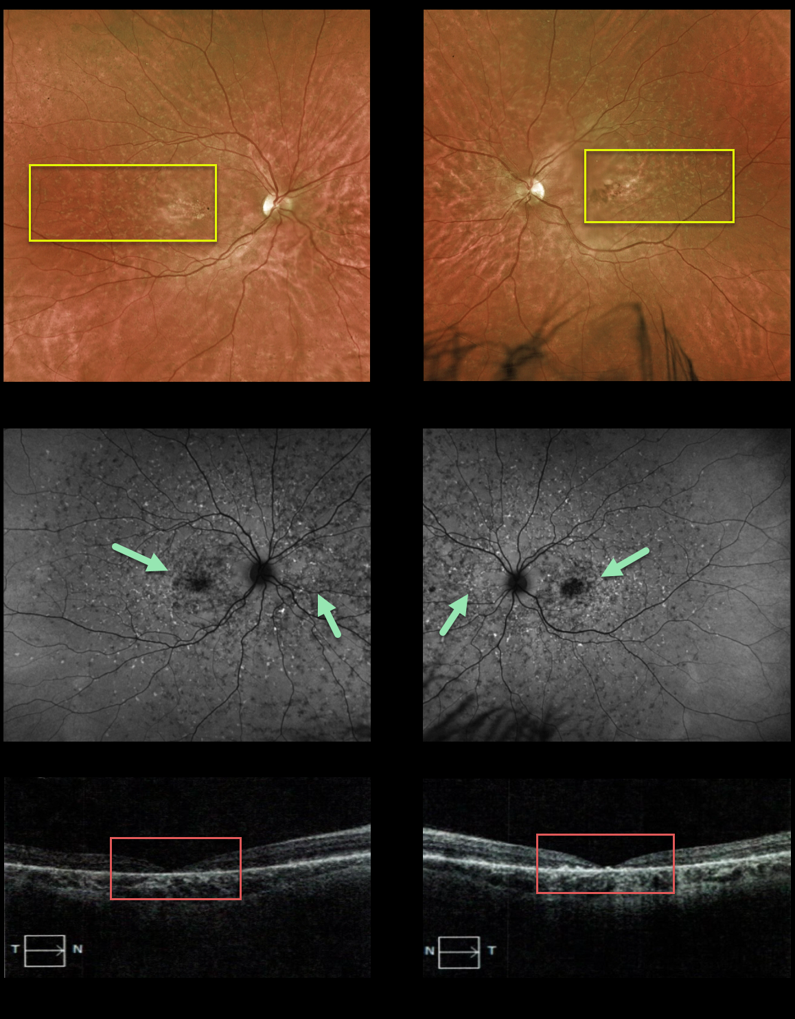
This 28YO female presented with 20/400 vision in both eyes, with classic findings of fundus flavimaculatus, including bilateral subretinal flecks extending from the macula to the midperiphery, variable outer retinal and RPE atrophy, and fundus autofluorescence changes. OCT scanning showed marked outer retinal atrophy, especially centrally. Her brother has similar findings.
Learning Points:
Fundus flavimaculatus is an autosomal recessive disorder caused by a mutation in the ABCA4 gene. The ABCA4 protein is located in the photoreceptor outer segments and is involved in the recycling of 11-cis-retinal.
Mutation of this gene causes the accumulation of lipofuscin in the RPE, which eventually leads to photoreceptor and RPE degeneration.
Other ABCA4 disorders include Stargardt’s disease, cone-rod dystrophy, and retinitis pigmentosa.
STARGARDT DISEASE
Originally posted on @retina.rocks April 12, 2021
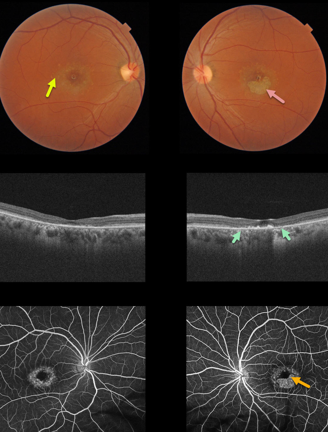
This 40YO female presented with vision of 20/400 in her right eye and counting fingers (CF) in her left eye due to classic findings from Stargardt’s disease, including perifoveal yellow subretinal flecks and variable outer retinal/RPE atrophy.
The OCT of her left eye best visualizes the bull’s-eye pattern of outer retinal and RPE atrophy. Loss of photoreceptors is evident as the outer plexiform layer (OPL) descends towards the RPE, eventually resulting in fairly complete atrophy of the outer retina and RPE.
Fluorescein angiography (FA) characteristically shows a ‘dark choroid’ from the lipofuscin-engorged RPE blocking the choroid (not present in this case).
Learning Points:
Stargardt’s disease is an autosomal recessive disorder caused by a mutation in the ABCA4 gene. The ABCA4 protein is located in the photoreceptor outer segments and is involved in the recycling of 11-cis-retinal.
Mutation of this gene causes accumulation of lipofuscin in the RPE, eventually leading to photoreceptor and RPE degeneration. Other ABCA4 disorders include fundus flavimaculatus and cone-rod dystrophy.
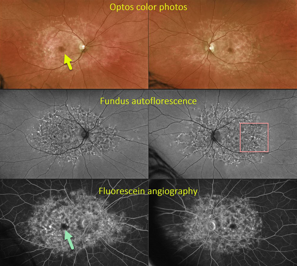
FUNDUS FLAVIMACULATUS
Originally posted on @retina.rocks January 7, 2021
This 44YO male presented with classic findings for fundus flavimaculatus, including perifoveal atrophy and variable hyper-autofluorescent subretinal yellow flecks extending from the macula to the midperiphery. Fluorescein angiography (FA) reveals a central dark choroid due to RPE cells becoming engorged with lipofuscin. Surprisingly, vision was 20/20 OU with a negative family history.
Learning Points:
Fundus flavimaculatus is an autosomal recessive disorder that is caused by mutations involving the ABCA4 gene.
This gene encodes one of a family of ATP-binding cassette (ABC) transmembrane proteins, which mediate the active transport of various substrates across cellular membranes. The ABCA4 protein is present in the photoreceptor outer segment disc membranes, where it participates in the recycling of 11-cis-retinal.
Stargardt’s disease is felt to be fundus flavimaculatus without peripheral findings. Vision in fundus flavimaculatus is often better than Stargardt’s due to later disease onset and less macular involvement. Both likely represent an RPE lipofuscin storage disease.
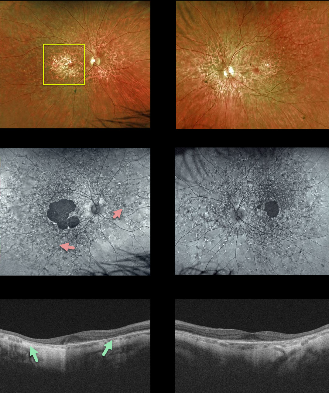
FUNDUS FLAVIMACULATUS
Originally posted on @retina.rocks July 17, 2020
Our patient shows fairly classic findings of fundus flavimaculatus. Central vision remains good at 20/40 OD and 20/30 OS, with preserved foveas.
Findings include bilateral macular atrophy and scattered yellow, subretinal, multifocal, and autofluorescent flecks extending to the mid-peripheral retina. OCT shows RPE and outer retinal atrophy.
Learning Points:
Fundus flavimaculatus is a genetic disorder, usually transmitted as an autosomal recessive trait, that is caused by mutations involving the ABCA4 gene.
ABCA4 encodes one of a family of ATP-binding cassette (ABC) transmembrane proteins that mediate the active transport of various substrates across cellular membranes. The ABCA4 protein is present in the photoreceptor outer segment disc membranes, where it participates in the recycling of 11-cis-retinal.
Stargardt’s disease is felt to be fundus flavimaculatus without peripheral findings. Vision in fundus flavimaculatus is often better than Stargardt’s due to later disease onset and less macular involvement.
Both likely represent an RPE lipofuscin storage disease. The RPE cells become engorged with lipofuscin, causing a dark choroid on fluorescein angiography.
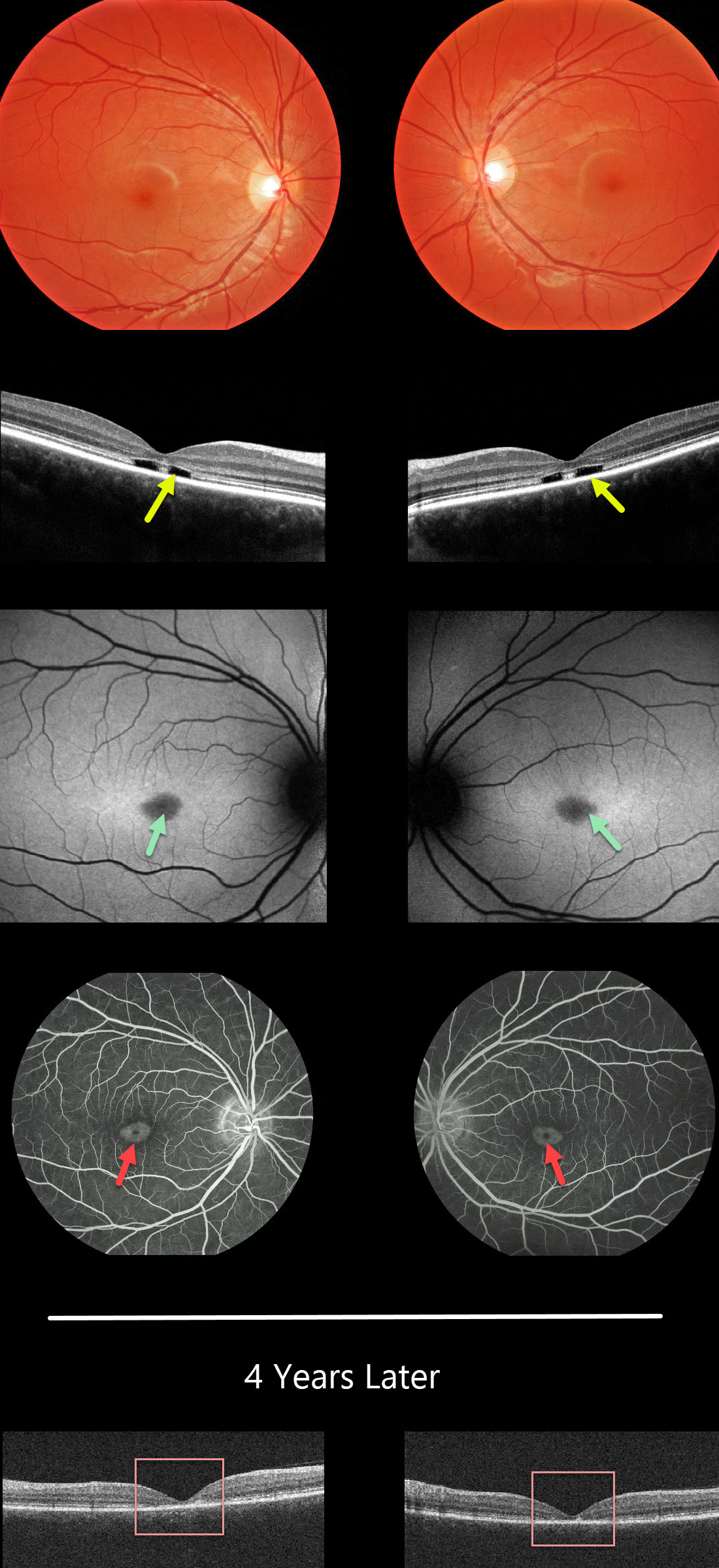
STARGARDT DISEASE
Originally posted on @retina.rocks May 26, 2020
This 14YO female presented with difficulty distinguishing colors and seeing the yellow softball when batting. Vision was 20/30 OD and 20/25 OS.
Her fundus examinations were normal. However, OCT scanning shows a pathognomonic loss of the photoreceptor layers outer to the external limiting membrane.
Fundus autofluorescence shows foveal hypofluorescence corresponding to the angiographic bull’s-eye lesions.
This patient tested positive for the ABCA4 gene, which is associated with both cone-rod dystrophy and Stargardt’s disease.
Four years later, vision and the clinical findings remain stable, but the OCT outer retinal defects have collapsed with increased outer retinal atrophy.
Learning Points:
Stargardt disease is an autosomal recessive disorder caused by a mutation in the ABCA4 gene. The ABCA4 protein is located in the photoreceptor outer segments and is involved in the recycling of 11-cis-retinal.
Mutation of this gene causes the accumulation of lipofuscin in the RPE, which eventually leads to photoreceptor and RPE degeneration. Other ABCA4 disorders include fundus flavimaculatus and cone-rod dystrophy.
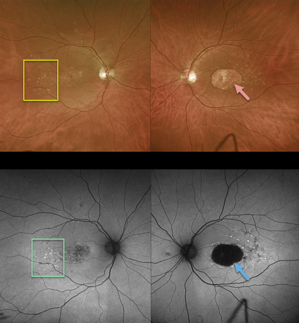
STARGARDT DISEASE
Originally posted on @retina.rocks May 5, 2020
This patient has bilateral 20/400 vision due to classic findings from Stargardt disease, including yellow subretinal flecks and outer retinal/RPE atrophy.
FAF primarily displays hyperfluorescence from RPE lipofuscin. Since Stargardt is essentially a lipofuscin storage disease, the subretinal flecks are hyperautofluorescent and the areas of RPE loss are hypoautofluorescent.
Learning Points:
Stargardt disease is an autosomal recessive disorder caused by a mutation in the ABCA4 gene. The ABCA4 protein is located in the photoreceptor outer segments and is involved in the recycling of 11-cis-retinal. The mutation of this gene causes accumulation of lipofuscin in the RPE, which eventually leads to photoreceptor and RPE degeneration.
Fluorescein angiography (not pictured) characterically shows a ‘dark choroid’ from the lipofuscin-engorged RPE blocking visualization of the underlying choroid.
Stargardt is one of the macular diseases where the vision loss is often much worse than expected from the fundus appearance.
STARGARDT DISEASE
Originally posted on @retina.rocks January 3, 2020
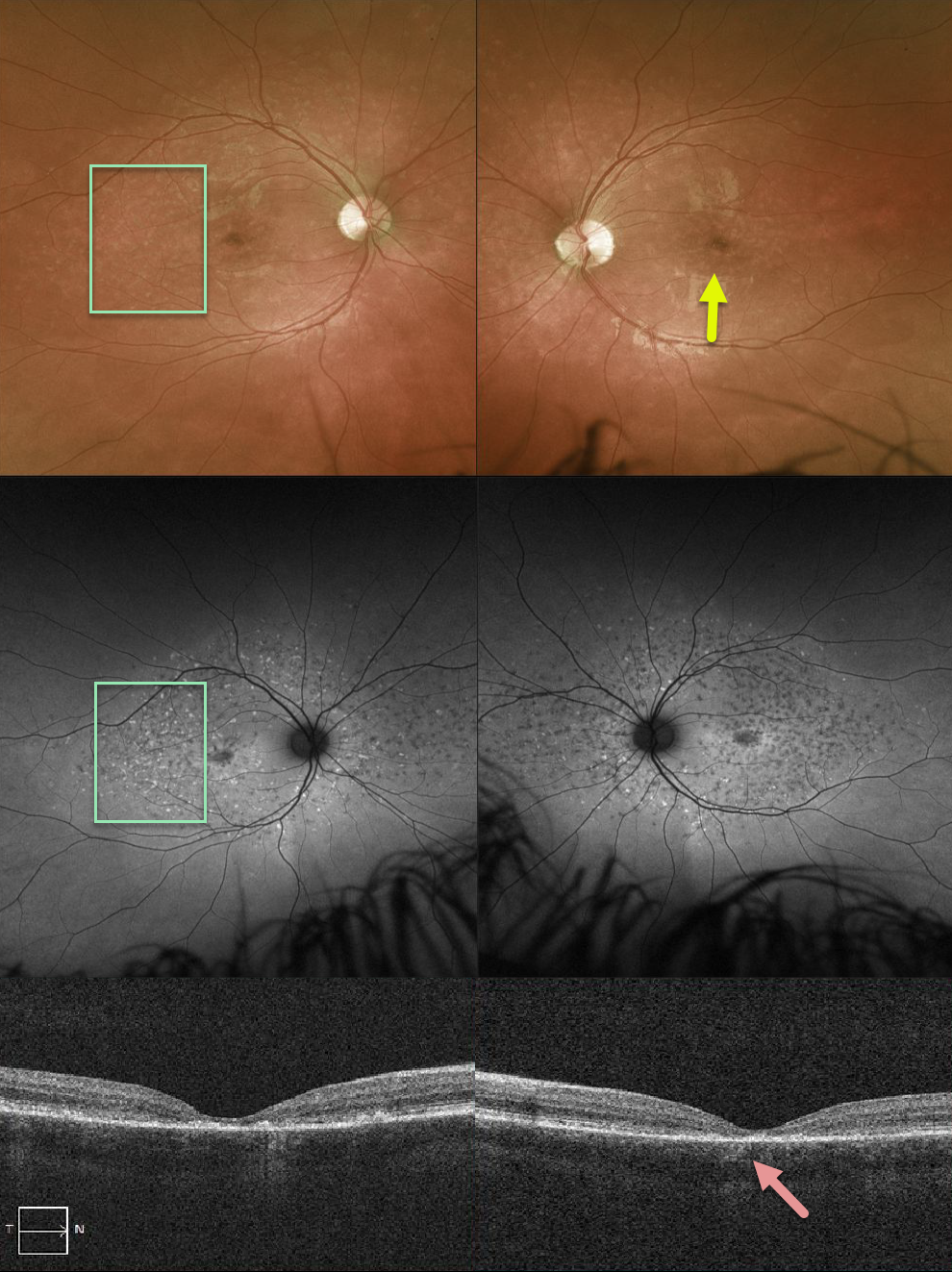
This patient has Stargardt’s disease with beaten-bronze macular atrophy and scattered pisciform flecks, which are more obvious on fundus autofluorescence. OCT scanning shows central outer retinal atrophy.
Learning Points:
Stargardt’s disease is caused by a mutation in the ABCA4 gene, which codes for a protein in the outer segments responsible for facilitating movement of retinoids across the disc membrane.
Lipofuscin accumulates in the RPE, eventually causing degeneration of the photoreceptors and RPE.


