

Steroids (Ozurdex + Iluvien + TriMoxi)
<< Back to Cases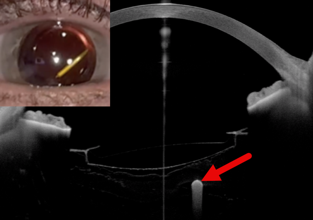
ANTERIORLY MIGRATED ILUVIEN IMPLANT
Fatma Shakarchi and Ahmed Sallam
Originally posted on @retina.rocks March 2, 2026
This 58YO female with panuveitis treated with bilateral fluocinolone acetonide intravitreal implants (Iluvien) and systemic adalimumab presented with an intermittent, moving floater near the visual axis in her right eye. She was pseudophakic in both eyes with posterior chamber intraocular lenses (IOLs) positioned within intact capsular bags.
External slit-lamp photography and anterior segment OCT show an Iluvien implant in the anterior vitreous cavity just behind the IOL.
Learning Points:
Migration of Iluvien implants into the visual axis has been reported and may cause visually disturbing symptoms, particularly when the implant assumes a vertical orientation within the vitreous cavity. In selected cases, Nd:YAG laser disruption of anchoring vitreous fibers (Moisseiev et al JAMA Ophthalmol 2016:134:1057-1058) or vitrectomy (Knight et al Retinal Cases & Brief Reports 2023;17:26-28) have been described as potential management strategies.
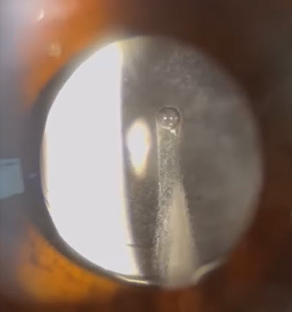
TRIAMCINOLONE-MOXIFLOXACIN (TriMoxi)
Adam Mueller, Benjamin Conkright, and Andrew Steele
Originally posted on @retina.rocks October 31, 2025
This 64YO female presented for her one-day post-dropless cataract surgery with some minor irritation and floaters. Vision without correction was 20/25+. Unbeknownst to our happy patient, a sinister ghoul was lurking behind her perfectly centered intraocular implant.
Our practice, along with many others, offers ‘no drop’ cataract surgery as a convenience to our patients. Instead of needing to take postoperative antibiotic and steroid drops, 0.2cc of TriMoxi (triamcinolone acetate 15mg/cc with moxifloxacin 1mg/cc) is injected into the anterior inferior vitreous at the end of the cataract surgery. Although this liquid usually remains well outside the visual axis, patients will often experience mild floaters perioperatively. Rarely, as in this patient, the liquid disperses throughout the vitreous cavity, causing significant transient floaters.
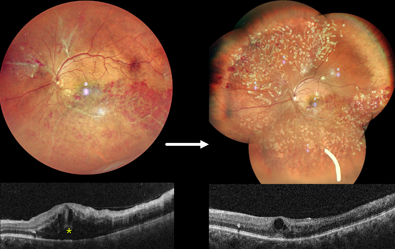
OCCLUSIVE RETINAL VASCULITIS
Anjana Mirajkar and Manish Nagpal
Originally posted on @retina.rocks July 23, 2024
This healthy 32YO male presented on 4/23/24 with 2 months of decreased vision in his left eye. Family history was negative. A prior workup by a prior doctor was negative, including protein electrophoresis and homocysteine levels. He failed a short trial of oral steroids. Vision was 20/20 in his healthy right eye and counting fingers in his left eye.
Fundus photography shows an ischemic inferotemporal branch retinal vein occlusion with moderate, mostly peripheral retinal hemorrhages. The superotemporal retinal vein is diffusely beaded, and an apparent independent ischemic BRVO is seen superonasally, with probable preretinal blood. Marked macular edema is seen on OCT. Unfortunately, fluorescein angiography is not available.
An Ozurdex injection was given for macular edema, and two sessions of panretinal photocoagulation were performed for proliferative disease. On 6/3/24, vision improved to 20/120, and the macular edema was improving.
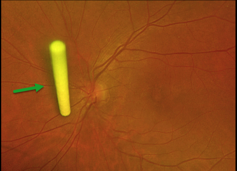
OZURDEX
S. Nazem Ibrahim, Joseph Touma, and Ahmed Sallam
Originally posted on @retina.rocks February 15, 2024
This 44YO female with a diagnosis of chronic anterior uveitis in the left eye presented with visual acuity of 20/60 and cystoid macular edema recalcitrant to topical and periocular steroids. We treated her with an intravitreal dexamethasone implant (Ozurdex).
Optos imaging one week later revealed an unusual vertical orientation of the implant, ‘standing proud’ near the optic disc within the posterior vitreous. There was no prior pars plana vitrectomy surgery.
Even in non-vitrectomized eyes, an Ozurdex implant may take a vertical position within the vitreous and not attach to the peripheral retina as it usually does. The patient was asymptomatic, and observation was recommended.
TRIAMCINOLONE-MOXIFLOXACIN (TriMoxi)
Originally posted on @retina.rocks October 30, 2023
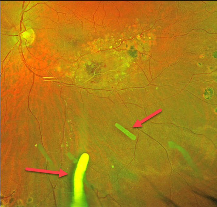
This 41YO male has been followed for years with Coats disease in his left eye. Numerous treatment modalities have been used, including thermal laser, intravitreal anti-VEGF, and Ozurdex (dexamethasone 0.7mg) injections. Most recently, he has received Ozurdex every 3-4 months for the past 2 years. Vision is stable at 20/30.
Optos color RG imaging shows multiple Ozurdex ghosts in variable stages of absorption.
Learning Points:
The Ozurdex implant slowly dissolves while releasing its dexamethasone. Although the clinical effect usually lasts about 2 to 3 months, remnants can remain in the eye for over a year (Kim et al, Retina 2020;40:2226-2231).
Approved for diabetic and retinal vein occlusion macular edema and posterior uveitis, it is also often used off-label for other causes of macular edema, including uveitic and pseudophakic CME. For our patient, Ozurdex has been the most successful modality at keeping his central macula dry.
TRIAMCINOLONE-MOXIFLOXACIN (TriMoxi)
Originally posted on @retina.rocks June 1, 2023
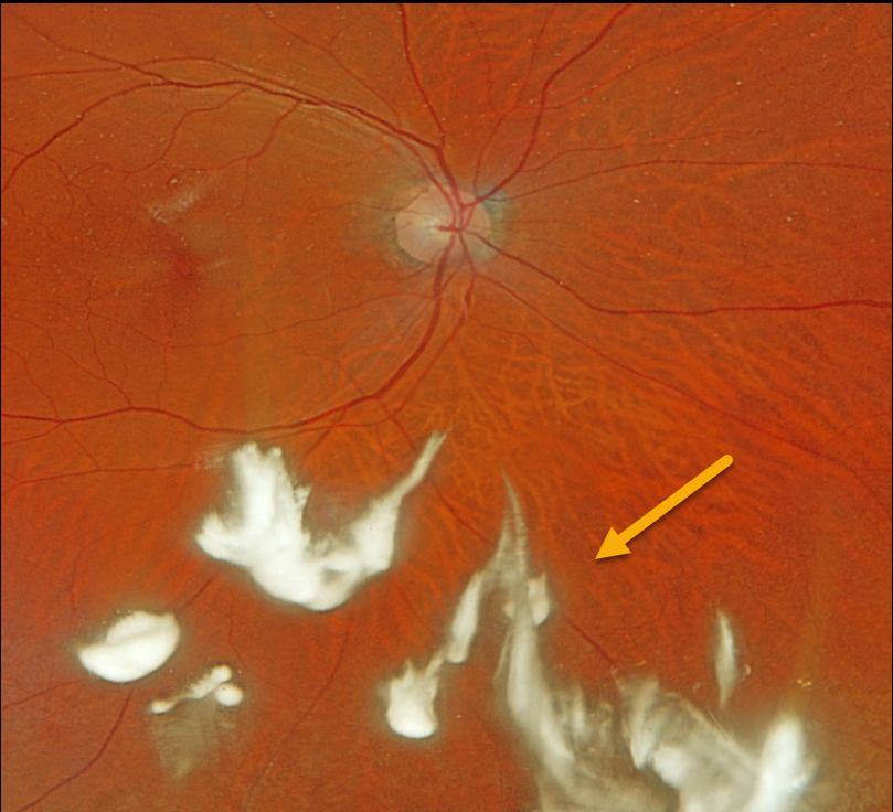
This 68YO male presented with symptomatic floaters 10 days following uneventful ‘no-drop’ cataract surgery with Trimoxi.
Optos color RGB imaging shows clumps of white triamcinolone suspended in the inferior vitreous.
Learning Points:
Our practice, along with many others, offers ‘no drop’ cataract surgery as a convenience to our patients. Instead of needing to take postoperative antibiotic and steroid drops, 0.2cc of Trimoxi (triamcinolone acetate 15mg/cc with moxifloxacin 1mg/cc) is injected into the anterior inferior vitreous at the end of the cataract surgery.
Although this liquid usually remains well outside the visual axis, patients will often experience mild floaters perioperatively. Rarely, as in this patient, the liquid disperses throughout the vitreous, causing significant transient floaters.
OZURDEX GHOST
Originally posted on @retina.rocks November 26, 2021
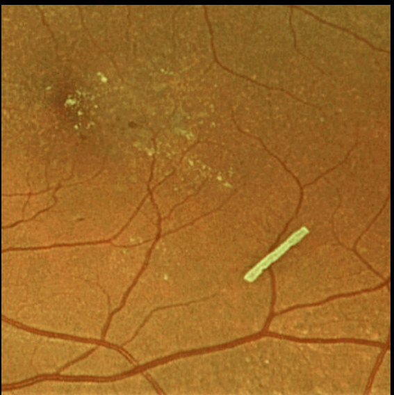
This 71YO female is getting Ozurdex intravitreal injections every 2 months to control her macular edema.
Learning Points:
The Ozurdex implant slowly releases the steroid and eventually totally absorbs, with the clinical effect usually lasting about 2 to 4 months.
The ghost-like remnants can persist for many months (see Kim et al, Retina 2020;40;2226-2231).
TRIAMCINOLONE-MOXIFLOXACIN (TriMoxi)
Originally posted on @retina.rocks May 11, 2021
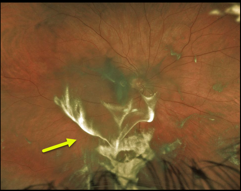
This patient experienced some superior visual field loss following cataract surgery. A stagnant blob of triamcinolone-moxifloxacin accumulated in the vitreous and subhyaloid space in the inferior macula.
The medication eventually dissolved as the patient’s symptoms resolved.
Learning Points:
TriMoxi is commonly used in dropless cataract surgery, so patients don’t need to use postoperative antibiotic and steroid drops. At the end of the procedure, 0.2cc of TriMoxi is injected into the anterior inferior vitreous. Usually, patients notice some large black floaters that subside within a few weeks.
OCULAR ISCHEMIA WITH OZURDEX IMPLANTS
Originally posted on @retina.rocks February 4, 2021
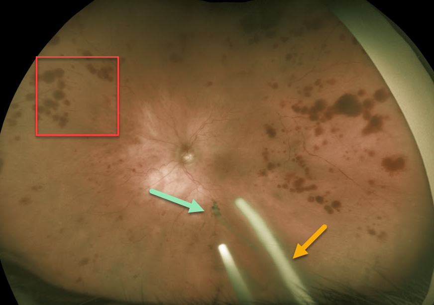
This 86YO patient was receiving Ozurdex (dexamethasone 0.7%) injections every 8 weeks in her left eye for macular edema from an inferior hemiretinal vein occlusion.
She subsequently developed ocular ischemic syndrome (OIS) like changes with scattered peripheral large outer retinal hemorrhages. Inferiorly, more recent Ozurdex implants are noted, along with another ghost-like remnant of an older implant. Although the Ozurdex implants are supposed to completely degrade, these remnants may persist beyond 1 year (see Kim et al Retina 2020;40;2226-2231).
Learning Points:
The retinal hemorrhages in ocular ischemic syndrome (OIS) are unique. Retinal vascular disorders like diabetic retinopathy, retinal vein occlusions, and hypertensive retinopathy usually have flame- and dot-shaped inner retinal hemorrhages concentrated around the posterior pole.
Our patient’s findings are classic for OIS, with hemorrhages in the deeper peripheral retina. The retinal periphery is literally the end of the line for the retinal arterial circulation. In an eye receiving less blood supply due to carotid occlusive disease, the more proximal retina has first dibs on the available oxygen. There are penetrating capillaries that dive radially from the nerve fiber and ganglion cell layers into the deeper retina. The deep vascular complex that supplies the inner and outer plexiform layers is thus furthest downstream, and these endothelial cells are likely damaged in OIS. Blood cells leaking into this space accumulate, causing the large outer blot to hemorrhage.
These patients need to have a carotid Doppler and ultrasound and be referred for medical or surgical intervention if they have a critical stenosis. They may also develop anterior or posterior segment neovascularization requiring photocoagulation and/or anti-VEGF injections. Iris neovascularization can develop, leading to neovascular glaucoma. Hypotony can also occur.
TRIAMCINOLONE-MOXIFLOXACIN (TriMoxi)
Originally posted on @retina.rocks July 21, 2020
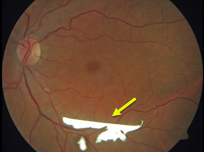
This patient experienced some superior visual field loss following cataract surgery. A stagnant blob of triamcinolone-moxifloxacin accumulated in the subhyaloid space in the inferior macula.
The medication eventually dissolved as the patient’s symptoms resolved.
Learning Points:
TriMoxi is commonly used in dropless cataract surgery, so patients don’t need to use postoperative antibiotic and steroid drops. At the end of the procedure, 0.2cc of TriMoxi is injected into the anterior inferior vitreous. Usually, patients notice large black floaters that subside within a few weeks.
TRIAMCINOLONE-MOXIFLOXACIN (TriMoxi)
Originally posted on @retina.rocks May 22, 2020
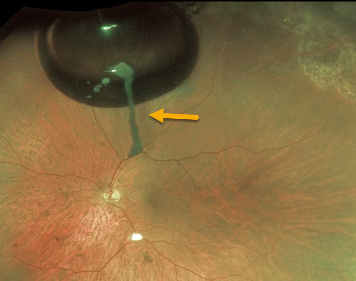
Six weeks prior to this retinal detachment repair, the patient underwent uncomplicated cataract surgery. This image shows TriMoxi (triamcinolone acetonide/moxifloxacin) medication literally “hanging on” the absorbing gas bubble!
Learning Points:
During cataract extraction, a commonly used combination of triamcinolone (15 mg/mL) and moxifloxacin (1mg/mL) is injected intracamerally. The medication eventually absorbs completely.


