

Susac Syndrome
<< Back to Cases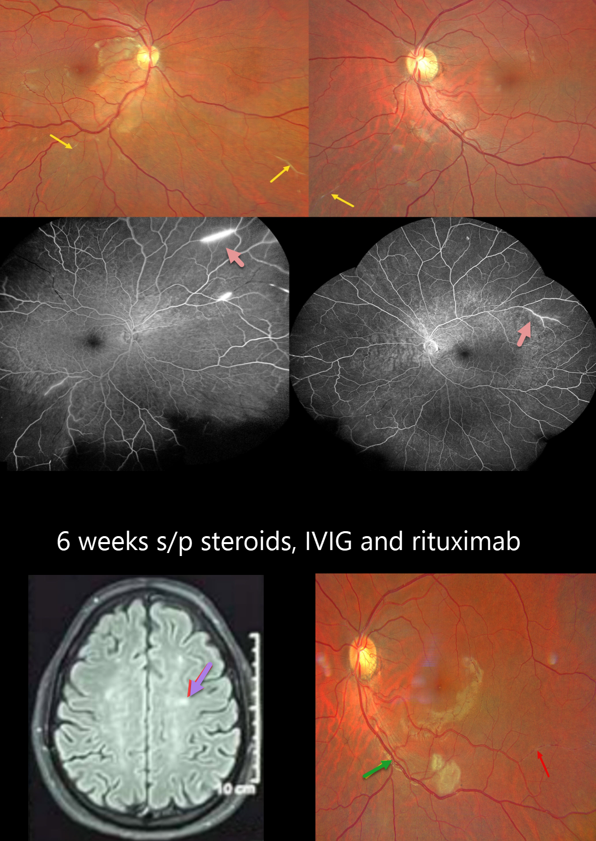
SUSAC SYNDROME
Ayushi Gupta and Vishal Agrawal
Originally posted on @retina.rocks October 15, 2024
This healthy 31YO male presented with about 2 weeks of vision loss in his right eye. He also complained of mild headaches over the prior month. Vision was counting fingers OD and 20/20 OS.
Fundus photography OD shows an opaque ischemic retina temporal to the disc, just sparing the fovea. Evidence of prior arterial occlusions is seen as bilaterally sclerosed vessels. Fluorescein angiography shows focal areas of scattered retinal ischemia with segmental areas of vascular staining.
MRI scanning shows multiple small, discrete, snowball-like lesions, and audiologic evaluation revealed bilateral mild sensorineural hearing loss, establishing the diagnosis of Susac syndrome. He was started on a 5-day course of intravenous immunoglobulins and steroids followed by maintenance rituximab therapy.
When he returned 6 weeks later, his vision was 20/80 OD and 20/20 OS. The nasal macular opacification is improving, but new areas of acute retinal ischemia are present in the inferior right and left maculae. Refractile deposits line an occluded arteriole along the left proximal inferotemporal arcade, and a new sclerosed arteriole is noted inferotemporally.
Learning Points:
Retinocochleocerebral vasculopathy, more commonly known as Susac syndrome, is a rare, microangiopathic disease of debated pathophysiology characterized by the classic triad of encephalopathy, sensorineural hearing loss, and branch retinal artery occlusions (Heng et al, Survey Ophthalmology 2019;64:477-485). The full triad may take months to evolve, frustrating the diagnosis. Retinal arterial wall atheromatous plaques, also known as Gass plaques, are often seen in later stages. They are a result of the occlusion, not the cause. The development of wide-field angiography has allowed for earlier detection and treatment. Although the findings are usually self-limiting while they wax and wane, systemic treatment with steroids and immunosuppression can be considered for more severe cases.
SUSAC SYNDROME
Originally posted on @retina.rocks July 20, 2021
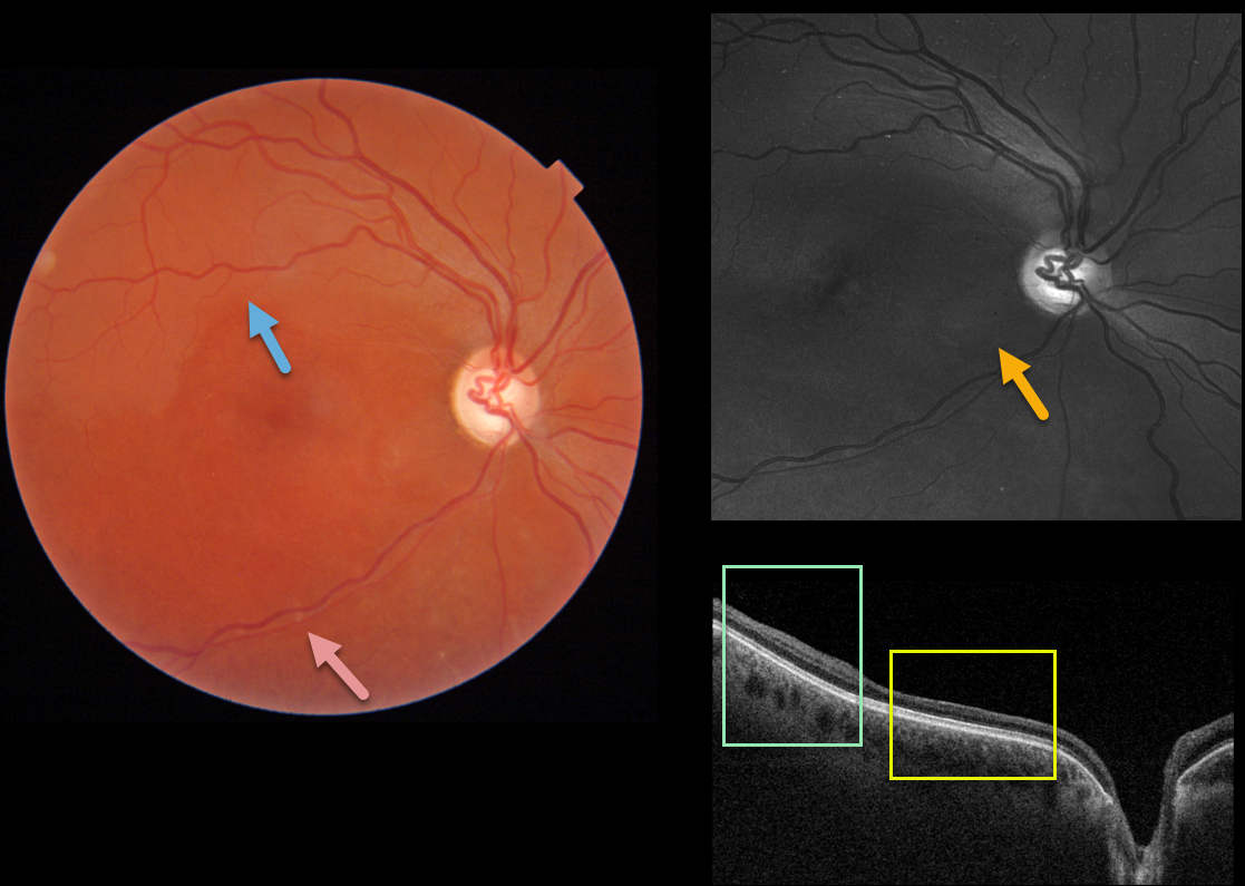
This healthy 30YO female was referred for poor vision in her right eye. She mentioned losing vision in this eye during high school, but couldn’t provide any further details. She also gave a history of hearing loss, which had never been evaluated. Vision was HM OD and 20/30 in her normal left eye.
Triton imaging shows faint retinal whitening in the superior macula. The retinal arterioles are narrowed and somewhat sheathed inferiorly.
Swept-source OCT shows normal inner retinal architecture temporally, with inner retinal atrophy and disorganization of the normal architecture more nasally. Right inferotemporal nerve fiber loss is quite dramatic on Optos green imaging.
We diagnosed her with a resolved inferior branch retinal artery occlusion (BRAO), although this may represent Susac’s syndrome. The involved retina appeared redder than the uninvolved superior retina, accounting for the whitish superior retina noted above.
Subsequent work-up, including hypercoagulable labs and MRI, was negative. She denied prior intravenous drug use. To complete the workup, we will be obtaining carotid Doppler and cardiac ultrasonography.
Learning Points:
Susac’s syndrome, also known as retinocochleocerebral vasculopathy, is a rare microangiopathic disease characterized by a triad of encephalopathy, sensorineural hearing loss, and branch retinal artery occlusions with a predilection for younger females.
In the presence of Susac’s syndrome, MRI findings should show corpus callosum involvement. However, the full triad commonly takes months to develop, complicating diagnosis.
SUSAC SYNDROME
Originally posted on @retina.rocks January 29, 2020
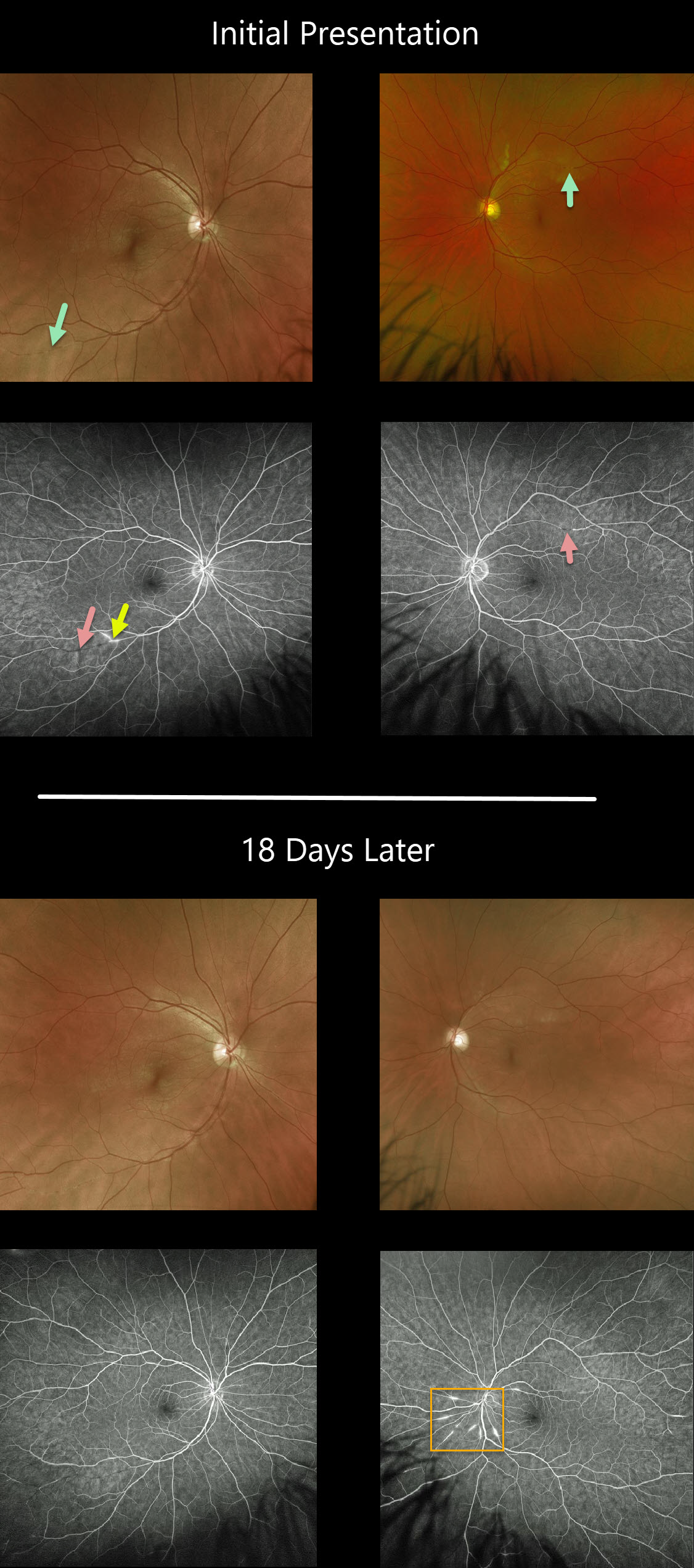
Our patient presented with silent ischemia inferotemporally in the right eye and new lesions superotemporally in the left. Note the areas of non-perfusion and leakage.
18 days later new multi-focal lesions OS are visualized dramatically on fluorescein angiography.
Fortunately, the patient has maintained excellent VA
Learning Points:
Retinocochleocerebral Vasculopathy, more commonly known as Susac Syndrome, is a rare, microangiopathic disease of debated pathophysiology characterized by the classic triad of encephalopathy, sensorineural hearing loss and branch retinal artery occlusions. The full triad may take months to evolve, frustrating the diagnosis.
Retinal arterial wall atheromatous plaques, also known as Gass plaques, are seen often in later stages. They are a result of the occlusion, not the cause.
The development of wide-field angiography has occasionally allowed for early detection which can be critical in order to minimize the often long-term, debilitating effects of the disease.


