

Syphilis
<< Back to Cases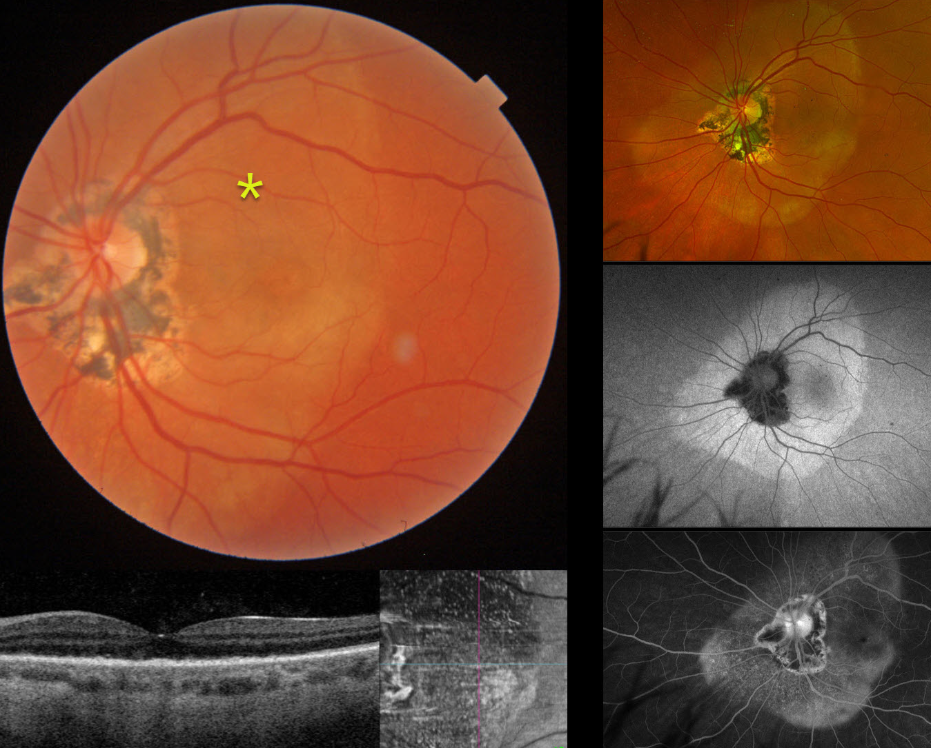
SYPHILIS
Originally posted on @retina.rocks December 9, 2025
This 61YO female presented with 1 week of vision loss in her left eye. Vision was 20/25 in her normal OD and counting fingers OS.
Triton color imaging shows a subretinal yellow-white placoid lesion extending beyond the macula, along with ocular histoplasmosis-related peripapillary scarring. Swept-source OCT shows replacement of the outer retinal bands with hyperreflective material, which is seen as innumerable hyperreflective dots on en face imaging. Optos color RGB imaging shows the full extent of the placoid lesion. Fundus autofluorescence (FAF) of this lesion shows homogenous hyper-FAF. It diffusely stains on fluorescein angiography. Laboratory testing was positive for syphilis and negative for HIV. She was referred to an infectious disease specialist and placed on oral doxycycline 100mg BID due to a penicillin allergy. When last seen two and a half months after her initial presentation, and after completing a full 1-month course of antibiotics, vision improved to 20/40 with near-complete resolution of the retinal findings (not shown).
Learning Points:
Ocular manifestations of syphilis vary widely and include virtually all variations of anterior and posterior segment inflammation. Originally described by Gass (Ophthalmology 1990;97:1288-1297), placoid chorioretinitis is one of the more common presentations. Inflammation is localized to the outer retina and RPE and appears on OCT as disruption of the outer retinal bands and hyperreflective pyramidal lesions as seen in our patient (Hu et al, Ophthalmology Retina 2022;6:172-178). The outer retinal damage unmasks the underlying RPE, accounting for the hyperfluorescent placoid lesions.
SYPHILIS
César Adrián Gómez Valdivia
Originally posted on @retina.rocks June 5, 2025
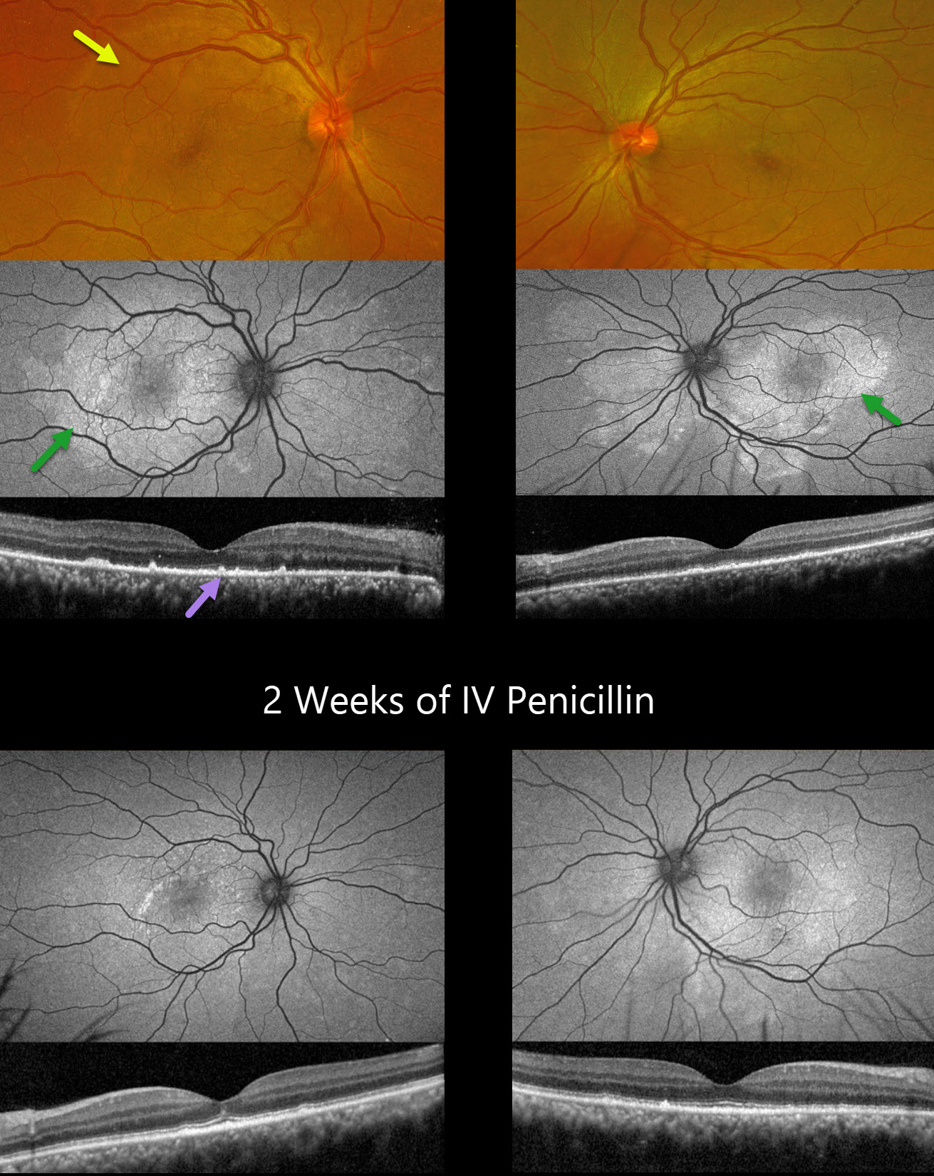
This 39YO HIV positive male presented with 5 days of bilateral decreased vision. Vision was 20/100 OD and 20/200 OS.
Optos color RG imaging shows bilateral whitish macular and peripapillary placoid lesions extending nasal to the nerves. Fundus autofluorescence (FAF) more dramatically shows these hyper-FAF placoid lesions, which resemble a butterfly in his left eye. OCT scanning shows variable loss of the outer retinal bands associated with hyperreflective deposits above the RPE.
VDRL was positive, and he was referred to his social security unit for systemic treatment. Seven weeks later, following two weeks of parenteral penicillin, vision improved to 20/20 OD and 20/20-2 OS, with improved funduscopic and OCT findings.
Learning Points:
Ocular manifestations of syphilis vary widely and include virtually all variations of anterior and posterior segment inflammation. Originally described by Gass (Ophthalmology 1990;97:1288-1297), placoid chorioretinitis is one of the more common presentations.
Inflammation is localized to the outer retina and RPE and appears on OCT as disruption of the outer retinal bands and hyperreflective pyramidal lesions as seen in our patient (Hu et al, Ophthalmology Retina 2022;6:172-178). The outer retinal damage unmasks the underlying RPE, accounting for the hyperfluorescent placoid lesions.
SYPHILIS
Fraser McKay
Originally posted on @retina.rocks April 21, 2025
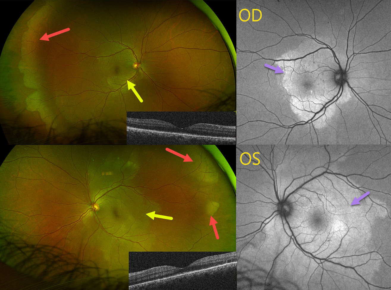
This 42YO female presented with 1 month of bilateral blurred vision. Vision was 20/30 OU.
Optos color RG imaging shows bilateral peripheral and posterior white areas without pressure. Correct??
WRONG! These posterior lesions are hyperautofluorescent. Macular OCT scanning shows variable loss of the outer retinal bands. Syphilis testing was positive, and she was immediately referred to an infectious disease specialist for treatment. When she returned 4 months later following a full course of parenteral penicillin, vision was 20/40 OD, 20/20 OS, with normalization of all fundus findings (not shown).
Learning Points:
Ocular manifestations of syphilis vary widely and include virtually all variations of anterior and posterior segment inflammation. Originally described by Gass (Ophthalmology 1990;97:1288-1297), placoid chorioretinitis is one of the more common presentations. Inflammation is localized to the outer retina and RPE and appears on OCT as disruption of the outer retinal bands and hyperreflective pyramidal lesions as seen in our patient (Hu et al, Ophthalmology Retina 2022;6:172-178). The outer retinal damage unmasks the underlying RPE, accounting for the hyperfluorescent placoid lesions.
SYPHILITIC PAPILLITIS
Navneet Mehrotra and Tamanna Patel
Originally posted on @retina.rocks March 12, 2025
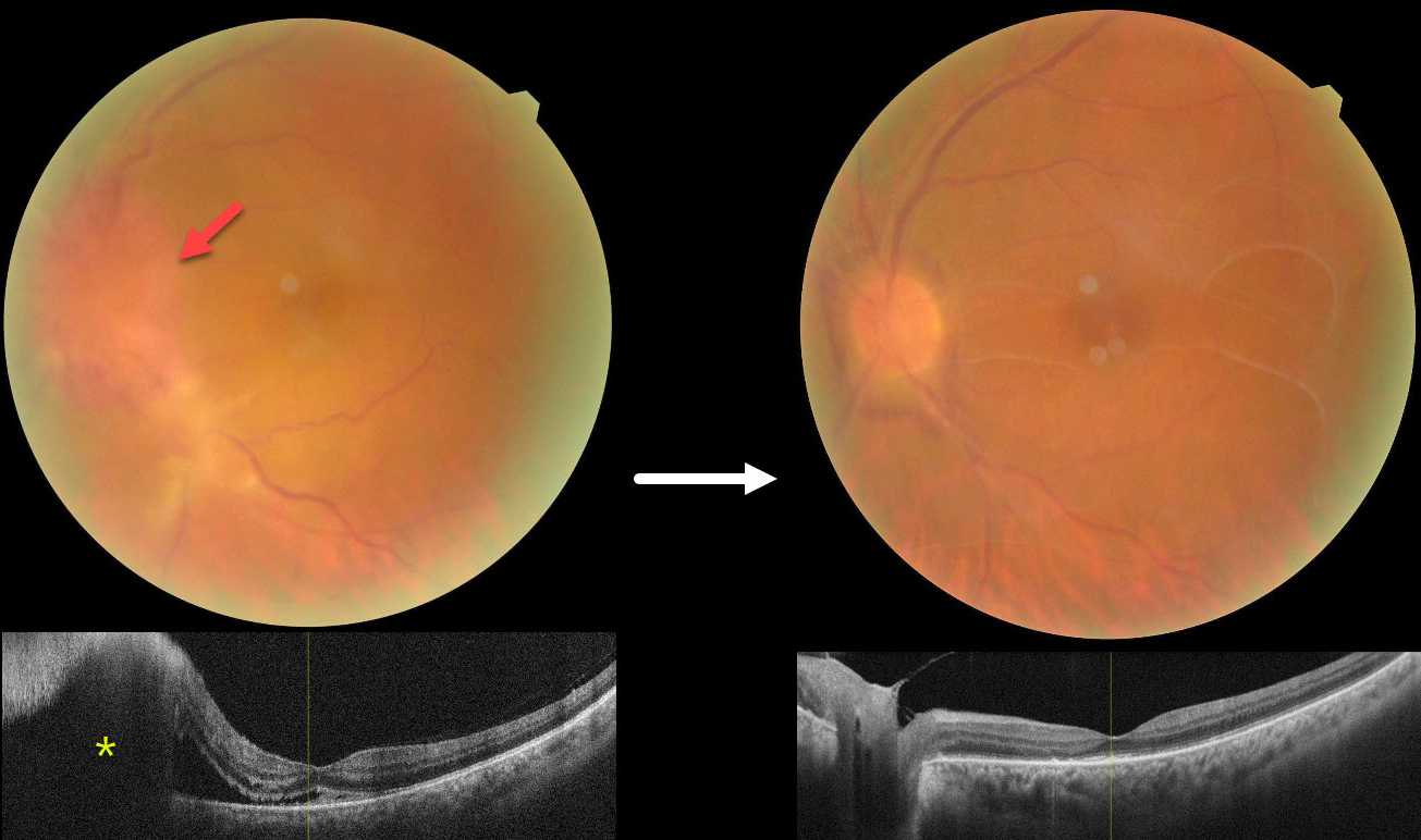
This 52YO male presented with 3 weeks of vision loss in his left eye. He has a history of diabetes, hypertension, and HIV. He was diagnosed elsewhere with optic neuritis and started on intravenous methylprednisolone with no improvement. Vision was 20/30 in his normal OD and counting fingers OS. There was a mild nongranulomatous anterior uveitis OS on slit lamp examination.
Color photography shows a markedly swollen nerve with possible retinitis extending inferotemporally. OCT scanning confirms the disc elevation with subretinal fluid extending beneath the nasal macula.
On further questioning, there was a recent history of a genital chancre, and the treponema pallidum hemagglutination assay was positive for syphilis. Following a 2-week course of parenteral penicillin, vision improved to 20/120 with marked improvement in the posterior segment findings. Unusual premacular vitreous membranes are noted.
Learning Points:
Ocular manifestations of syphilis vary widely and include virtually all variations of anterior and posterior segment inflammation. In our experience, placoid syphilis is the most common posterior segment presentation, although a recent systematic review and meta-analysis found papillitis to be the most reported finding in syphilitic uveitis (Zhang et al, J of Ophthalmology 2017;1; 6594849). Papillitis can be isolated or associated with other findings.
Any patient who tests positive for syphilis should always be checked for HIV since coinfection, as in our patient, is common.
SYPHILIS
Originally posted on @retina.rocks December 2, 2024
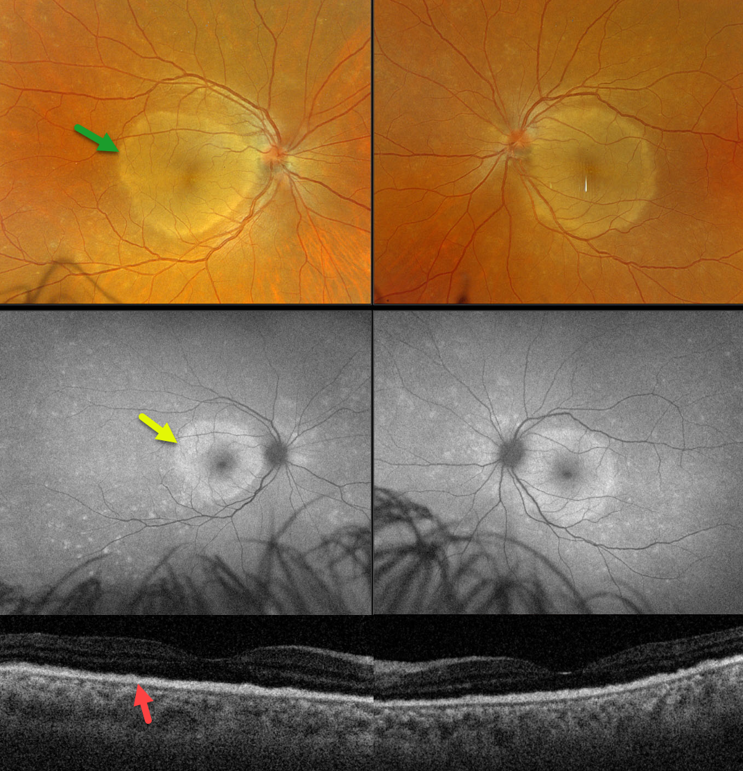
This 45YO female presented with one week of bilateral vision loss. Vision was counting fingers in each eye.
Optos color RGB imaging shows symmetrical, discrete subretinal yellow-white placoid lesions occupying each macula. Fundus autofluorescence (FAF), however, shows multifocal hyper-FAF spots extending peripherally to the hyper-FAF macular lesions. Swept-source OCT shows replacement of the outer retinal bands with hyperreflective material.
Laboratory testing was positive for syphilis and negative for HIV. She was referred to an infectious disease specialist for systemic penicillin treatment. Unfortunately, she was immediately lost to follow-up.
Learning Points:
Ocular manifestations of syphilis vary widely and include virtually all variations of anterior and posterior segment inflammation. Originally described by Gass (Ophthalmology 1990;97:1288-1297), placoid chorioretinitis is one of the more common presentations. The discrete placoid lesions were quite dramatic on our patient’s multimodal imaging.
SYPHILIS
Mattie Adams
Originally posted on @retina.rocks October 22, 2024
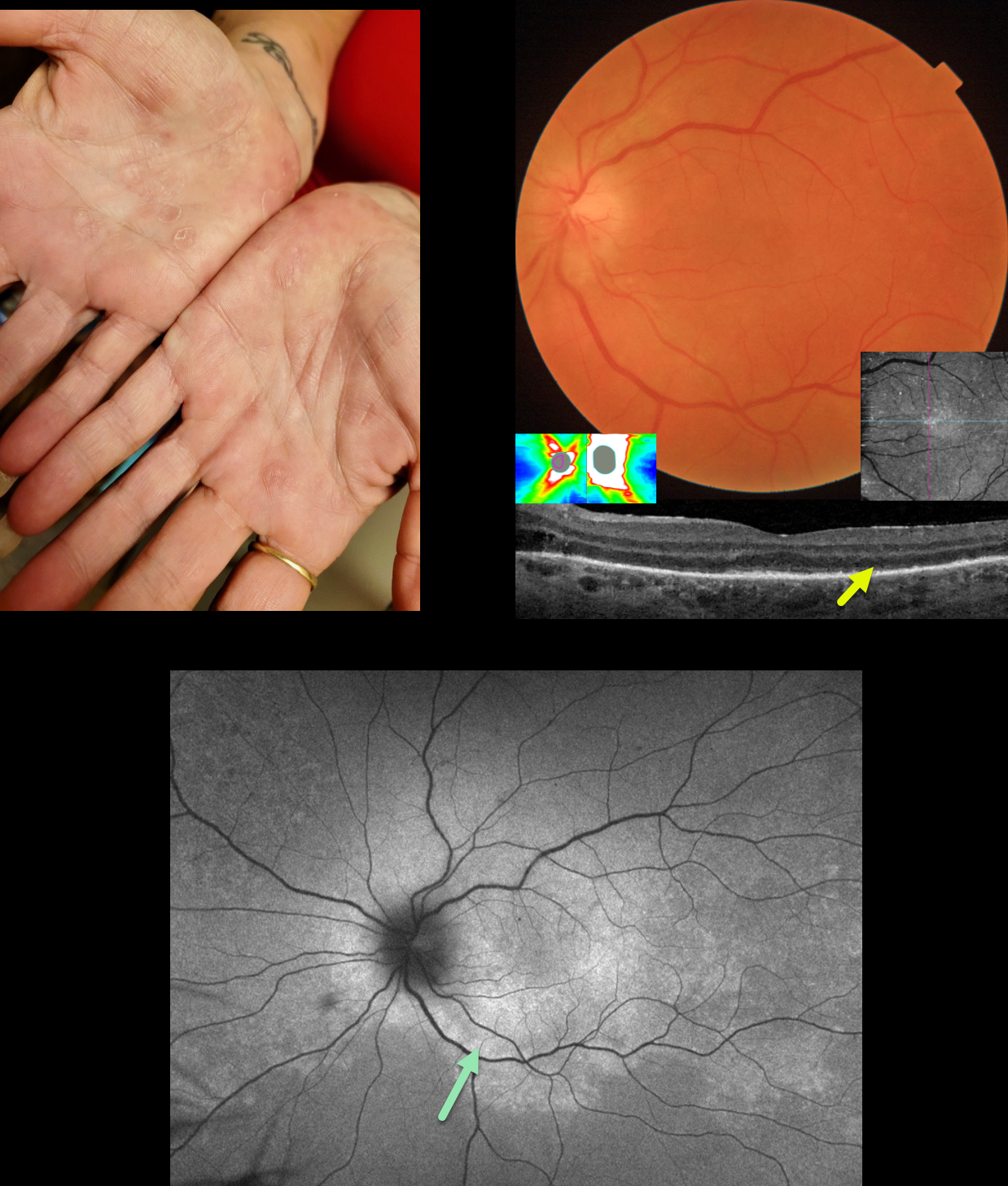
This 51YO female presented with one month of flashes and vision loss in her left eye. Vision was 20/20 in her normal OD and counting fingers OS.
When asked about any rashes, she showed us her palms. What’s your diagnosis??
Triton color imaging of the left posterior pole shows mild optic nerve swelling, with some subtle subretinal whitish pigmentary changes. The nerve swelling was confirmed on OCT. Swept-source OCT shows variable loss of the outer retinal bands and ellipsoid zone with hyperreflective material above the RPE. En face imaging of the outer retina shows this hyperreflective material as numerous bright dots.
Fundus autofluorescence (FAF) shows confluent macular and peripapillary hyper-FAF with more patchy areas of hyper-FAF extending peripherally. Laboratory testing was positive for syphilis and negative for HIV. She was referred to an infectious disease specialist for systemic penicillin treatment and was subsequently lost to follow-up.
This case was submitted by Mattie Adams.
Learning Points:
Ocular manifestations of syphilis vary widely and include virtually all variations of anterior and posterior segment inflammation, including a recently described presentation as the multiple evanescent white dot syndrome phenotype (Russel et al Int Ophthalmol 2020;40:627-738). Hence, the reason it’s been dubbed the great imitator or masquerader. One of the more common presentations is placoid chorioretinitis (Eandi et al, Retina 2012;32:1915-1941), which was evident in our patient’s multimodal imaging.
The rates of syphilis in the US have steadily climbed since 2000, and according to the CDC, its incidence rose nearly 80% between 2018 and 2022 (https://www.cdc.gov/std/statistics/2022/default.htm). Syphilis is prevalent in men having sex with men, among heterosexuals, and in both sexes. These numbers are reflected in how often we are now finding and diagnosing syphilis in our practice. We must always remember that as eye doctors, we are uniquely privileged to not only treat eye problems, but also diagnose systemic diseases. In our case, the painless palmar rash and macular placoid findings were both pathognomonic and led to a rapid diagnosis with referral for treatment.
SYPHILIS
Anjana Mirajkar, Navneet Mehrotra and Manish Nagpal
Originally posted on @retina.rocks May 15, 2024
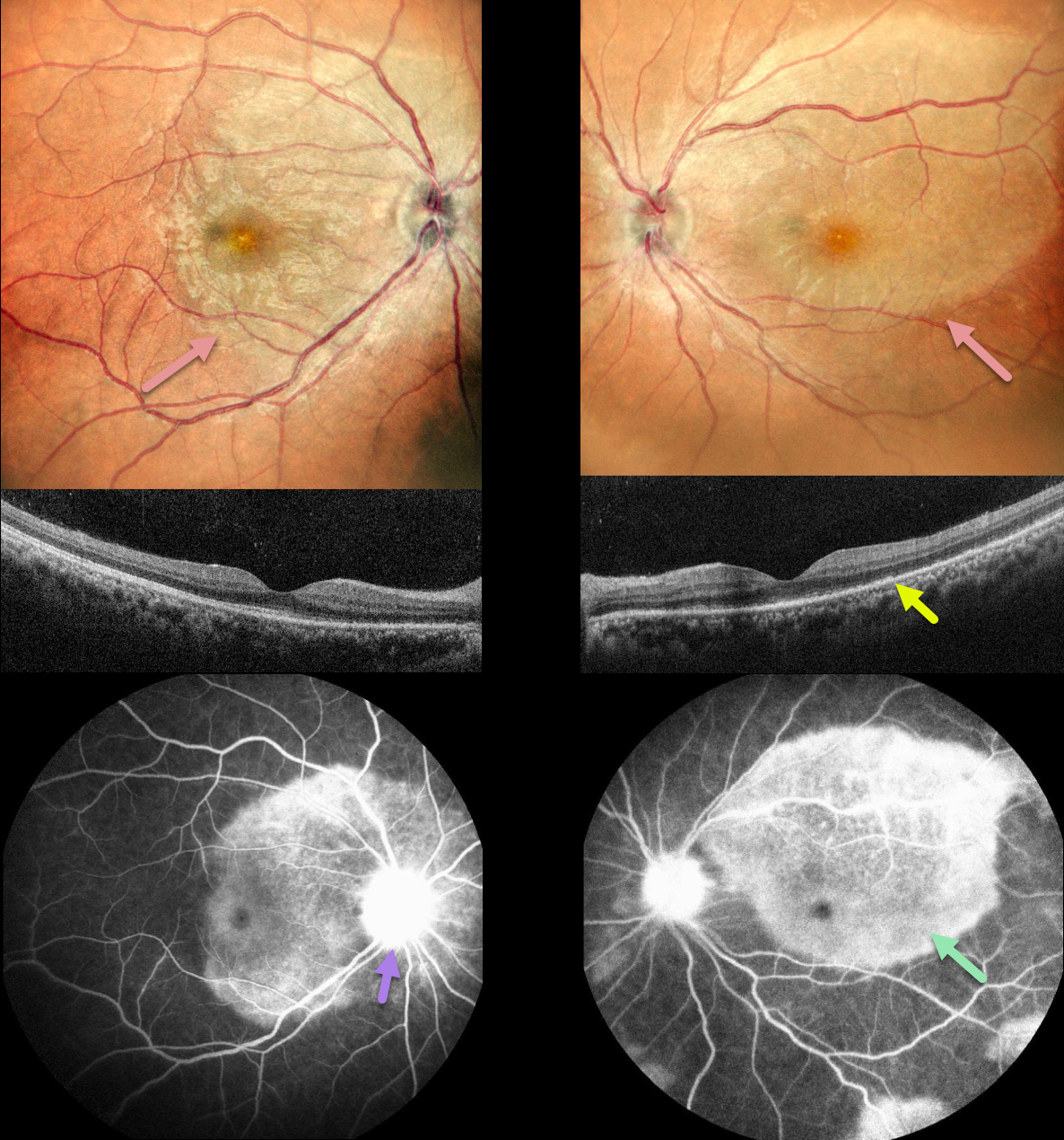
This 36YO female presented with a month of bilateral vision loss, which got worse over the prior 2 days. She also complained of 3 months of headaches and painful urination. Vision was 20/30 OD and 20/120 OS.
Multicolor imaging shows bilateral creamy placoid lesions. OCT scanning shows variable ellipsoid zone loss with small hyperreflective material above. Some vitreous cells are also noted. Fluorescein angiography shows late staining of these placoid lesions with optic nerve leakage.
Bloodwork was positive for syphilis, and she was referred to an infectious disease specialist for further management.
Learning Points:
Ocular manifestations of syphilis vary widely and include virtually all variations of anterior and posterior segment inflammation, including a recently described presentation as the multiple evanescent white dot syndrome phenotype (Russel et al Int Ophthalmol 2020;40:627-738). Hence, the reason it’s been dubbed the great imitator or masquerader.
One of the more common presentations is placoid chorioretinitis (Eandi et al, Retina 2012;32:1915-1941), which was evident in our patient’s multimodal imaging.
SYPHILIS
Originally posted on @retina.rocks March 7, 2024
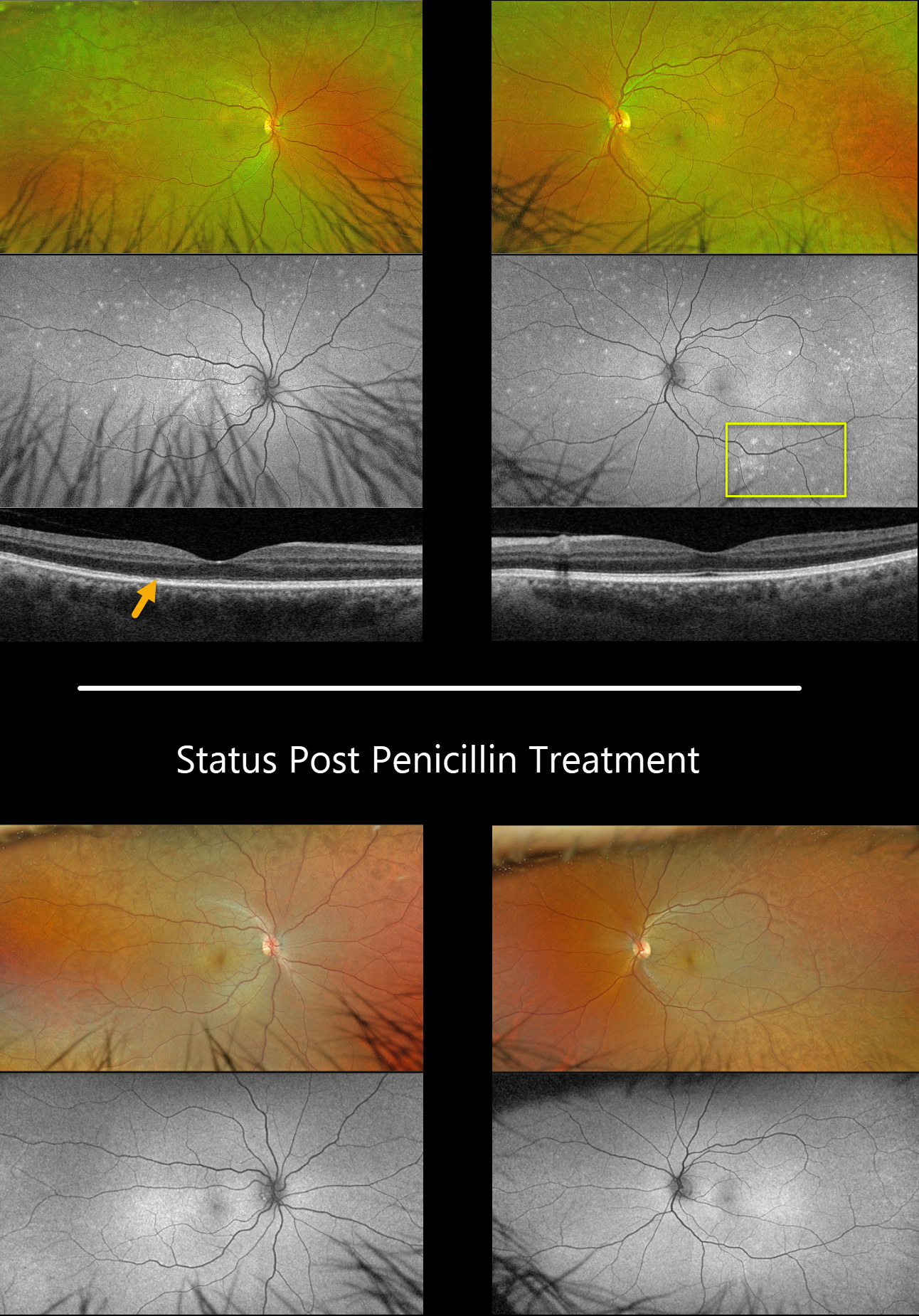
This 57YO male presented with 3 weeks of blurred vision in his right eye. Vision was 20/40 OD and 20/25 OS.
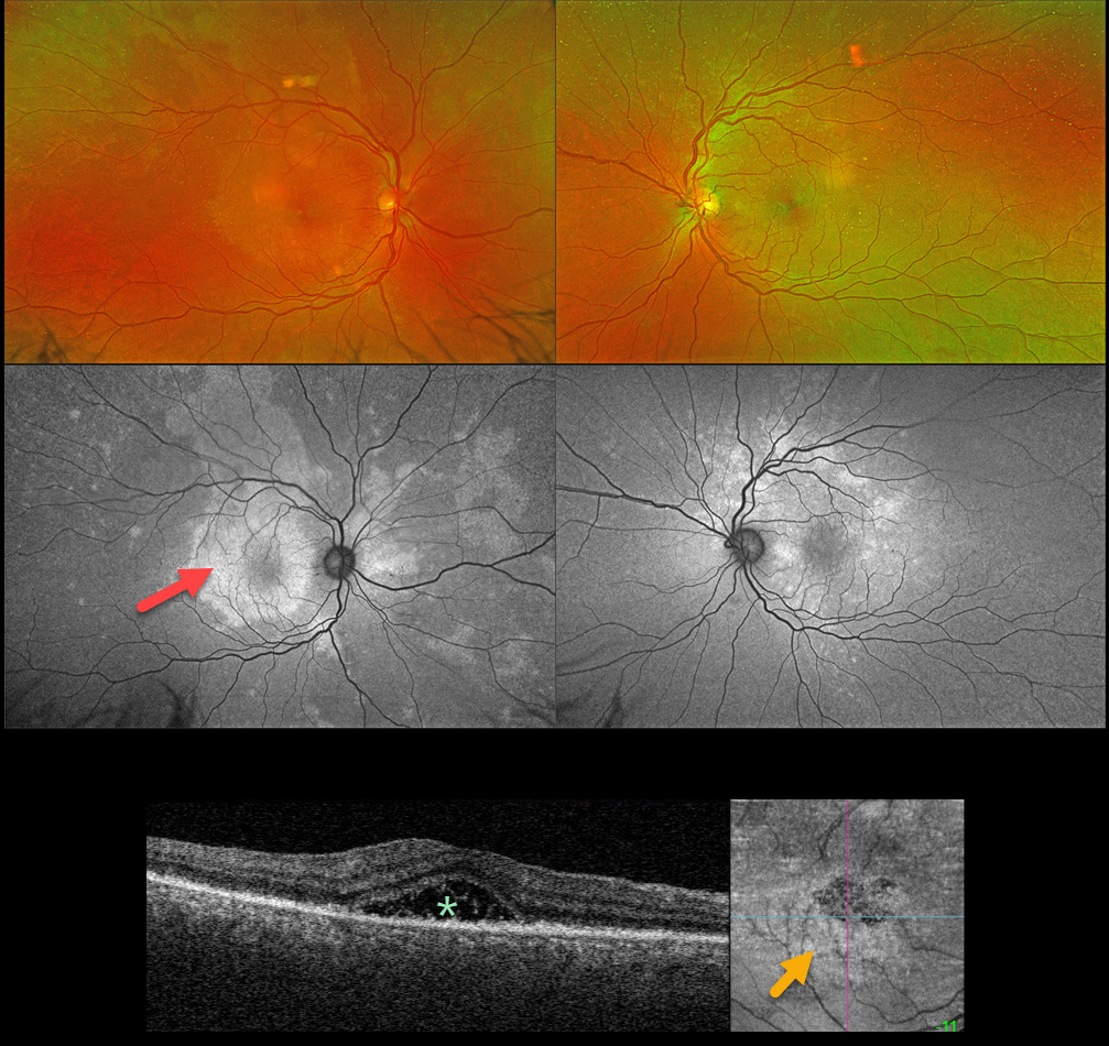
Optos color RG imaging shows bilateral plaques of whitish deep retinal discoloration. Fundus autofluorescence (FAF) shows multifocal MEWDS-like hyper-FAF spots scattered throughout each fundus. Triton swept-source OCT shows an indistinct temporal foveal ellipsoid zone OD and a fairly normal B-scan OS.
RPR testing was positive, and the patient was started on parenteral penicillin treatment. The autofluorescent findings dramatically resolved seven weeks later.
Learning Points:
Ocular manifestations of syphilis vary widely and include virtually all variations of anterior and posterior segment inflammation, including a recently described presentation as the multiple evanescent white dot syndrome phenotype (Russel et al Int Ophthalmol 2020;40:627-738). Hence, the reason it’s been dubbed the great imitator or masquerader.
SYPHILIS
Originally posted on @retina.rocks November 16, 2023
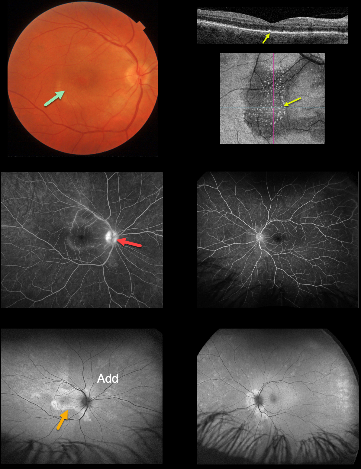
This 52YO male presented with a one-week history of blurred central vision in his right eye. There was no past medical history. Vision was 20/200 OD and 20/25 OS. Anterior segments were normal.
An oval, whitish submacular lesion is noted OD. Fundus autofluorescence (FAF) reveals much more extensive findings than those noted funduscopically. In particular, the placoid lesion seen in the right macula is markedly hyper-FAF and extends into the right superior midperiphery.
Triton swept-source OCT of the right eye shows variable hyperreflective subfoveal fluid, and en face OCT shows fine scattered hyperreflective dots in the outer retina.
Treponema pallidum antibodies were reactive with a high quantitative RPR (1:64). He was started on parenteral penicillin.
Learning Points:
Ocular manifestations of syphilis vary widely and include virtually all variations of anterior and posterior segment inflammation, including a recently described presentation as the multiple evanescent white dot syndrome phenotype (Russel et al Int Ophthalmol 2020;40:627-738). Hence, the reason it’s been dubbed the great imitator or masquerader.
One of the more common presentations is placoid chorioretinitis (Eandi et al, Retina 2012;32:1915-1941), which was evident in our patient’s multimodal imaging.
SYPHILIS
Originally posted on @retina.rocks October 14, 2022
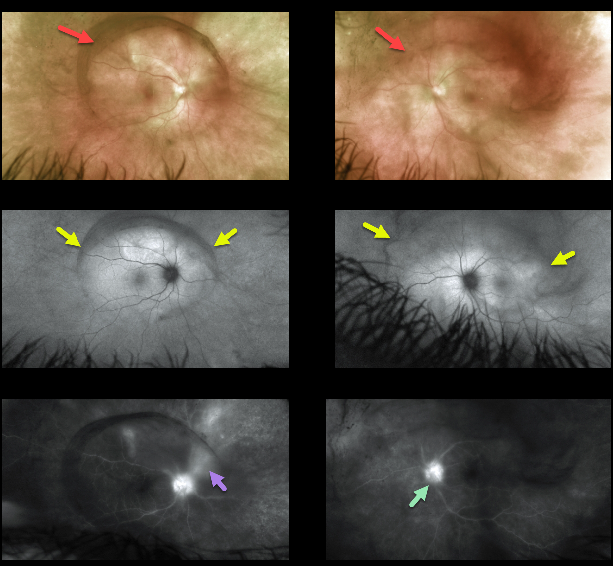
This 49YOM presented with a one-week history of blurred central vision in his right eye. There was no past medical history.
Vision was 20/400 OD and 20/25 OS. Anterior segments were normal. An oval, whitish submacular lesion was noted, and the optic nerve also appeared mildly swollen in the right eye.
OCT scanning of the right macula shows numerous, needle-like projections extending from the RPE into the outer retina. En face imaging best shows the hyperreflective outer retinal opacities.
Fluorescein angiography shows staining of the right nerve, mild peripapillary subretinal staining OU, and macular staining OD.
Fundus autofluorescence (FAF) shows much more extensive findings than those noted clinically, with peripapillary hyper-FAF OU, macular hyper-FAF OD corresponding to the placoid lesion, and peripheral hyperreflective dots bilaterally.
Treponema pallidum antibodies were reactive, with a high quantitative RPR (1:32). He was started on parenteral penicillin.
Learning Points:
Ocular manifestations of syphilis vary widely and include virtually all variations of anterior and posterior segment inflammation, including a recently described presentation as the multiple evanescent white dot syndrome phenotype (Russel et al Int Ophthalmol 2020;40:627-738). Hence, the reason it’s been dubbed the great imitator or masquerader.
One of the more common presentations is placoid chorioretinitis (Eandi et al., Retina 2012;32:1915-1941), which was evident on FAF imaging in our patient.
SYPHILIS
Originally posted on @retina.rocks June 2, 2022
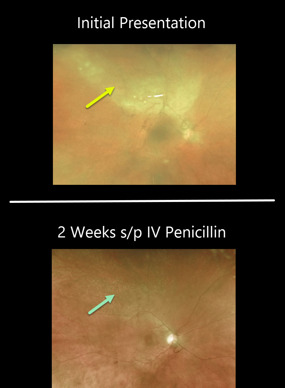
This 49YO female presented with a 1-month history of bilateral vision loss and eye pain. Vision was 20/200 OU. There was a mild-moderate, bilateral nongranulomatous anterior uveitis.
Vitreous debris was centered over each posterior pole, which was grossly normal.
Fundus autofluorescence (FAF) shows an oval region of placoid hyper-FAF in each posterior pole. Fluorescein angiography shows bilateral nerve leakage and vasculitis.
RPR and treponema pallidum antibody testing were positive, and she was immediately started on intravenous penicillin.
Learning Points:
Ocular manifestations of syphilis vary widely and include virtually all variations of anterior and posterior segment inflammation, including a recently described presentation as the multiple evanescent white dot syndrome phenotype (see Russel et al Int Ophthalmol 2020;40:627-738). Hence, the reason it’s been dubbed the great imitator or masquerader.
One of the more common presentations is placoid chorioretinitis, which was evident on FAF imaging in our patient.
Patients also need to be evaluated for HIV since syphilis and HIV infections often coexist. Fortunately, our patient was HIV negative.
SYPHILIS
Originally posted on @retina.rocks December 2, 2020
This 46YO female presented with worsening pain and vision loss in her left eye despite being treated two days earlier by the emergency room for conjunctivitis. In our office, vision was counting fingers OS with a granulomatous panuveitis.
There is a creamy-white area of confluent retinitis superiorly. The image is hazy due to vitritis.
Stat bloodwork revealed a positive RPR and FTA-ABS. She denied any other symptoms or known history of syphilis.
Patients also need to be evaluated for HIV since syphilis and HIV infections often coexist. Fortunately, our patient was HIV negative.
After 2 weeks of parenteral penicillin, the panuveitis had improved markedly, and the retinitis had completely resolved.
Learning Points:
Ocular manifestations of syphilis vary widely and include virtually all variations of anterior and posterior segment inflammation, including a recently described presentation as the multiple evanescent white dot syndrome phenotype (see Russel et al Int Ophthalmol 2020;40:627-738).
Hence, the reason it’s been dubbed the great imitator or masquerader. One of the more common presentations is placoid chorioretinitis.
SYPHILIS
Originally posted on @retina.rocks May 27, 2020
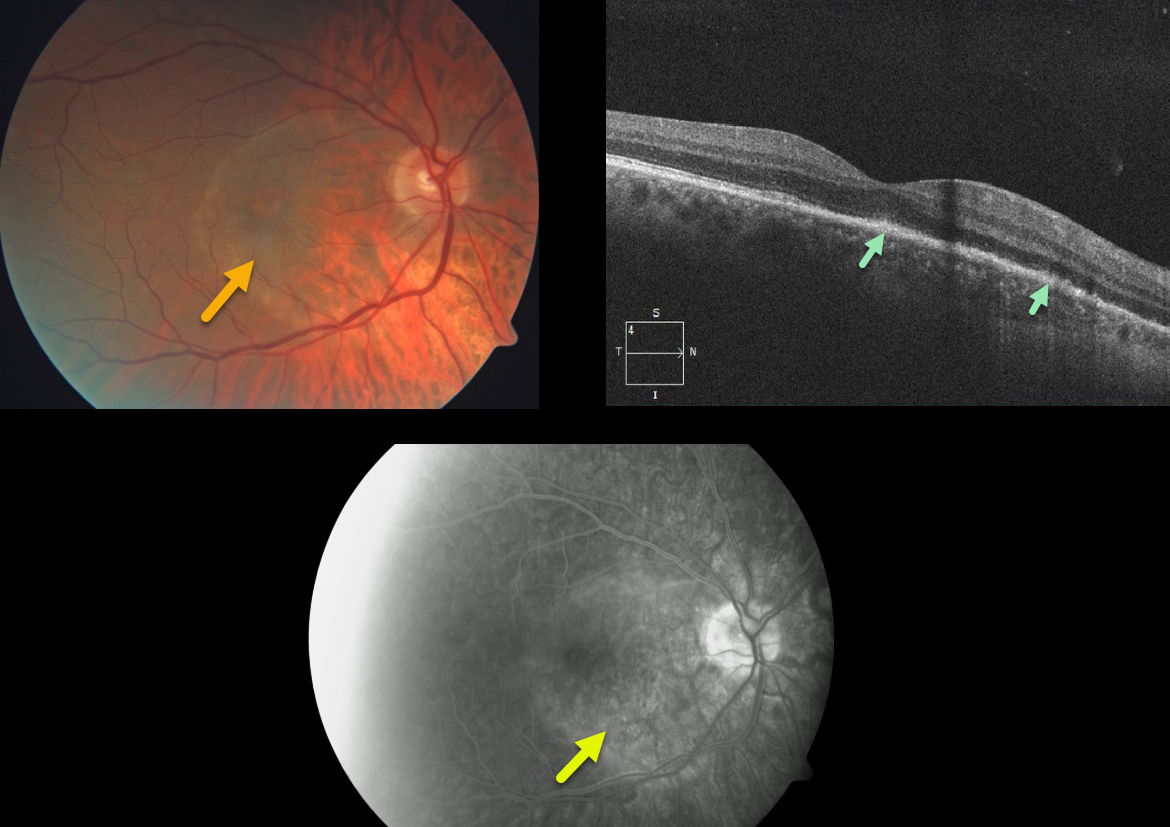
This patient has classic findings for acute placoid syphilis in the right eye.
The fundus photo demonstrates an orange-yellow subretinal lesion. OCT shows attenuation of the outer retinal layers (including the ellipsoid zone) and a thickening of the RPE.
Angiographically, this inflammatory lesion blocks early and stains late. The patient was referred to the infectious disease specialist for parenteral antibiotics.
Learning Points:
In internal medicine, syphilis has been called “the great imitator” since it can masquerade as so many systemic disorders. The ocular manifestations are legion as well.
Originally described by Gass (Ophthalmology 1990;97;1288-1297), placoid syphilis affects the outer retina, RPE, and choriocapillaris and clinically manifests as an orange-yellow subretinal lesion.
Placoid syphilis has a fairly good visual prognosis with treatment. Patients also need to be evaluated for HIV since syphilis and HIV infections often coexist.


