

Systemic Lupus Erythematosus (SLE)
<< Back to Cases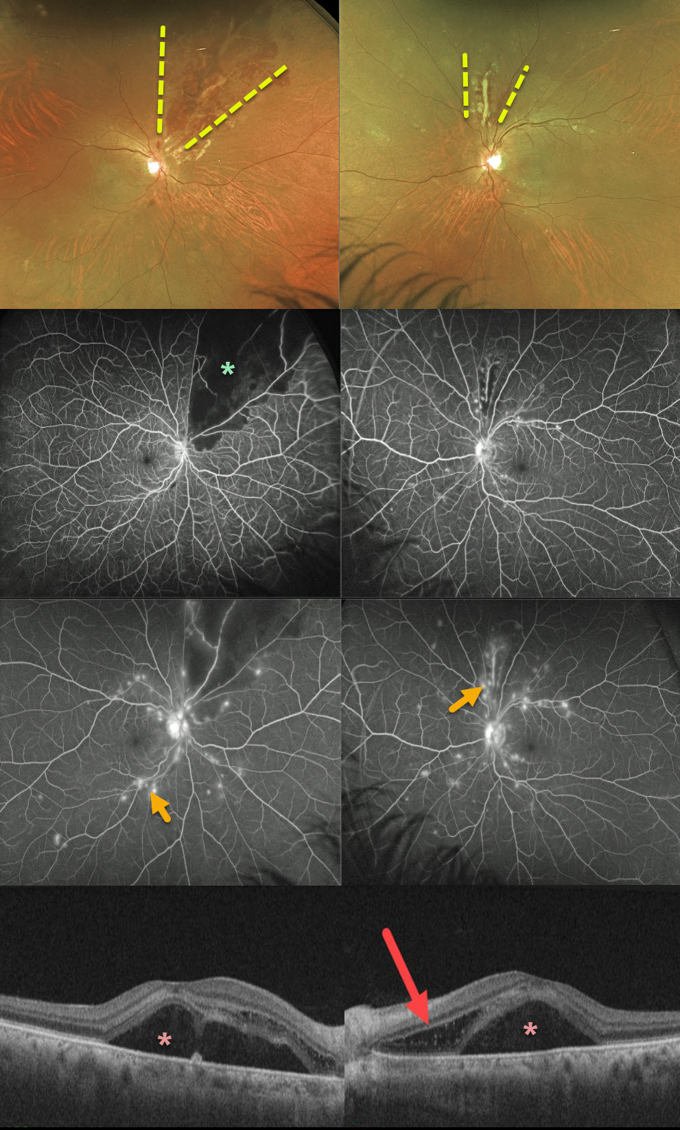
SYSTEMIC LUPUS ERYTHEMATOSUS (SLE)
Originally posted on @retina.rocks December 29, 2020
This 52YO female presented with severe bilateral vision loss (20/400 OD, counting fingers OS) following a several-week gastrointestinal illness with 60-pound weight loss.
She has bilateral areas of hemorrhagic retinal vasculitis, with areas of ischemia and leakage seen on fluorescein angiography. OCT shows macular subretinal fluid and a bacillary detachment in the left macula.
She had been hospitalized recently for hypertension and kidney failure and was diagnosed with a previously asymptomatic stroke. Extensive blood work was negative, and she denied symptoms of Behcet’s.
We suspected an underlying rheumatologic cause, and she was subsequently diagnosed with severe systemic lupus erythematosus (SLE) vasculitis.
She was started on high-dose oral prednisone followed by Cytoxan and hydroxychloroquine. Her ocular findings rapidly improved, and at her last examination 4 months later, vision had improved to 20/40 OD and 20/100 OS.
Learning Points:
SLE-associated retinopathy more typically appears as hypertensive retinopathy (bilateral nerve fiber layer infarcts and retinal hemorrhages), often in patients with cerebral vasculitis. Purtscher-like retinopathy can also be seen.


