

Talc Retinopathy
<< Back to Cases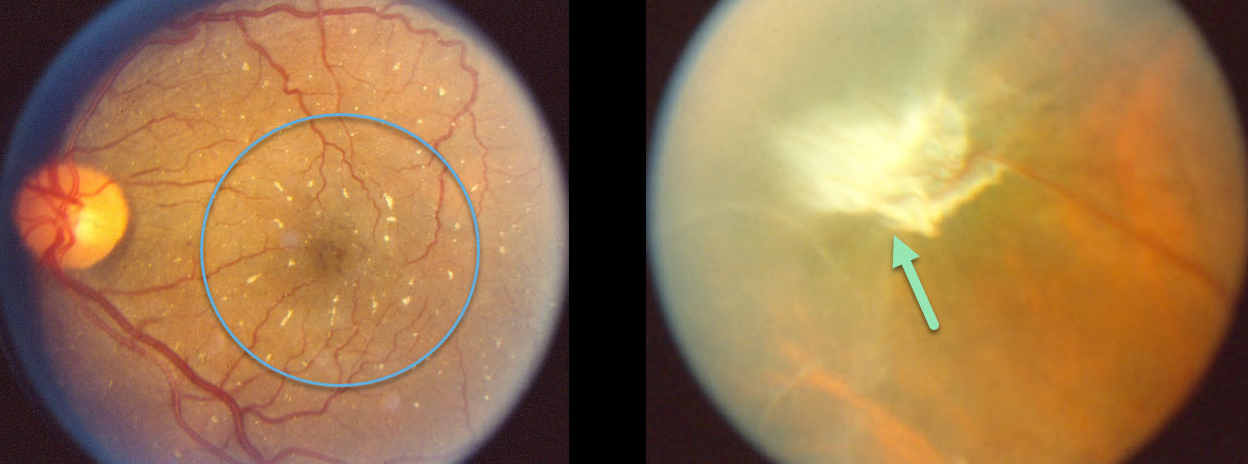
TALC RETINOPATHY
Originally posted on @retina.rocks June 18, 2020
Glistening crystals in the posterior pole are prevalent in this intravenous drug user with talc retinopathy.
There is also a large fibrosed area of neovascularization along the border between the perfused retina to the right and the ischemic retina to the left, a neovascularization pattern somewhat similar to that seen in Eale’s disease.
Learning Points:
Intravenous drug users may prepare a suspension of crushed tablets that include inadequately filtered insoluble fillers, including talc and cornstarch.
Talc emboli lodge in the pulmonary circulation and eventually enter the arterial circulation through pulmonary collaterals. These talc emboli can eventually enter the retinal arterial circulation, leading to retinal ischemia and neovascularization.


