

Tay-Sachs
<< Back to Cases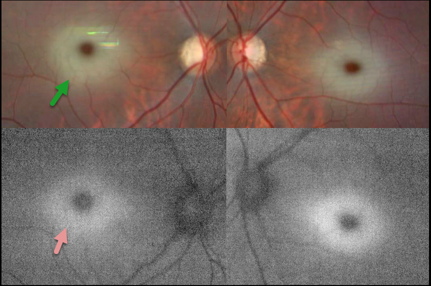
TAY-SACHS DISEASE
The European VitreoRetina Society (EVRS) and David Aggarwal
Originally posted on @retina.rocks July 18, 2025
This 15-month-old girl was brought to a multi-specialty hospital by her parents with complaints of fever, seizures, and progressive weakness in her limbs for two days. She was admitted to the pediatric intensive care unit, and symptomatic treatment was started. She was the firstborn of a third-degree consanguineous marriage.
Fundus imaging is blurred due to constant eye movement, but shows a symmetric central macular whitish appearance surrounding an orange- brown foveal center. On fundus autofluorescence (FAF), this white material is hyper-FAF. Whole genome sequencing revealed GM2-gangliosidosis, AB variant (Tay-Sachs disease).
Learning Points:
A cherry-red spot refers to a reddish-brown appearance of the normal macular center, which is surrounded by opaque white parafoveal retina. This is usually caused by an acute central retinal artery occlusion (CRAO), but it is also a common feature of sphingolipidoses, a group of lysosomal metabolic disorders that cause abnormal lipid accumulation in retinal ganglion cells. As the foveal pit lacks the ganglion cell layer, it retains its normal transparency, giving a cherry-red appearance.
In Tay-Sachs disease, GM2 trihexosylceramide accumulates due to a deficiency of the A component of hexosaminidase. This material is hyper-FAF (Abalem et al, JAMA Ophthalmology 2014;132:876), unlike a CRAO, which appears hypo-FAF due to blockage of the underlying RPE.


