

04 Retinal Vascular Disorders: Diabetes, RAO and RVO
<< Back to Cases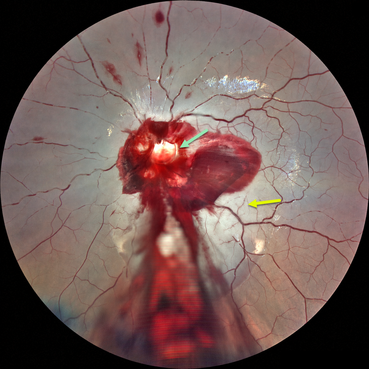
ACUTE OPTIC NERVE AVULSION WITH SECONDARY CRAO
Ayushi Gupta and Vishal Agrawal
Originally posted on @retina.rocks September 8, 2026
This 6YO boy developed sudden and complete loss of vision in his left eye immediately following blunt trauma. He was riding a bicycle as a pillion passenger when the bike stopped suddenly, throwing his face against the handlebar. Vision was no light perception. Anterior segments were normal.
Fundus photography shows preretinal blood surrounding the nerve, extending through the macula and into the inferior periphery. A pale-yellow cavity is noted where the nerve head should be. A fresh central retinal artery occlusion (CRAO) with a stagnant blood column is noted, along with some scattered inner retinal hemorrhages. There were no other physical or neurologic injuries.
Learning Points:
Traumatic optic nerve head avulsion is a rare and visually devastating traumatic event in which the optic nerve is forcibly separated from the globe at its junction with the sclera, typically resulting in immediate, severe, and irreversible vision loss. The mechanism involves sudden forceful rotation or anterior displacement of the globe following blunt periorbital trauma, including projectiles, falls, or finger-poke injuries, whereby the globe moves anteriorly or rotationally while the optic nerve remains relatively fixed, causing violent separation. Fundoscopic examination may reveal a partial or complete tear of the optic nerve head from the sclera (Foster et al, Arch Ophthalmol 1997;115:623-630). Avulsion can be partial or complete, and may be associated with a CRAO, as was noted in our patient.
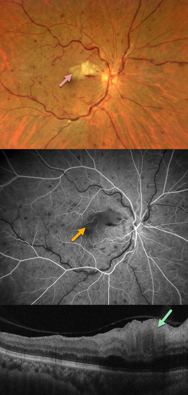
CRVO WITH SECONDARY CILIORETINAL ARTERY OCCLUSION
Francis Ezra Laxamana, Bianca Teresa Suarez, Jesus Jacinto Bautista and Carlo Ladores.
Originally posted on @retina.rocks September 7, 2026
This 58YO female with a history of hypertension and diabetes presented with sudden painless vision loss in her right eye. Vision was 20/25 OD and 20/20 in her normal OS.
Fundus photography shows a non-ischemic central retinal vein occlusion (CRVO) with dilated tortuous veins, mild scattered retinal hemorrhages, and a superonasal macular branch retinal artery occlusion (BRAO). The BRAO shows inner retinal hyperreflectivity on OCT. Fluorescein angiography shows good perfusion throughout the distribution of the CRVO, with a filling defect from the BRAO.
Learning Points:
Some patients with CRVO will develop a secondary non-embolic cilioretinal artery occlusion caused by either compression from optic nerve edema or from a sudden rise in intraluminal retinal capillary bed pressure (Hayreh et al, Retina 2008;28;581-594).
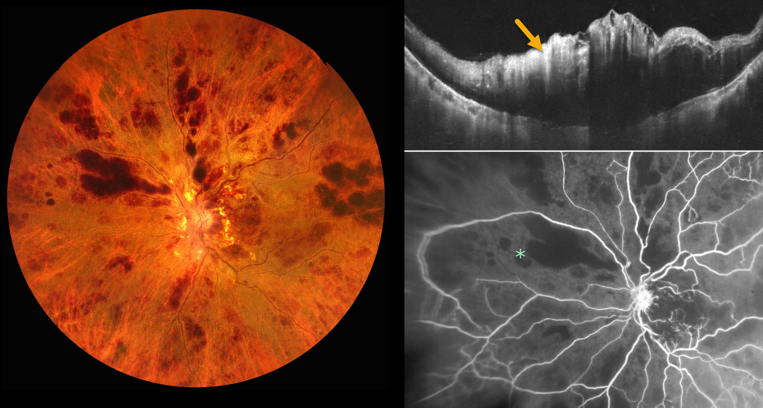
ISCHEMIC CRVO
Ayushi Gupta and Vishal Agrawal
Originally posted on @retina.rocks August 18, 2026
This 78YO male presented with 1 week of sudden, painless decreased vision in his left eye. Vision was 20/20 in his normal OD and light perception OS. Despite iris neovascularization, intraocular pressure was normal.
Color photography shows a hemorrhagic central retinal vein occlusion (CRVO) with variably confluent inner retinal hemorrhages and scattered nerve fiber layer infarcts. OCT scanning shows marked macular thickening with a hyperreflective inner retina. Fluorescein angiography shows profound diffuse retinal capillary loss. An intravitreal anti-VEGF injection was given. One week later, the rubeosis completely regressed, and full panretinal photocoagulation was performed.
Learning Points:
Ischemic CRVO, which accounts for 20% of eyes, is characterized by severe visual loss (typically worse than 20/200), a relative afferent pupillary defect, extensive retinal hemorrhages, cotton-wool spots, marked venous dilation and tortuosity, optic disc edema, and large areas of angiographic nonperfusion of the retinal capillaries.
Untreated iris neovascularization usually leads to neovascular glaucoma (NVG). Treatment involves an immediate anti-VEGF injection to allow for more rapid regression of neovascularization, followed by full panretinal photocoagulation. NVG is much less common in the modern anti-VEGF era, since the majority of these eyes also have macular edema that requires ongoing anti-VEGF therapy.
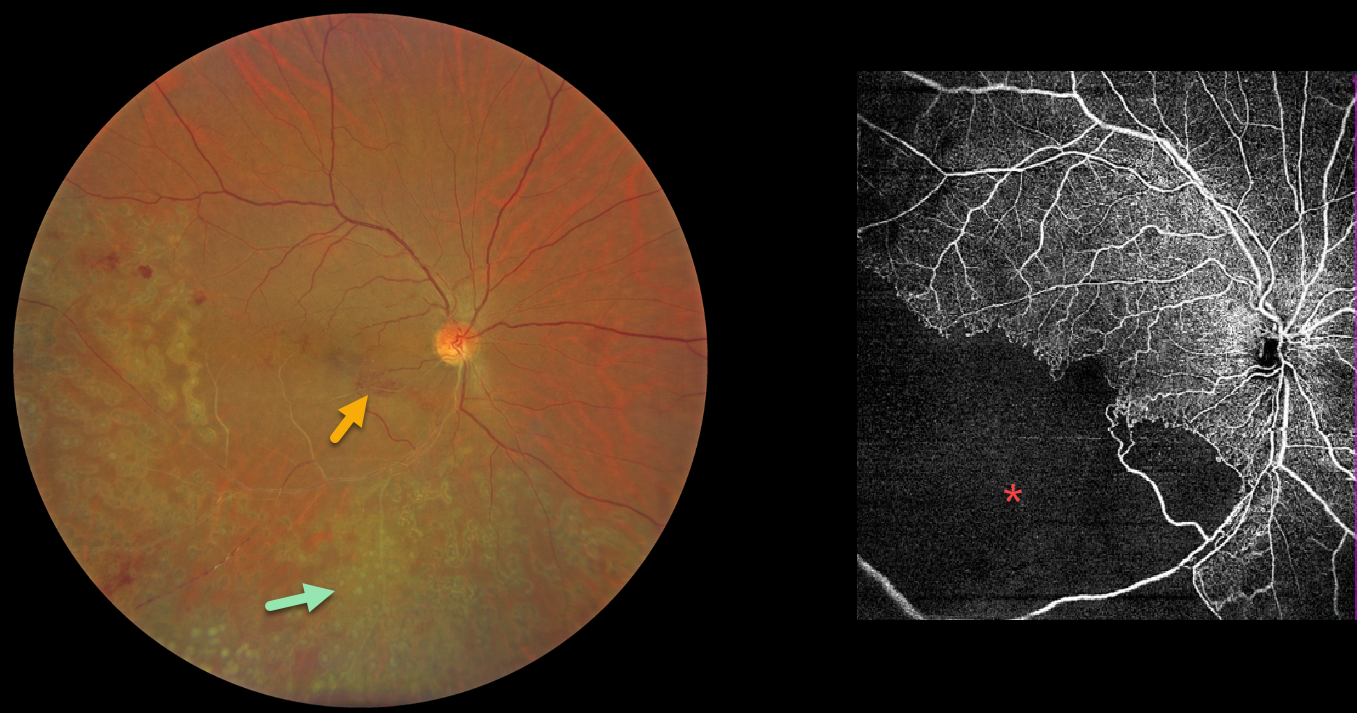
BRVO WITH RETINAL NEOVASCULARIZATION
Abhishek Karra and Ravindra Karra
Originally posted on @retina.rocks August 10, 2026
This 42YO male presented with a 3-month history of decreased vision in his right eye. He had received laser treatment in this eye elsewhere. Vision was 20/60 OD.
Color photography shows an old ischemic inferotemporal branch retinal vein occlusion (BRVO) with heavy scatter laser throughout its distribution. Collateral vessels are noted in the inferonasal macula. OCT angiography shows profound capillary loss. There was no macular edema, and observation was recommended.
Learning Points:
In 1986, the Branch Vein Occlusion Study Group reported that scatter laser decreased the risk of vitreous hemorrhage from retinal neovascularization in BRVO and suggested that laser should be applied once neovascularization develops (Arch Ophthalmol 1986;104:34-41). Thirty-five years later, scatter laser remains the gold standard, although some may prefer to stabilize the neovascularization with anti-VEGF therapy before laser.
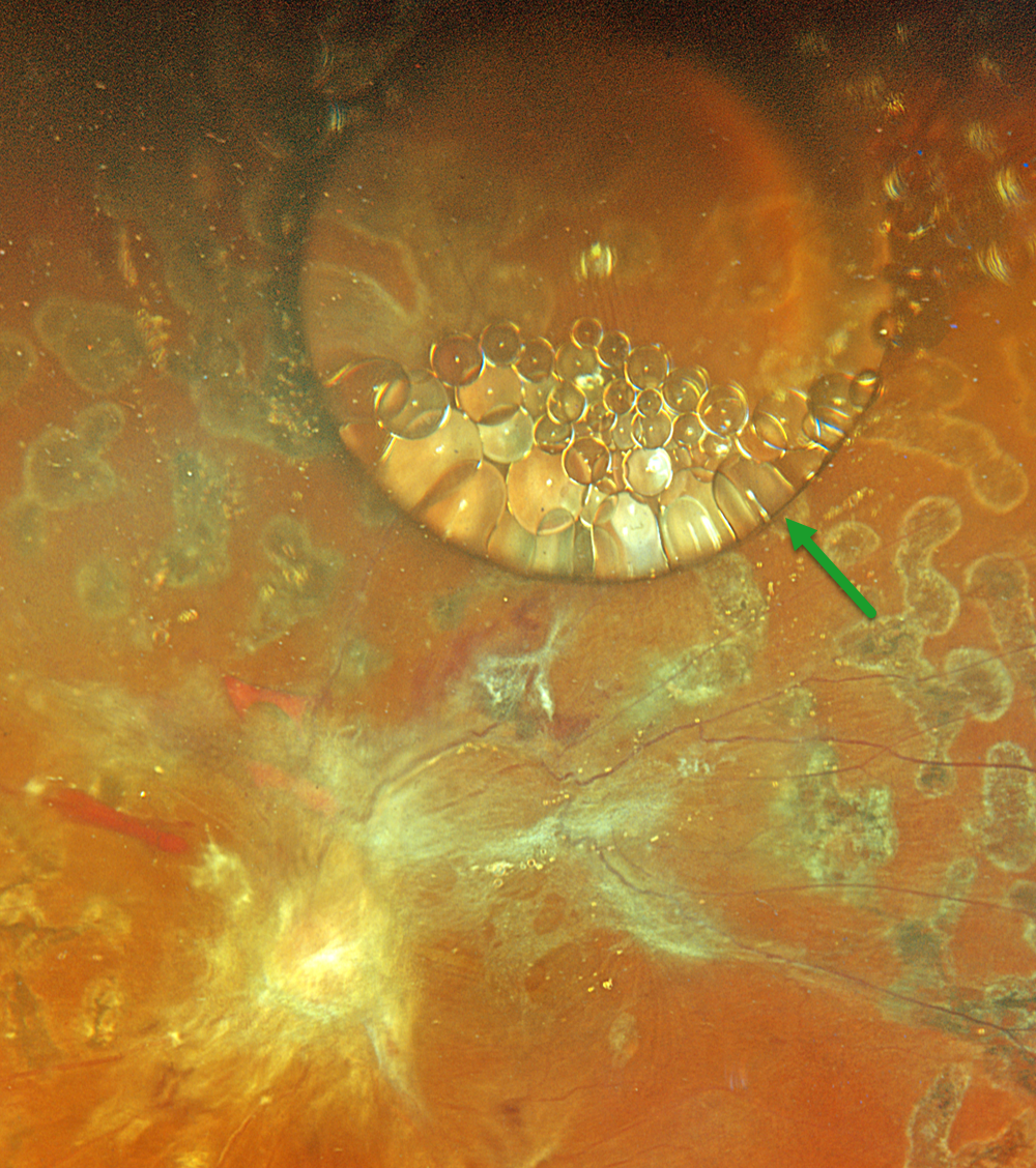
SILICONE FISH EGGS
Originally posted on @retina.rocks July 21, 2026
This 43YO male underwent a vitrectomy elsewhere for a diabetic traction retinal detachment about 5 months earlier. Vision was 20/400.
Optos color RGB imaging shows a large, round silicone oil droplet in the superior midperiphery. This bubble acts like a convex lens, magnifying the details of the underlying retina. Suspended within and along the edges of this droplet are numerous fish egg droplets. Residual fibrotic membranes extend from the nerve into the macula and along the superotemporal arcade. Repeat vitrectomy was scheduled.
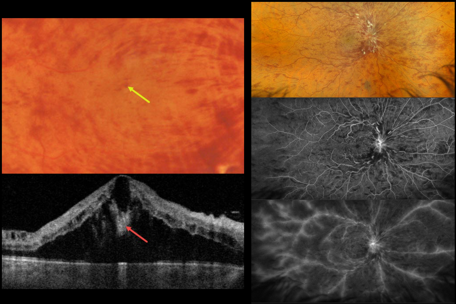
CRVO WITH CENTRAL BOUQUET HEMORRHAGE
Originally posted on @retina.rocks June 26, 2026
This healthy 52YO male presented with recent severe vision loss in his right eye. Vision was 20/400 OD and 20/20 in his normal OS.
Color photography and Optos RGB imaging show a central retinal vein occlusion (CRVO) with marked retinal hemorrhages and macular edema. Inner blood-filled retinal cysts are noted on funduscopy and swept-source OCT. On fluorescein angiography, the occlusion is fairly well perfused with late leakage from the nerve, macula, and retinal veins. Monthly anti-VEGF therapy was started.
Learning Points:
Cicinelli et al recently reported that central bouquet hemorrhages in retinal vein occlusion are associated with poor visual outcomes due to localized effects of elevated venous pressure and macular ischemia (IOVS 2025;66(12):62).
Prior to the introduction of intravitreal injections in the early 2000s, there were essentially no treatments for CRVO-related macular edema. Anti-VEGF injections are now generally the initial treatment of choice, with about 50-60% of eyes gaining at least 3 lines of vision. Intravitreal steroids, usually Ozurdex, are more often used as secondary treatment. Macular grid laser photocoagulation is not performed because it has been shown to confer no visual benefit in the Central Vein Occlusion Study Group (Ophthalmology 1995;102:1425-1433).
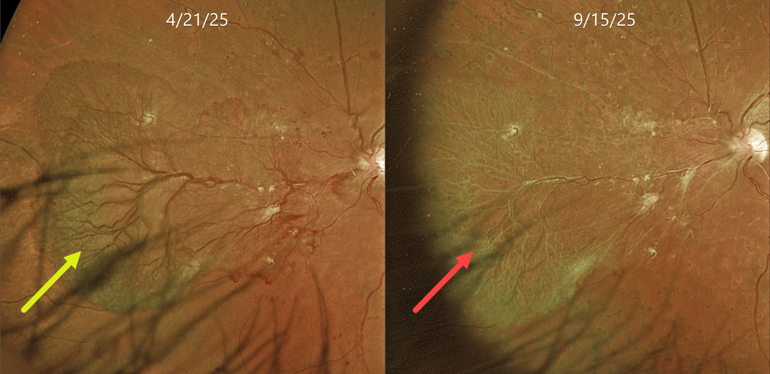
PROLIFERATIVE DIABETIC RETINOPATHY
Originally posted on @retina.rocks June 24, 2026
This 53YO female with a known history of type 2 diabetes was referred for asymptomatic proliferative disease (PDR) in her left eye. Vision was 20/50.
Optos color RG imaging shows a large sea fan-shaped area of succulent and flat neovascularization extending from the optic nerve into the nasal midperiphery. Panretinal photocoagulation (PRP) was applied in three sessions over several months.
Learning Points:
An attached vitreous provides the scaffold for neovascularization to grow onto and spread. Retinal neovascularization grows from the inner retinal surface and proliferates along the outer posterior hyaloid face (Vaz-Pereira et al, Retina 2017;37:1287-1296). Following PRP or anti-VEGF therapy for PDR, retinal neovascularization ‘involutes’, often morphing from a fine capillary network into larger vessels with fibrosis. In the process, the contracting neovascularization and vitreous can cause increasing traction. Rapid tissue contraction with traction retinal detachment is an uncommon but well-described phenomenon known as the ‘crunch’ syndrome (Survey of Ophthalmology 2021;66:926-932).
We were quite concerned about the possibility of a crunch in our patient and discussed it prior to starting treatment. Five months following her initial exam, the neovascularization completely fibrosed without traction. Vision was 20/40. We are continuing to follow her closely.
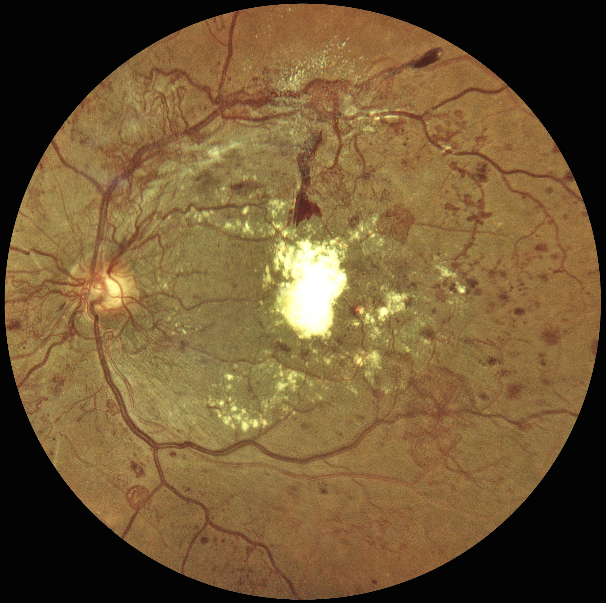
VERY SEVERE PDR AND DME FOLLOWING PATIENT LOST TO FU
Sowparnika Basavaraju
Originally posted on @retina.rocks June 16, 2026
This 48YO male with a history of poorly controlled type 2 DM was seen 4 years earlier for an eye examination elsewhere but was immediately lost to follow-up. He presented to us with hand-motion vision in his left eye.
Color fundus photography shows florid disc and peripheral neovascularization variably coating the posterior pole. A large plaque of subfoveal lipid is also noted.
Learning Points:
These findings would have been caught earlier if he had not been lost to follow-up, which, unfortunately, is quite common among diabetic patients (Obeid et al, Ophthalmology 2018;125:1386-1392; Gao et al, Ophthalmology Retina 2019;3:230-236; Green et al, AJO 2020;216:18-27; Ophthalmology Retina 2025;9:1167-1174, etc, etc).
At this stage, our patient will not recover central vision due to the subfoveal lesion, and our hope is to prevent further vision loss. Anti-VEGF therapy, panretinal photocoagulation, and macular photocoagulation were suggested. He is at significant risk for traction retinal detachment as the neovascularization involutes (crunch), and we will therefore follow him very closely.

CRAO FROM ATRIAL MYXOMA
Ayushi Gupta and Vishal Agrawal
Originally posted on @retina.rocks June 15, 2026
This previously healthy 29YO female presented with 2 days of sudden vision loss in her left eye. Vision was 20/20 in her normal OD and light perception OS.
Color photography shows an acute central retinal artery occlusion (CRAO) with diffuse retinal opacification and a prominent foveal cherry-red spot. The retinal arterioles are markedly attenuated with segmentation of the arterial and venous blood columns (box-carring). OCT shows inner retinal thickening and opacification except in the foveal center. Cardiac imaging revealed a left atrial myxoma, and she underwent immediate surgical excision.
Learning Points:
Atrial myxoma, the most common primary cardiac tumor, is typically attached to the interatrial septum at the fossa ovalis and can shed tumor fragments, thrombi, or surface debris into the systemic circulation (Griborio-Guzman et al, Heart 2022;108:827-833). Embolism occurs in 30-40% of patients, and the cerebral and retinal arteries are the most affected vascular beds. The vast majority of CRAOs occur in a more elderly population from carotid or valvular disease. Atrial myxomas are a rare but known cause, especially in younger patients.
Half of patients have an elevated erythrocyte sedimentation rate (ESR) due to tumor-produced interleukin-6 (Acebo et al, Chest 2003;123:1379-1385). These cytokines drive the systemic inflammatory response that produces constitutional symptoms (fever, fatigue, weight loss, arthralgias, myalgias) and laboratory abnormalities (elevated ESR/CRP, anemia, leukocytosis, elevated globulins). Elevated inflammatory markers can be a diagnostic pitfall, as they frequently lead to misdiagnosis of endocarditis, rheumatic fever, vasculitis, collagen vascular disease, or even polymyalgia rheumatica.
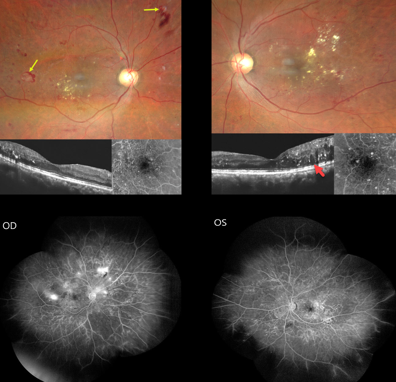
ETHAMBUTOL TOXICITY WITH CONCOMITANT DIABETIC RETINOPATHY
Ayushi Gupta and Vishal Agrawal
Originally posted on @retina.rocks May 4, 2026
This 54YO male presented for a second opinion with several weeks of progressive bilateral painless vision loss. He gave a history of active pulmonary tuberculosis, diagnosed about 6 months earlier, which was being treated with antitubercular therapy (ATT) including ethambutol. There was also a 5-year history of well-controlled type 2 diabetes. Vision was 20/200 OU.
Fundus photography shows bilateral non-central diabetic macular edema (DME) with mostly temporal lipid. The central macula looks fairly normal bilaterally on OCT, and fundus fluorescein angiography (FFA) shows relatively preserved foveal avascular zones. Retinal neovascularization (NV) with some associated preretinal blood is noted OD. Widefield FFA confirms scattered retinal NV OD with moderate bilateral capillary nonperfusion. Disc OCT showed a normal retinal nerve fiber layer, and 30-2 visual fields showed severe generalized depression (not shown).
Learning Points:
Our patient was previously seen by other specialists who felt that diabetic retinopathy was the sole reason for his complaints and findings, despite the lack of central foveal fluid or ischemia. Our patient reminds us of 2 dictums in medicine: 1) we try to explain all findings by a single disease, and 2) that a patient can have as many diseases as they so desire. In our patient’s case, we felt that ethambutol better explained his clinical picture.
Ethambutol is one of the agents used as part of ATT, a multi-drug regimen for treating active tuberculosis. About 1-3% of patients can develop drug-induced optic neuropathy, most commonly following 2 months of therapy but rarely developing within days of starting treatment. Symptoms include decreased visual acuity, scotoma, color blindness, and visual defects in one or both eyes.
Although the FDA recommends baseline visual acuity and color vision testing, followed by monthly color discrimination testing during therapy, there are no studies demonstrating any benefit of such screening. Ethambutol should be discontinued if toxicity is suspected. Although vision loss is often thought to be permanent, a recent systematic review showed significant improvement in vision, color vision, and visual field loss with discontinuation of the drug (Sabhapandit et al., Indian J Ophthalmol 2023;71:729-735).
We contacted our patient’s pulmonologist, who stoppe therapy immediately. Anti-VEGF therapy followed by panretinal photocoagulation was recommended.
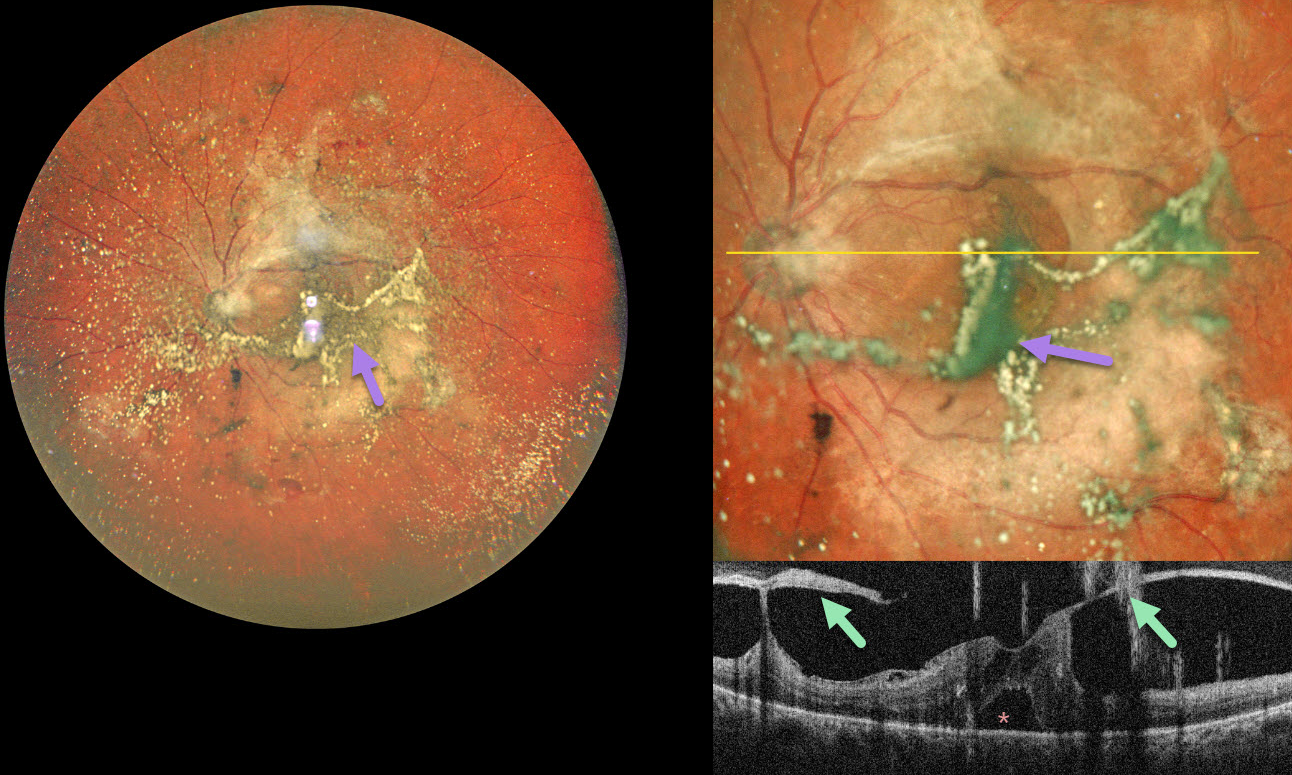
PROLIFERATIVE DIABETIC RETINOPATHY WITH ASTEROID HYALOSIS
The European VitreoRetinal Society (EVRS), Akansha Sharma, Manish Nagpal, and Vaidehi Sathaye
Originally posted on @retina.rocks April 27, 2026
This 55YO diabetic male presented with 2 months of bilateral vision loss. Vision was 20/80 OD and 20/120 OS.
Pseudocolor SLO ultrawidefield imaging of his left eye shows moderate asteroid hyalosis, with larger condensed stands of material obscuring the underlying macula. Spectral domain OCT scanning shows complex variable vitreous traction on the disc and macula with a small foveal detachment. The asteroid particles appear as hyperreflective vertical lines resembling falling rain.
Learning Points:
Examining the fundus in a patient with dense asteroid, as in this diabetic patient, can be extremely challenging. Fluorescein angiography and OCT are great modalities because they can visualize the retina when the clinical exam can’t. In our patient’s case, the OCT revealed foveal traction, indicating the need for vitrectomy surgery.
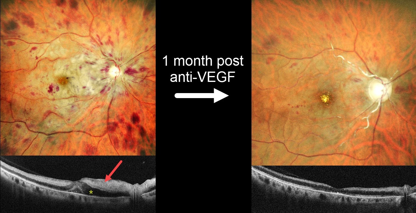
COMBINED CRAO-CRVO
Shraddha Raj Shrivastava and Manish Nagpal
Originally posted on @retina.rocks April 20, 2026
This 57YO female with a history of severe hypertension presented with one day of severe vision loss in her right eye. Blood pressure was 200/100 mmHg. Vision was hand motion OD and 20/20 in her normal OS.
MultiColor SLO imaging shows inner retinal opacification sparing the macular center, consistent with a fresh central retinal artery occlusion (CRAO). A central retinal vein occlusion (CRVO) is also noted with a swollen nerve and retinal hemorrhages in all quadrants. OCT scanning shows marked hyperreflectivity with loss of the band detail throughout the inner and mid-retinal layers. The peripapillary retina is edematous with fluid in the nasal outer nuclear layer and mild foveal subretinal fluid.
She was diagnosed with a combined CRAO-CRVO. An intravitreal anti-VEGF injection, and 1 month later, vision improved to 20/400. Repeat imaging shows marked improvement in the retinal hemorrhages, retinal opacification, and macular fluid, although new foveal lipid is noted. The nerve is flat and pale, and the major proximal arterioles are white without a blood column. The inner retina remains hyperreflective on OCT but is thinning with loss of the normal architecture. Panretinal photocoagulation was scheduled.
Learning Points:
Combined CRAO-CRVO is thankfully an extremely rare event, accounting for 0.3% of all retinal vascular occlusions (Raval et al, Indian J Ophthalmology 2020;68:2136-2142). Risk factors appear similar to those associated with isolated occlusions. The CRAO may be embolic or secondary to compression from a swollen optic nerve.
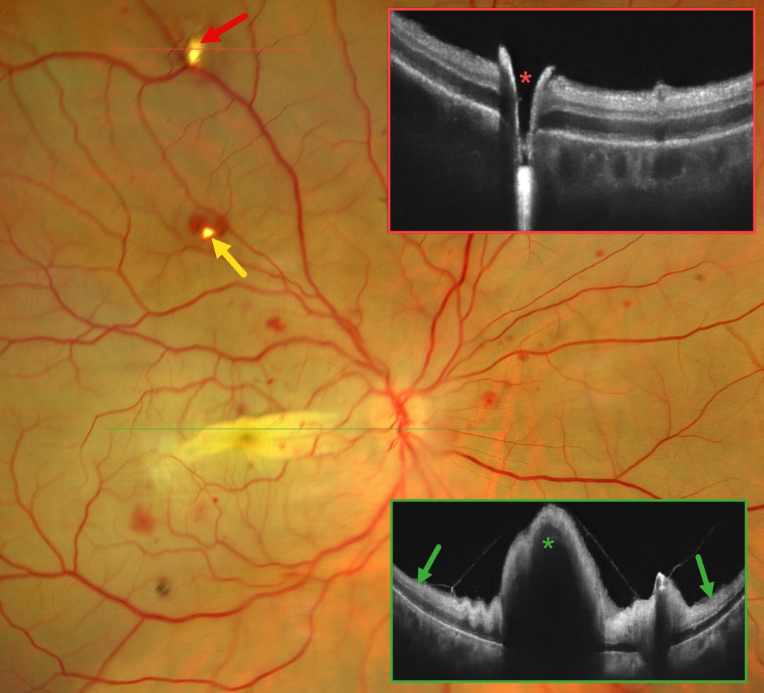
CRAO FOLLOWING DOUBLE PERFORATION FROM PERIBULBAR INJECTION
Ayushi Gupta and Vishal Agrawal
Originally posted on @retina.rocks April 6, 2026
This 42YO female immediately lost vision in her right eye 4 days earlier following pterygium excision surgery performed under peribulbar anesthesia elsewhere. Vision in our office was light perception.
Fundus photography shows an opaque retina throughout the posterior pole consistent with an acute central retinal artery occlusion (CRAO). There is a horizontal retinal fold throughout the macula with scattered blot retinal hemorrhages. Entry and exit needle penetration sites are noted. OCT scan through the superior entry site confirms a full-thickness retinal break. Macular OCT shows diffuse inner retinal hyperreflectivity with elevation through the fold.
Learning Points:
Peribulbar anesthesia is considered to have a lower risk of inadvertent globe perforation, although this risk is not zero (Lim et al., J Clin Ophthalmol 2021;5:414-416). CRAO after such an injury is exceedingly rare and was reported by Lake et al. (J Cataract Refract Surg 2003;29:2234-2235), who felt the occlusion might have resulted from increased IOP due to intraocular anesthetic. A CRAO was also reported by Gyasi et al. following peribulbar anesthesia for pterygium excision, although they found no evidence of globe perforation (Ghana Medical Journal 2012;46:48-50).
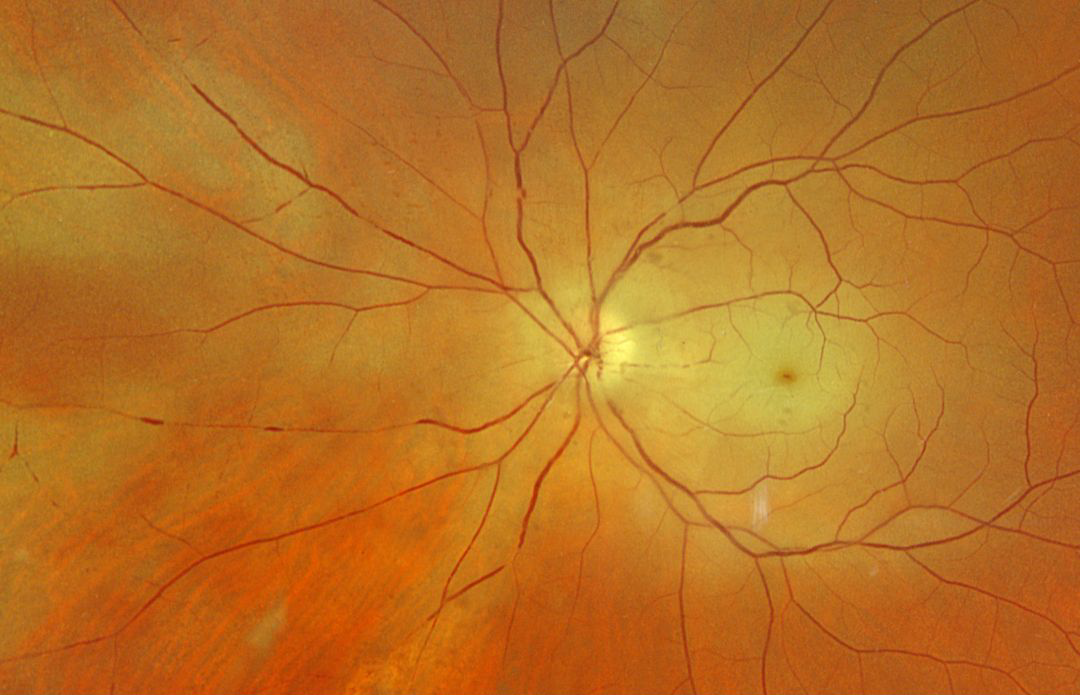
CRAO FOLLOWING CRANIOTOMY
Fraser McKay
Originally posted on @retina.rocks March 17, 2026
This 62YO male became hospitalized following a motor vehicle accident on 3/10/25. He underwent craniotomy on 4/16/25 and on 4/22/25. He woke from this last surgery blind in his right eye.
Optos color RGB imaging shows a fresh central retinal artery occlusion (CRAO) with a macular cherry red spot and patches of peripheral inner retinal whitening. Both the arteries and veins show focal areas of an absent blood column, indicating stagnant blood flow. Observation was recommended.
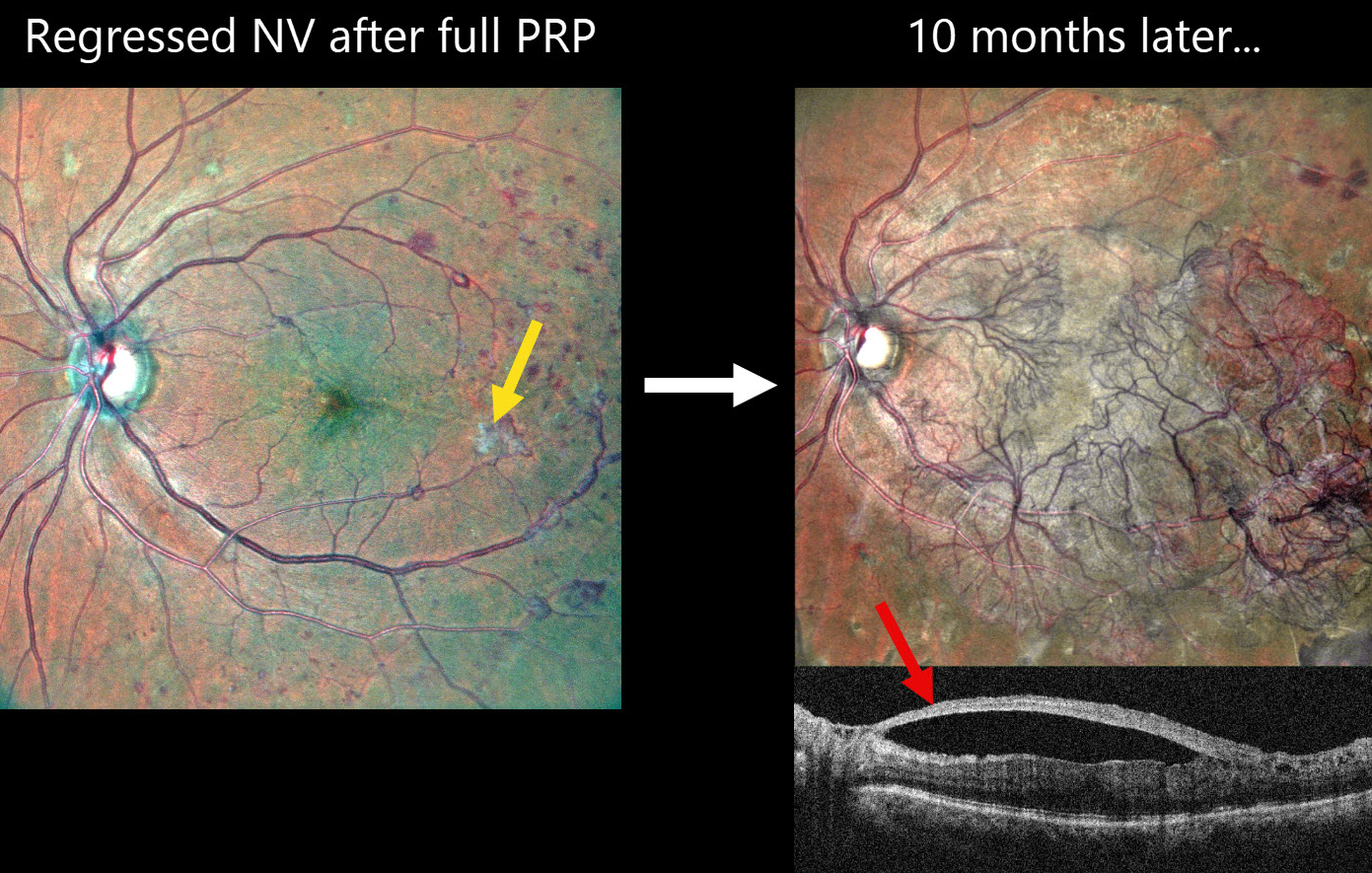
PROLIFERATIVE DIABETIC RETINOPATHY (PDR) WITH PROGRESSIVE NV DESPITE FULL PRP
Shishir Verghese
Originally posted on @retina.rocks March 4, 2026
This 60YO female with a history of type 2 diabetes underwent full panretinal photocoagulation (PRP) in her left eye. Following laser, MultiColor SLO imaging shows small residual areas of variably fibrosed retinal neovascularization. Vision was 20/30.
Ten months later, she presented complaining of 2 months of decreased vision in this eye. Vision was 20/60. MultiColor SLO imaging shows retinal neovascularization covering the posterior pole. On OCT, the fovea is remarkably attached with the hyperreflective neovascularization elevated along the partially detached posterior hyaloid.
Learning Points:
Although PRP is often considered a ‘one and done’ procedure, this is not always the case (Gonzalez et al, Ophthalmology 2021;128:1448-1457). Our patient is a perfect example of why these patients require lifelong monitoring even if their disease initially appears quiescent.
Given the aggressive neovascular proliferation, intravitreal anti-VEGF was given, followed by vitrectomy 5 days later. Although PRP is often considered a ‘one and done’ procedure, this is not always the case (Gonzalez et al, Ophthalmology 2021;128:1448-1457). Our patient is a perfect example of why these patients require lifelong monitoring even if their disease initially appears quiescent.
Given the aggressive neovascular proliferation, intravitreal anti-VEGF was given, followed by vitrectomy 5 days later.
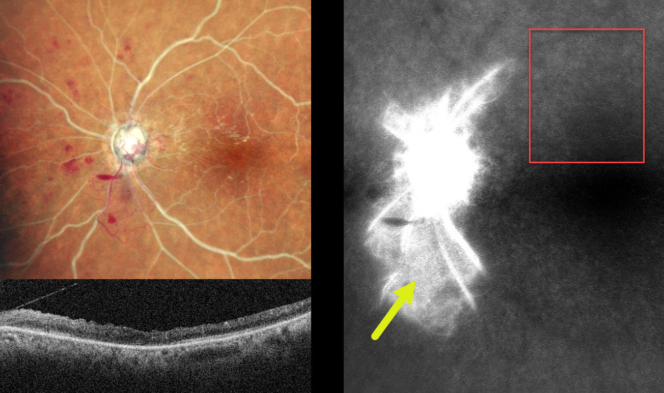
COMBINED CRAO-CRVO
Malvika Singh and Manish Nagpal
Originally posted on @retina.rocks March 3, 2026
This 60YO male presented with sudden vision loss in his left eye 1 month earlier. Vision was 20/20 in his normal OD and no light perception OS.
MultiColor SLO imaging shows a combined central retinal artery occlusion (CRAO) and central retinal vein occlusion (CRVO) with white arteries and veins with no blood column. Disc neovascularization is present inferotemporally, and a small preretinal hemorrhage is noted just inferonasal to the nerve. OCT shows a disorganized hyperreflective inner retina. Fluorescein angiography shows profound panretinal ischemia with a small island of preserved capillary bed inferior to the nerve.
Vasculitis workup was negative. An emergent stroke workup, including carotid Doppler, MRI, and neurologic consultation, was ordered. Given our patient’s vision, comfort measures for his left eye were recommended.
Learning Points:
Combined CRAO-CRVO is thankfully an extremely rare event, accounting for 0.3% of all retinal vascular occlusions (Raval et al, Indian J Ophthalmology 2020;68:2136-2142). Risk factors appear similar to those associated with isolated occlusions. The CRAO may be either embolic or secondary to compression from a swollen optic nerve.
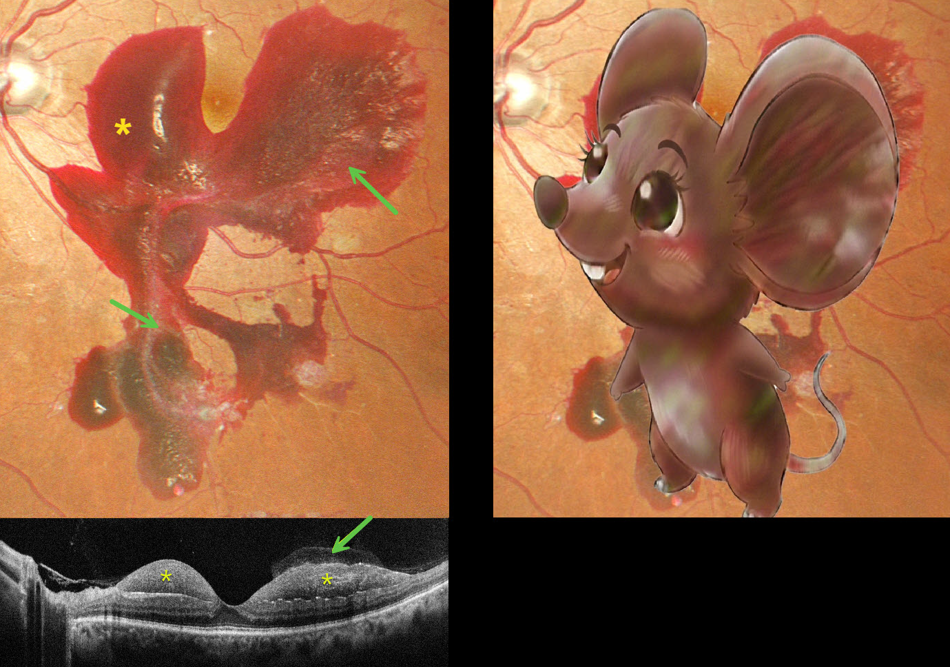
MOUSE MACULOPATHY AKA PROLIFERATIVE DIABETIC RETINOPATHY (PDR) WITH SUBHYALOID HEMORRHAGE
Originally posted on @retina.rocks February 24, 2026
This 48YO male with a 15-year history of poorly controlled type 2 diabetes presented with one day of blurred vision in his left eye. Vision was 20/20 bilaterally.
Pseudocolor SLO imaging shows a large macular hemorrhage, sparing the foveal center, which extends into the midperiphery. The hemorrhage is mostly subhyaloid with some breakthrough blood into the vitreous. The blood compartments are well visualized on OCT.
Learning Points:
The normal vitreous undergoes a decades-long process of orchestrated degeneration, including liquefaction and gradual separation from the retinal surface. The posterior vitreous detachment (PVD) begins in the perifoveal retina early in life, followed by a foveal PVD, then a peripheral PVD, and then finally a complete PVD when the vitreous separates from the optic nerve (Johnson AJO 2010;149:371-382). Although a diabetic subhyaloid hemorrhage can extend throughout the macula, our patient’s findings illustrate the normal increase in vitreofoveal adhesion.
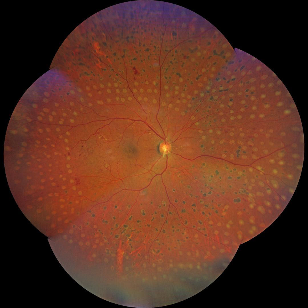
FILL-IN PANRETINAL PHOTOCOAGULATION
Giacomo De Rosa
Originally posted on @retina.rocks February 23, 2026
This 63YO male with type 2 diabetes presented to us with a history of recent panretinal photocoagulation (PRP) OD elsewhere. Vision was 20/20 OU.
Color photography shows irregularly sized and variably confluent laser scars mostly in the right inferior, nasal, and inferior midperiphery. Fill-in laser was applied at the slit lamp to complete the PRP.
Learning Points:
PRP should begin at least 1 disc diameter from the optic nerve and outside the major temporal arcades. Treatment is performed with a wide-angle lens or an indirect ophthalmoscope and should be applied from the posterior retina towards the periphery to prevent accidental macular burns. The laser spots should be 200-500 microns in size at the retina, be spaced about one burn-width apart, and should extend as far anteriorly as possible.
We generally do not recommend titrating the PRP based on involution of neovascularization, primarily due to the significant risk of the patient being lost to follow-up (Obeid et al, Ophthalmology 2018;125:1386-1392). Once the decision is made for PRP, we complete treatment in several sessions within a few months. Postoperative ultra-widefield imaging is helpful for assessing the adequacy of laser coverage, as areas of retinal ischemia are often undertreated (Sowmya L et al., Retina 2023;43:1922-1927).
We aren’t certain whether the initially treating laser surgeon planned further laser treatment, but the treatment was not performed in a controlled enough manner to control for burn size and spacing.
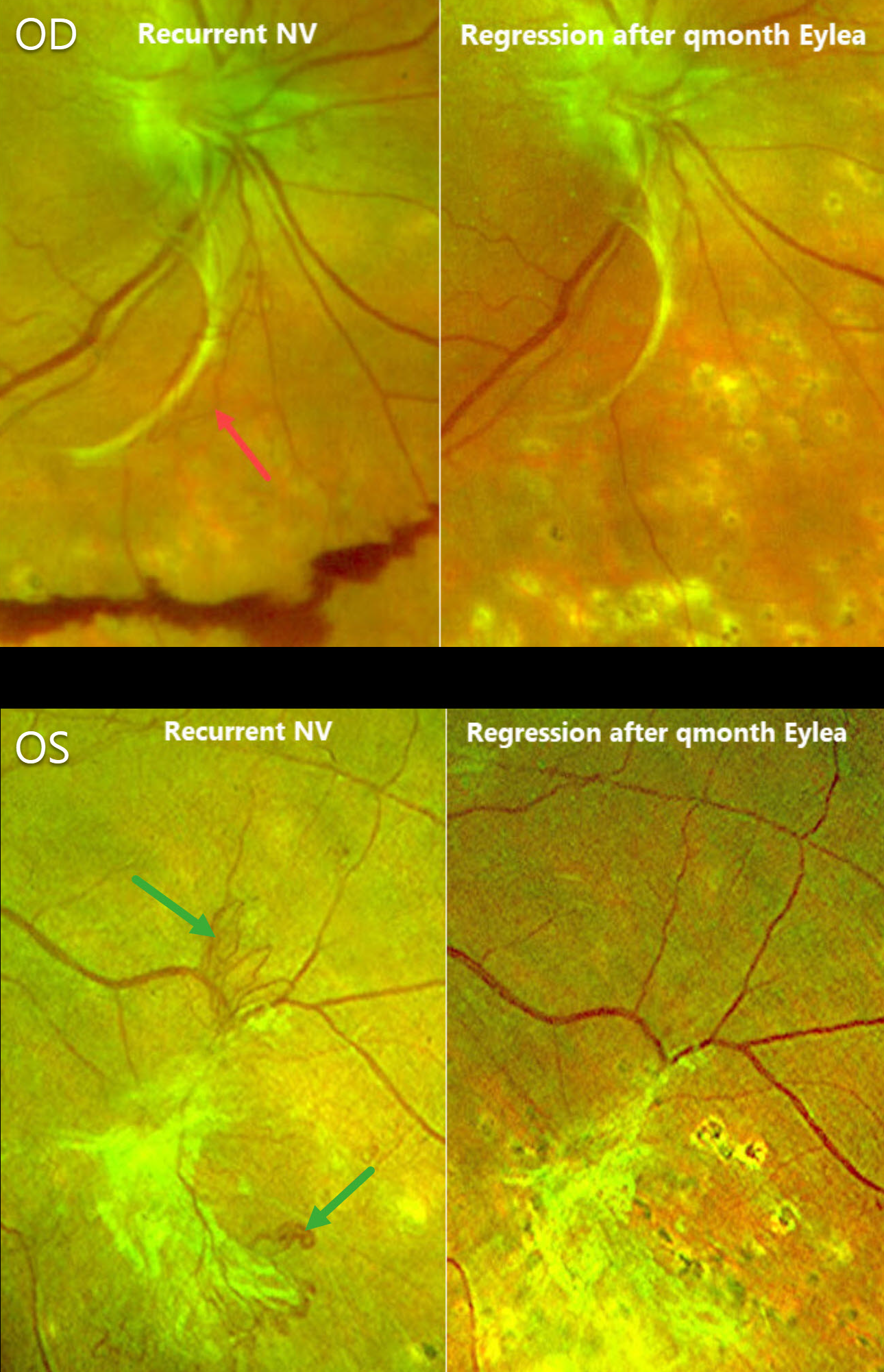
RECURRENT DIABETIC NEOVASCULARIZATION TREATED WITH SERIAL EYLEA
Michael Colucciello
Originally posted on @retina.rocks February 16, 2026
This 71YO female underwent prior full panretinal photocoagulation (PRP) bilaterally for proliferative diabetic retinopathy (PDR). She initially responded well with involution of all neovascularization. She presented for a routine follow-up with 20/25 vision OU.
Optos color RG imaging OD shows fibrosed disc neovascularization extending inferiorly with areas of active red neovascularization and inferior preretinal blood. The left eye shows fibrotic NV in the inferotemporal midperiphery along with some adjacent active vessels. Serial monthly bilateral Eylea injections were given, and 16 weeks later, the active NV fully regressed bilaterally.
Learning Points:
Although PRP is often considered a ‘one and done’ procedure, this is not always the case (Gonzalez et al, Ophthalmology 2021;128:1448-1457). Our patient is a perfect example of why these patients require lifelong monitoring even if their disease initially appears quiescent.
Prior to anti-VEGF therapy, our patient would have been treated with either additional fill-in PRP (putting her at risk for iatrogenic peripheral field loss) or possibly vitrectomy for increasing blood. Few currently advocate anti-VEGF injections as the sole treatment for PDR per the DRCR Protocol S. However, regarding adjuvant therapy to laser, we find these injections helpful for stabilizing macular edema or NV before starting PRP (Alsoudi et al, JAMA Ophthalmology 2024;142:972-975) and for those eyes needing supplemental treatment for active disease despite prior laser (Park et al, Eye 2021;35:3056-3063).
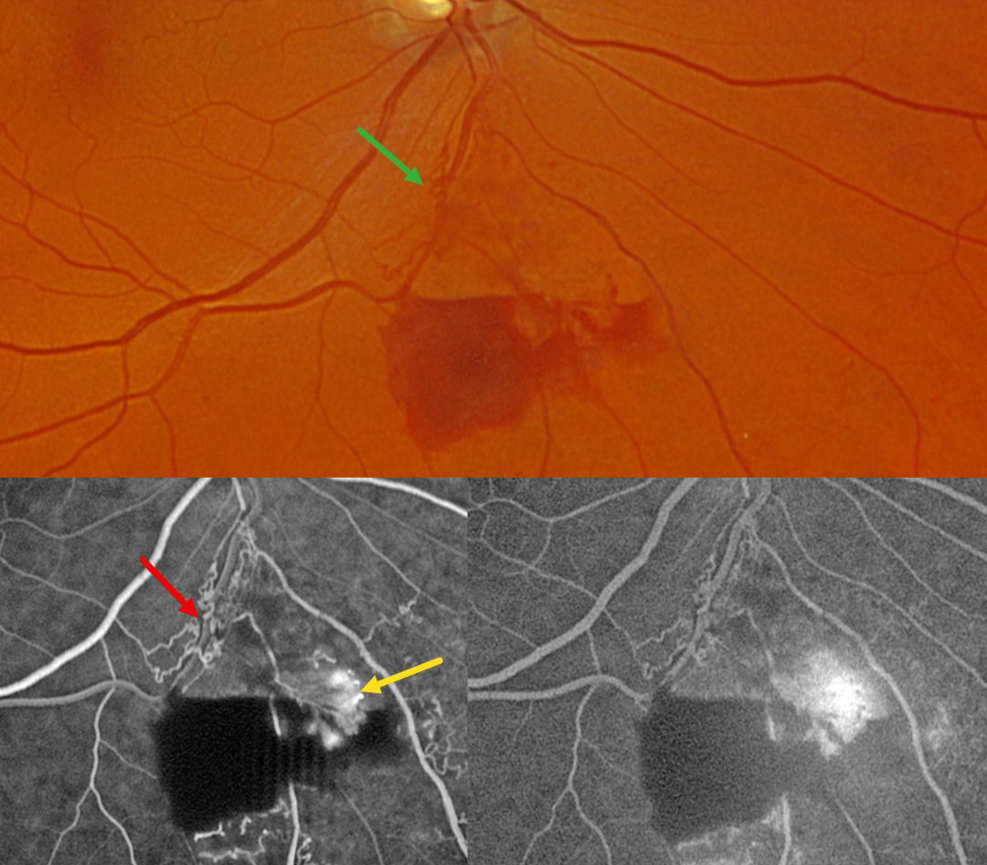
BRVO WITH RETINAL NEOVASCULARIZATION AND COLLATERALS
Originally posted on @retina.rocks February 11, 2026
This 59YO male was referred for asymptomatic retinal findings in his right eye. Vision was 20/30.
Optos color RG imaging shows an inferior branch retinal vein occlusion with midperipheral preretinal blood. Collateral vessels are noted at the site of the occlusion. These collaterals show no leakage angiographically, although the adjacent retinal neovascularization does leak.
Learning Points:
Collateral vessels are dilated, pre-existing vessels that bypass an occlusion. These are most commonly seen in the posterior pole following retinal venous occlusions and are exceedingly rare with arterial occlusions. The endothelial tight junctions are intact, so they don’t leak angiographically. This helps differentiate them from neovascularization, which does leak. Our patient’s eye shows us a wonderful example of both.
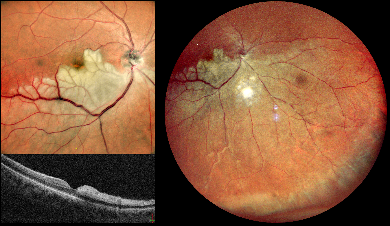
HEMI-CRAO
Rohan Jain and Manish Nagpal
Originally posted on @retina.rocks January 26, 2026
This is a healthy 42YO male presented with 7 days of blurred vision in his left eye. Vision was 20/90 OD and 20/20 in his normal OS.
Pseudocolor SLO imaging shows a white, opaque retina in the inferior retinal hemisphere, which spares the central macula. The vertical B-scan contrasts the normal superior retina with the ischemic inferior retina with marked inner retinal hyperreflectivity.
Learning Points:
Once the central retinal artery passes anteriorly through the lamina cribrosa, it branches into superior and inferior branches, which then further subdivide to supply the inner two-thirds of the neurosensory retina. Hemi-CRAO is a rare CRAO variant caused by the blockage of one of these superior or inferior branches. As with more typical CRAO, emboli or giant cell arteritis are the most common etiologies.
Our patient had acute retinal ischemia (defined as acute BRAO, central retinal artery occlusion, or transient monocular vision loss) and was immediately referred to the nearest stroke center. Unfortunately, he was immediately lost to follow up.
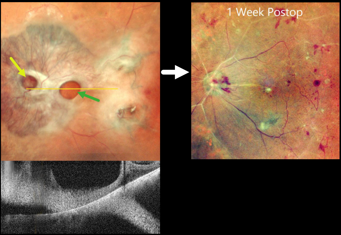
TRACTIONAL RETINAL DETACHMENT WITH PSEUDO-GIANT MACULAR HOLE
The European VitreoRetinal Society (EVRS) and Shishir Verghese
Originally posted on @retina.rocks January 16, 2026
This 65YO diabetic female presented with counting fingers bilaterally from proliferative diabetic retinopathy with macular tractional retinal detachments.
Preoperative pseudocolor SLO imaging of her left eye shows a complex fibrovascular membrane covering the posterior pole. Pseudo giant-macular holes are noted overlying the disc and macula. OCT scanning confirms the lack of a true macular hole. One week following vitrectomy, membrane peeling, and air injection, vision improved to 20/200, and the retina was completely attached with no residual traction.
Learning Points:
A giant macular hole, also reported in Alport syndrome (Shah and Weinberg Ophthalmic Genetics 2010;31(2):93-97), is an exceedingly rare traumatic event (Hernandez-Da Mota Case Rep Ophthalmol 2011;2:283-286). The unusual anatomy of our patient’s fibrovascular diabetic membrane simulated a giant macular hole.
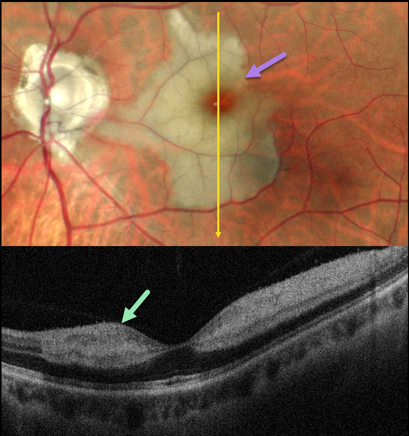
CILIORETINAL ARTERY OCCLUSION
Malvika Singh and Manish Nagpal
Originally posted on @retina.rocks December 30, 2025
This healthy 46YO male presented with an 8-day history of a central scotoma in his left eye. Vision was 20/20 in his normal OD and 20/400 OS.
Pseudocolor SLO imaging shows an acute cilioretinal artery occlusion with foveal sparing. OCT scanning shows marked inner retinal hyperreflectivity with posterior shadowing.
Learning Points:
The inner two-thirds of the neurosensory retina is supplied by the central retinal artery, with the choroid supplying the RPE and photoreceptor layer. Cilioretinal arteries, which arise from the posterior choroidal circulation, are present in up to 50% of individuals, and bilateral in about 25% (Schneider et al, Acta Ophthalmologica 2021;99:e310-e318).
Their clinical significance usually arises from foveal preservation in eyes with concomitant retinal artery occlusions (Brown and Shields, Arch Ophthalmology 1979;97:84-92). However, they can also be a rare cause of primary branch retinal artery occlusion, either in isolation (our patient) or in association with other entities, including central retinal vein occlusions (Hayreh et al, Retina 2008;28:581-594).
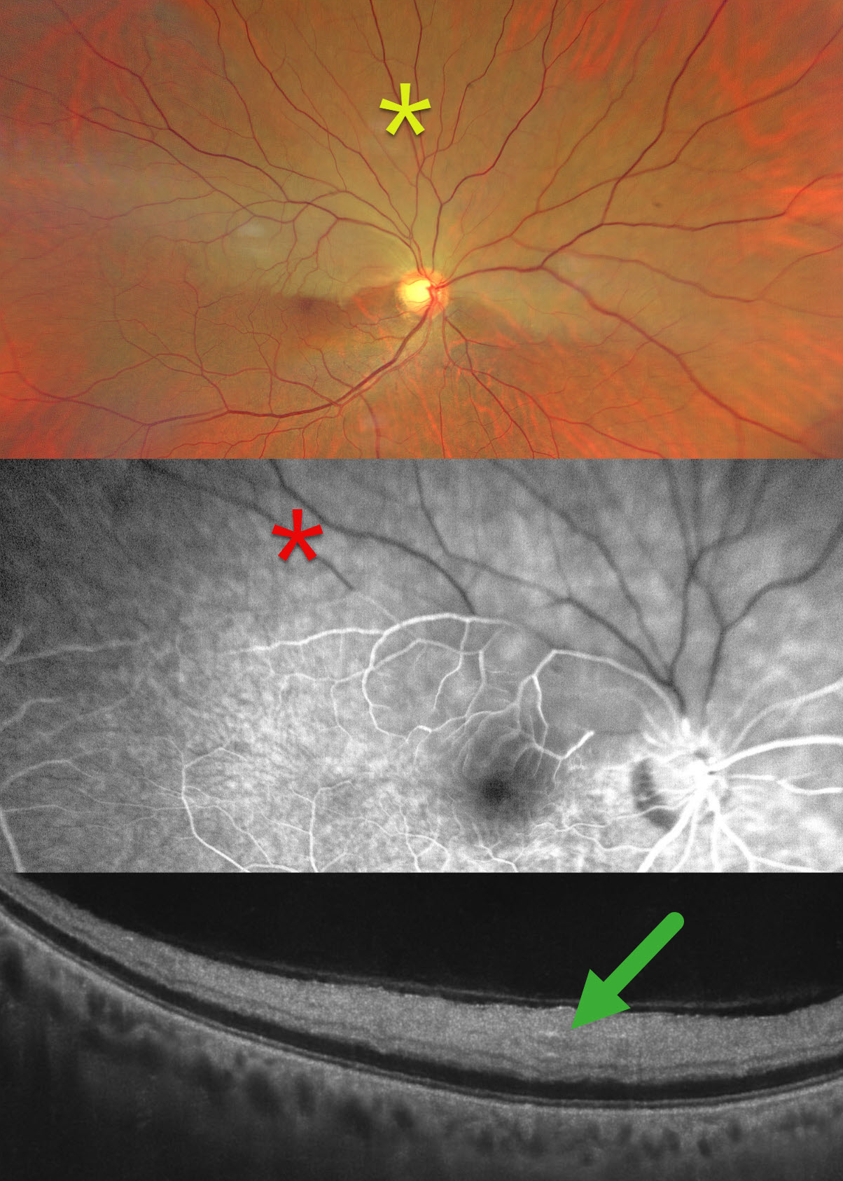
HEMI-CRAO
Ayushi Gupta and Vishal Agrawal
Originally posted on @retina.rocks December 2, 2025
This healthy 65YO male presented with 2 days of sudden painless decreased vision in his right eye. Vision was 20/80 OD and 20/20 in his healthy OS.
Color photography shows an acute superior hemi-central retinal artery occlusion (CRAO) with opaque white retina. On fluorescein angiography, there is a marked absence of perfusion in the superior hemiretina, which persists 4 minutes into the study. On OCT, the retina is thickened and hyperreflective from the outer plexiform layer anterior to the internal limiting membrane.
Our patient was immediately referred to cardiology for a stroke protocol evaluation.
Learning Points:
Once the central retinal artery passes anteriorly through the lamina cribrosa, it branches into superior and inferior branches, which then further subdivide to supply the inner two-thirds of the neurosensory retina. Hemi-CRAO is a rare variant of CRAO caused by the blockage of one of these superior or inferior branches. As with more typical CRAO, emboli or giant cell arteritis are the most common etiologies.
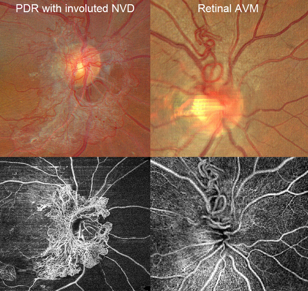
PROLIFERATIVE DIABETIC RETINOPATHY NVD VS RETINAL ARTERIOVENOUS MALFORMATION
Ayushi Gupta and Vishal Agrawal.
Originally posted on @retina.rocks November 12, 2025
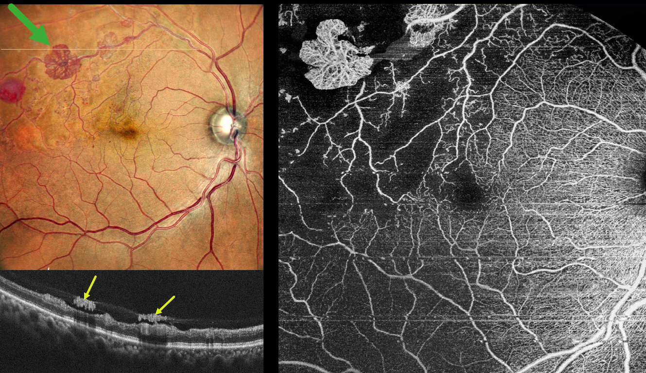
A patient with disc neovascularization (NVD) due to proliferative diabetic retinopathy (PDR) following full panretinal photocoagulation is compared with a patient with an asymptomatic retinal arteriovenous malformation (AVM).
Color photography of the PDR patient shows diffuse flat NVD with variable tight capillary networks of new vessels, admixed with larger vessels and fibrosis. OCT angiography (OCTA) confirms flow signals within these vessels, along with diffuse capillary loss superior, nasal, and inferior to the nerve. Color photography and OCTA of the retinal AVM show a tangle of arterioles and veins extending superiorly from the nerve. Fluorescein angiography, if performed, would show leakage from the NVD but not from the AVM.
BRVO WITH RETINAL NEOVASCULARIZATION
Originally posted on @retina.rocks November 3, 2025
This 55YO hypertensive male presented for a routine eye examination. He gave a history of a resolved superotemporal branch retinal vein occlusion (BRVO) with macular edema in his right eye that was successfully treated with a single anti-VEGF injection in 2022. Vision was 20/15 OD and 20/15 in his normal OS.
Pseudocolor SLO imaging shows an ischemic superotemporal BRVO with several fronds of active red and variably fibrosed retinal neovascularization. A round deep retinal hemorrhage is noted distally. On OCT, the neovascularization appears as hyperreflective plaques along the posterior edge of the partially detached posterior hyaloid. OCT angiography confirms diffuse capillary nonperfusion, a relatively preserved foveal avascular zone, and flow signals within the areas of neovascularization.
Learning Points:
An attached vitreous provides the scaffold for neovascularization to grow onto and spread. Retinal neovascularization grows from the inner retinal surface and proliferates along the outer posterior hyaloid face (Vaz-Pereira et al, Retina 2017;37:1287-1296). This tethering to a partially detached and contracting hyaloid is what causes tractional retinal detachment and vitreous hemorrhage in proliferative retinopathies.
In 1986, the Branch Vein Occlusion Study Group reported that scatter laser decreased the risk of vitreous hemorrhage from retinal neovascularization in BRVO and suggested that laser should be applied once neovascularization develops (Arch Ophthalmol 1986;104:34-41). Almost 40 years later, scatter laser remains the gold standard, although some may prefer to stabilize neovascularization with anti-VEGF therapy before laser. An anti-VEGF injection, followed by a superotemporal scatter laser, was therefore recommended for our patient.
BRVO WITH RED AND YELLOW DEVITALIZED BLOOD
Originally posted on @retina.rocks September 29, 2025
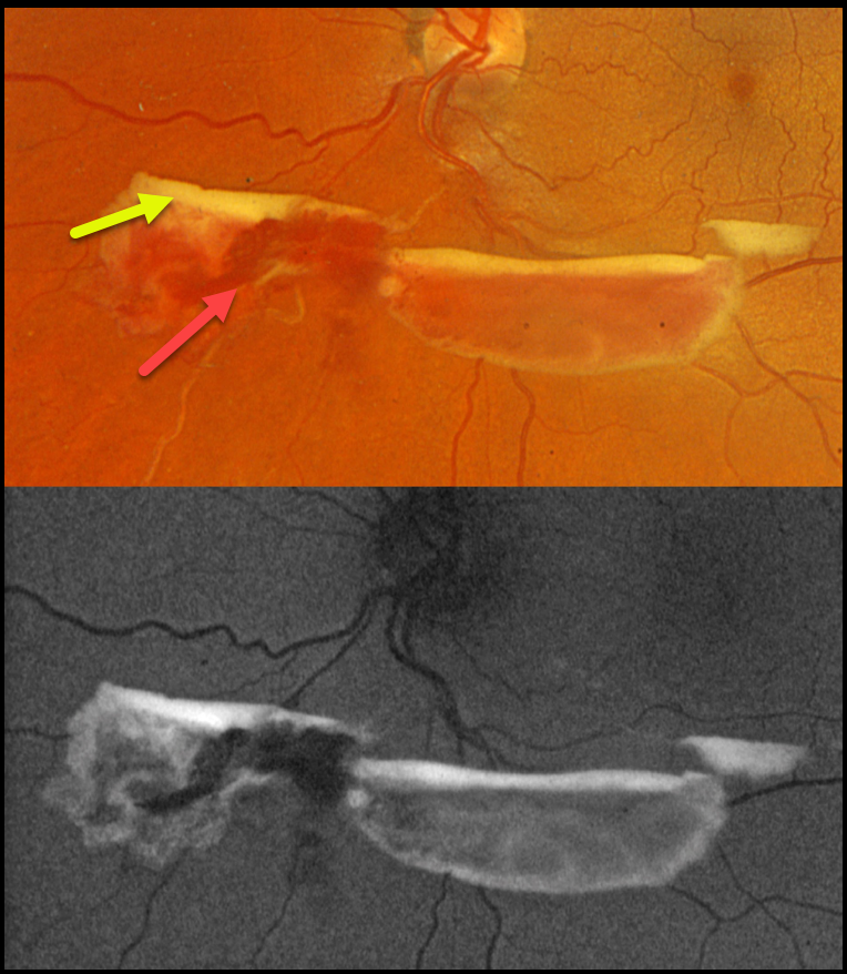
This 48YO female was referred for asymptomatic retinal findings. Vision was 20/20 in the normal OD and 20/20 OS.
Optos color RGB imaging shows red and yellow preretinal blood just inferior to the nerve due to an ischemic extramacular inferonasal branch retinal vein occlusion (BRVO). On fundus autofluorescence (FAF), the red blood is hypo-FAF, and the devitalized yellow blood is markedly hyper-FAF. Intravitreal Avastin was given, and scatter laser was scheduled, although she was immediately lost to follow-up.
Learning Points:
Porphyrins are organic molecules that are part of the heme molecule in hemoglobin. When these compounds break down, the devitalized blood appears yellow and becomes intensely hyperautofluorescent (Bloom and Spaide, Retinal Cases & Brief Reports 2022;16:401-402).
COMBINED TRACTIONAL-RHEGMATOGENOUS RETINAL DETACHMENT
The European VitreoRetina Society (EVRS) and Shishir Verghese
Originally posted on @retina.rocks September 19, 2025
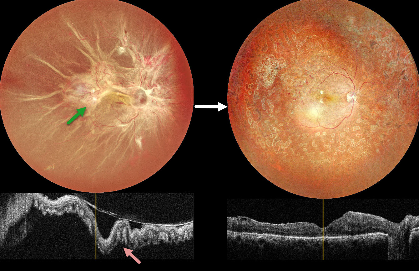
This 40YO female presented with vision loss in her right eye. There was a 15-year history of type 1 diabetes. Vision was hand motion OD.
Color photography shows a total combined traction-rhegmatogenous retinal detachment (RRD). There is a complex pattern of fibrovascular tissue covering the posterior pole with radiating retinal folds extending peripherally. OCT scanning shows a variably undulated detached retina with outer retinal corrugations.
Five days following an intravitreal anti-VEGF injection, she underwent pars plana vitrectomy with membrane peeling, endolaser, and silicone oil injection. Three months later, the silicone oil was removed. Postoperatively, vision improved to 20/80 with complete retinal reattachment.
Learning Points:
Outer retinal corrugations, also known as hydration outer retinal folds, are one of the key findings that help differentiate a RRD from exudative and traction retinal detachments. The cause of these folds remains uncertain but may result from the hydration of glycosaminoglycans in the interphotoreceptor matrix (Dalvin et al, Retinal Cases & Brief Reports 2020;14:355-359). This OCT biomarker confirms the rhegmatogenous component of our patient’s detachment.
CRAO
Tejaswita Verma and Manish Nagpal
Originally posted on @retina.rocks September 1, 2025
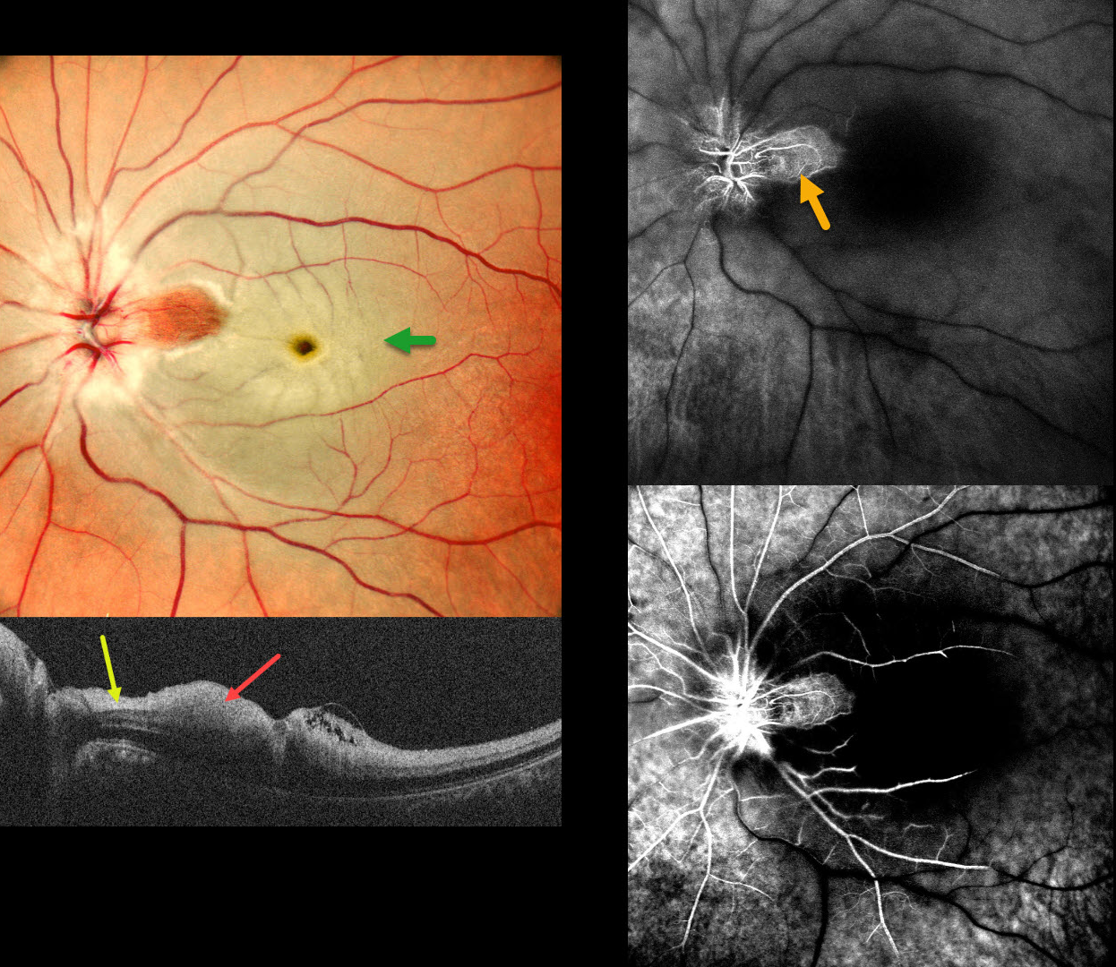
This 37 YO male presented with acute vision loss in his left eye 3 days earlier. Vision was 20/20 in his normal OD and 20/200 in his OS.
Pseudocolor SLO imaging shows a fresh central retinal artery occlusion (CRAO) with sparing of a small area of temporal peripapillary retina supplied from a patent cilioretinal artery. OCT scanning shows opaque, hyperreflective, thickened inner retinal layers, except for a small area of more normal retina within the cilioretinal sparing.
Fluorescein angiography shows profound diffuse retinal nonperfusion and delayed filling except for the nasal macula. Workup elsewhere included an MRI showing left ICA and left MCA chronic infarcts.
Learning Points:
The inner two-thirds of the neurosensory retina is supplied by the central retinal artery, with the choroid supplying the RPE and photoreceptor layer. Acute CRAO, therefore, presents with a white edematous inner retina that spares the fovea. Cilioretinal arteries, which arise from the posterior choroidal circulation, are present in up to 50% of individuals, and bilateral in about 25% (Schneider et al, Acta Ophthalmologica 2021; 99:e310-e318).
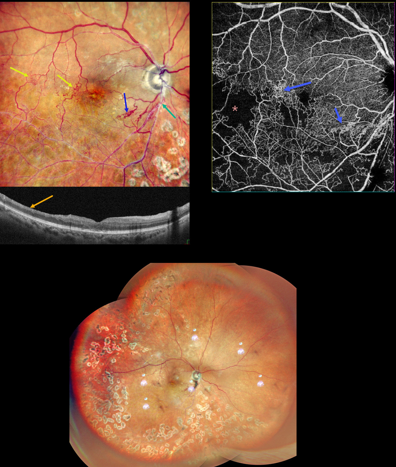
BRVO WITH COLLATERALS AND DRIL
Malvika Singh, Akansha Sharma and Manish Nagpal
Originally posted on @retina.rocks August 19, 2025
This 65YO male presented with a branch retinal vein occlusion (BRVO) in his right eye and 20/40 vision. He was treated elsewhere about a year ago with 3 intravitreal injections and scatter laser 7 years earlier.
Pseudocolor SLO shows an old major inferotemporal ischemic BRVO, which arises from an arteriovenous crossing just inferior to the disc. Extensive collateral vessels bypass the occlusion and cross the temporal horizontal raphe. OCT scanning shows thinned, featureless inner retina (disorganization of retinal inner layers, DRIL). Scatter laser is noted temporally and inferiorly. OCT angiography shows variable capillary loss throughout the BRVO distribution and highlights the collateral vessels.
Learning Points:
Collateral vessels are dilated, pre-existing vessels that bypass an occlusion. These are most commonly seen in the posterior pole following retinal venous occlusions and are exceedingly rare with arterial occlusions. The endothelial tight junctions are intact, so they don’t leak angiographically. This helps differentiate them from neovascularization, which does leak.
DRIL is seen in various retinal vascular disorders, including diabetic macular edema and retinal vein occlusions. It is generally associated with worse vision and increased anti-VEGF treatment burden (see Babiuch et al, JAMA Ophthalmol 2019;137:38-46).
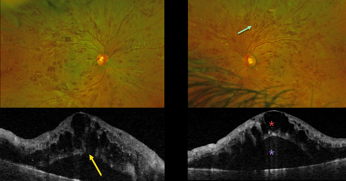
LEUKEMIA WITH PSEUDO-BILATERAL CRVO
Mattie Adams
Originally posted on @retina.rocks July 28, 2025
This previously healthy 38YO male presented with 1 month of bilateral blurriness. Vision was 20/60 OD and 20/80 OS.
Optos color RG imaging shows severe, bilateral, mostly white-centered retinal hemorrhages extending from the maculae into the retinal peripheries. Triton swept-source OCT shows marked bilateral cystic foveal swelling with subretinal fluid. An outer macular hole is probably present OD. His white blood cell count was markedly elevated at 206,400. Subsequent emergent oncologic workup led to the diagnosis of chronic myelogenous leukemia.
Learning Points:
The term “Roth spot” was named after Dr. Moritz Roth, who, in 1872, noted white-centered hemorrhages in patients with bacterial endocarditis. He felt that these white spots were septic emboli that originated from an infected cardiac valve. However, similar lesions can be seen in many other disorders, including leukemia, hyperviscosity, anemia, diabetes, and hypertension (Duane et al Ophthalmology 1980;87:66-69). The white material may represent a variety of causes, including fibrin. So, these lesions are best called white-centered hemorrhages, unless one is specifically referring to those found associated with bacterial endocarditis.
In our patient’s case, the severity of the retinal hemorrhages gave an appearance resembling bilateral central retinal vein occlusions (CRVO). However, CRVO hemorrhages are located predominantly in the nerve fiber layer (our patients’ hemorrhages were deep and white-centered), and the optic nerve is usually swollen with this degree of hemorrhage (our patients’ nerves were flat).
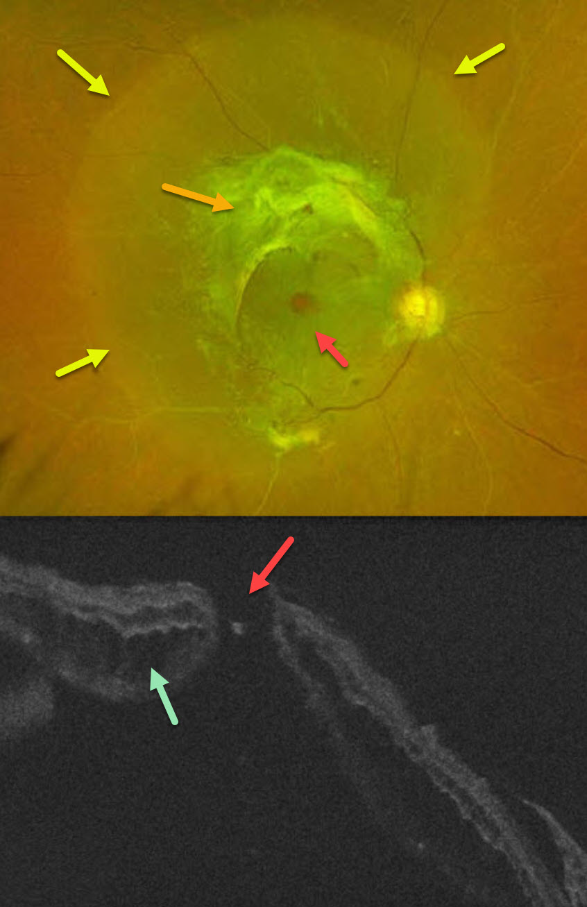
COMBINED TRACTIONAL-RHEGMATOGENOUS RETINAL DETACHMENT WITH MACULAR HOLE
Sucheta Kulkarani, Anita Gaikwad and Anjana Mirajkar
Originally posted on @retina.rocks July 24, 2025
This 49YO male with type 2 diabetes noticed decreased vision in his right eye for 4 months. A single anti-VEGF injection was given elsewhere in his right eye around the time of his vision loss. Vision was 20/400 OD and 20/30 OS.
Optos color RG imaging OD shows a combined tractional-rhegmatogenous retinal detachment from dense fibrovascular proliferation and a full-thickness macular hole. OCT shows diffuse cystic edema within the detached retina, predominantly in the outer nuclear layer, and a macular hole.
Learning Points:
Tractional macular holes as a cause of combined tractional-rhegmatogenous diabetic retinal detachments are a rare event in our experience. Unfortunately, our patient was lost to follow-up immediately. We expect him to lose central vision in his left eye soon if he does not seek care.
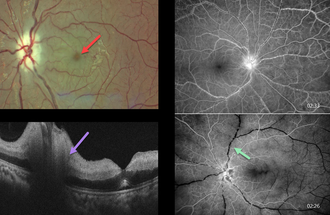
TAKAYASU VASCULITIS WITH CRAO
Ayushi Gupta and Vishal Agrawal
Originally posted on @retina.rocks April 29, 2025
This previously healthy 14YO girl presented with 10 days of vision loss in her right eye. She also gave a history of recurrent migraine-type headaches. She saw an outside eye doctor, where blood pressure was not checked, and spectacles were prescribed. At presentation, vision was 20/100 OD and 20/20 OS.
Funduscopic examinations revealed mild bilateral disc edema with venous dilation (not shown). Blood pressure could not be detected in any limb. We suspected Takayasu arteritis and urgently referred her to a rheumatologist and neurologist. Unfortunately, she presented 2 weeks later with sudden vision loss in her left eye. Vision was 20/100 OD and no light perception OS.
Color photography shows a cherry red spot and pale disc edema in her left eye. Fluorescein angiography shows markedly delayed venous filling in both eyes at 2.5 minutes following injection. OCT scanning shows a swollen disc and marked inner retinal hyperreflectivity with posterior shadowing. An anterior chamber paracentesis was performed with no change in her funduscopic appearance.
Medical workup between our initial and subsequent examinations revealed no pulses in the radial and dorsalis pedis arteries, with reduced left femoral and left carotid pulses. Renal artery Doppler revealed high-resistance flow bilaterally in the main renal and segmental arteries with 80-90% lumen narrowing in the infrarenal aorta. Brain MRI revealed an old infarct with encephalomalacia and gliosis in the left cerebellar hemisphere. Immediately following her second exam with us, she was emergently admitted by rheumatology and given a methylprednisolone infusion. She was immediately lost to follow-up.
Learning Points:
Takayasu arteritis is an idiopathic granulomatous vasculitis involving medium- to large-sized vessels throughout the body. Involvement of the aortic arch and its branches can cause ocular ischemia, including anterior segment ischemia with rubeosis and neovascular glaucoma.
Posterior segment findings include retinal ischemia, neovascularization, and vascular occlusions. Surprisingly, over 50% of patients present with ocular findings as the initial manifestation of their disease. For a wonderful review of the posterior segment findings in Takayasu, see Sharma et al, Indian J Ophthalmol 2024;72:637-647.
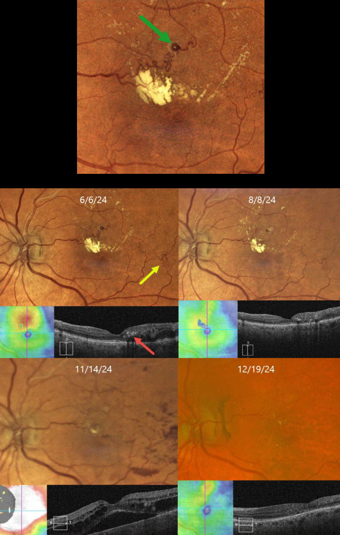
BRVO WITH RETINAL VENOUS MACROANEURYSM
Originally posted on @retina.rocks April 14, 2025
This 72YO female has been followed since 2017 for a branch retinal vein occlusion in her left eye. She has required ongoing anti-VEGF therapy for macular edema and failed macular laser for treatment burden several times. Although her eye was stable for years, on 6/6/24, there was new macular lipid with increased superior macular edema. Vision was stable at 20/25.
Color imaging shows a major superotemporal branch retinal vein occlusion with extensive collateral vessels and superior macular edema. Confluent lipid exudates threaten the macular center and appear as hyperreflective deposits in the mid-retina on OCT. A retinal venous macroaneurysm (RVM) is noted in the superior macula.
An intravitreal Eylea injection was administered, followed by focal thermal laser treatment of the RVM. Two months later, on 8/8/24, vision was 20/30. The lipid was decreasing, and the edema resolved. On 11/14/24, vision decreased to 20/60. Although the lipid was almost resolved, recurrent macular fluid persisted. Following a single Avastin injection, vision improved to 20/30, and the edema resolved. Macular grid laser was applied in hopes of reducing the treatment burden. We will continue to follow her closely and treat her as needed.
Learning Points:
In 1990, Cousins et al reported that RVMs can be an additional finding in occlusive venous disease, always in association with collateral vessels or capillary or arterial macroaneurysms (AJO 1990;109:567-570). These vascular abnormalities, also known as retinal capillary macroaneurysms or telangiectatic capillaries (TelCaps), tend to have a higher rate of intraretinal lipid (40%) but are less likely to affect central vision since they are usually outside the macula. They tend to have a higher association with retinal ischemia, thus being at a higher rate for developing retinal neovascularization (32%). Although these lesions will usually involute spontaneously, we treated our patient out of concern that the lipid might extend into the foveal center.
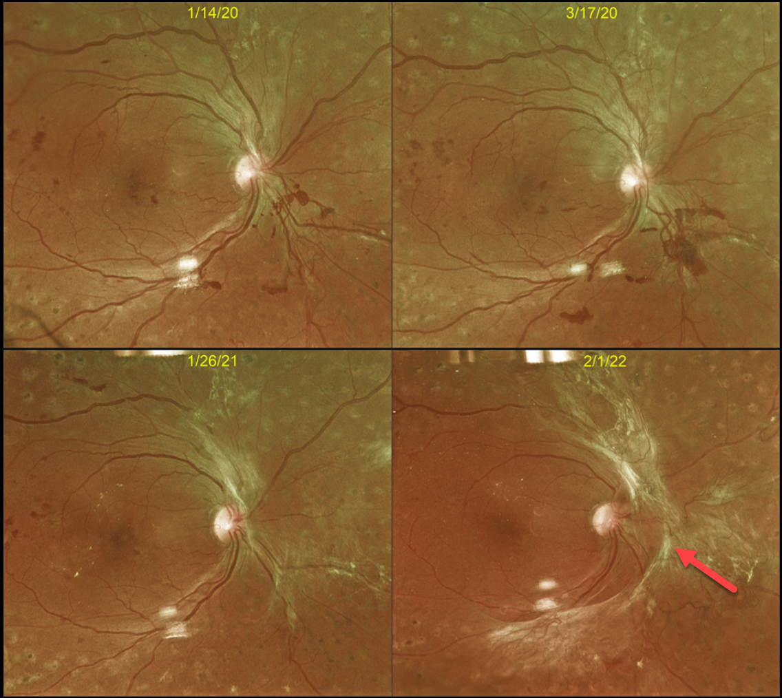
TRACTIONAL RETINAL DETACHMENT
Originally posted on @retina.rocks January 29, 2025
This 43YO male with type 2 diabetes initially responded beautifully in 2016 to full panretinal photocoagulation (PRP) for proliferative diabetic retinopathy (PDR). However, he presented on 3/17/20 with some new preretinal blood (Optos color RG image), and fill-in PRP was applied.
Over the course of 2 years, progressive fibrotic neovascularization with extramacular traction developed. Despite the traction, vision remained at 20/40, and careful observation was recommended. We were hesitant to add further laser or anti-VEGF therapy for fear of causing increased traction and ‘crunch’ (Tan et al, Surv Ophthalmology 2021;66:926-932). When last examined on 4/3/24, the retinal findings remained unchanged, and vision was 20/30 (not shown).
Although PRP is often a ‘one and done’ procedure, this is not always the case (Gonzalez et al, Ophthalmology 2021;128:1448-1457). Our patient is a perfect example of why these patients require lifelong monitoring even if their disease appears quiescent.
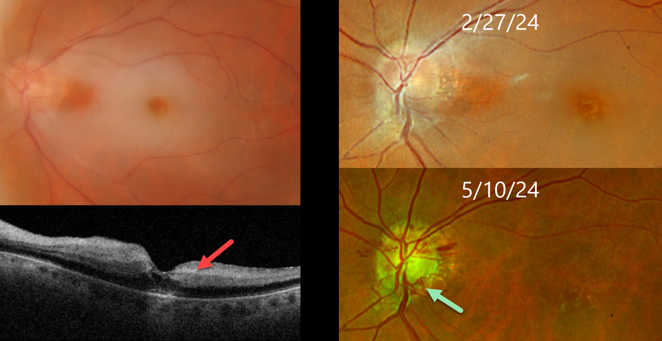
CRAO
Originally posted on @retina.rocks December 23, 2024
This 75YO female presented with an acute counting fingers central retinal artery occlusion. Triton color imaging shows a small area of papillomacular sparing, and swept-source OCT shows inner retinal hyperreflectivity. The retinal opacification completely resolved 10 weeks later with the development of extensive disc collaterals. Vision at counting fingers.
Learning Points:
Collateral vessels are dilated, pre-existing vessels that bypass an occlusion. These are seen in the posterior pole most commonly following retinal venous occlusions. Disc collaterals are exceedingly rare with arterial occlusions and were first described by Nettleship in 1891 (Ragge and Hoyt, Br J Ophthalmology 1992;76:186-188). The endothelial tight junctions are intact so they don’t leak angiographically. This helps to differentiate them from neovascularization which does leak.
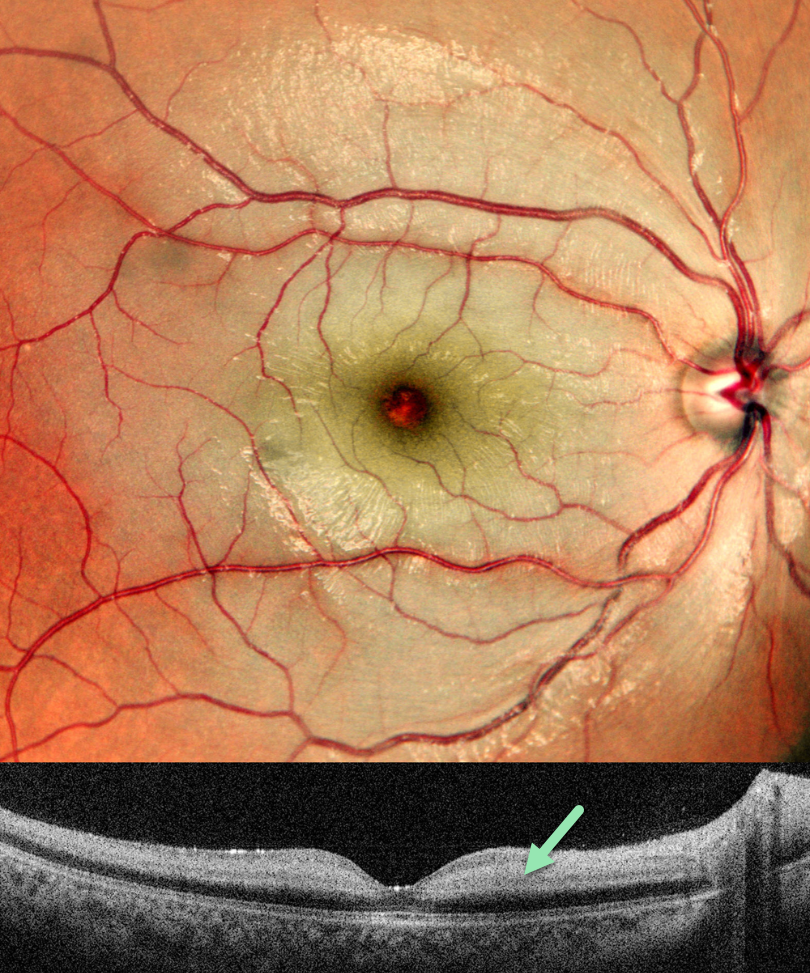
CRAO
Ankit Jain and Manish Nagpal
Originally posted on @retina.rocks December 11, 2024
This previously healthy 8YO boy presented with sudden vision loss in his right eye 10 days earlier. Vision was counting fingers.
Pseudocolor SLO imaging shows diffuse inner retinal whitening, sparing the central macula. OCT scanning shows a somewhat thickened and markedly hyperreflective inner retina. Cardiac 2D Echo and MRA of the head and neck were normal. Anticardiolipin antibodies were elevated, and he was started on oral clopidogrel and aspirin.
Learning Points:
Central retinal artery occlusion (CRAO) in the pediatric population is exceedingly rare and requires an aggressive search for underlying clotting abnormalities. The antiphospholipid antibody syndrome, also known as anticardiolipin antibody syndrome or lupus anticoagulant syndrome, is caused by autoantibodies directed against phospholipid-bound proteins. These predispose patients to venous and arterial thrombi and increase the risk for pregnancy complications and miscarriage. The syndrome is more common in women and in those with other autoimmune disorders, including systemic lupus.
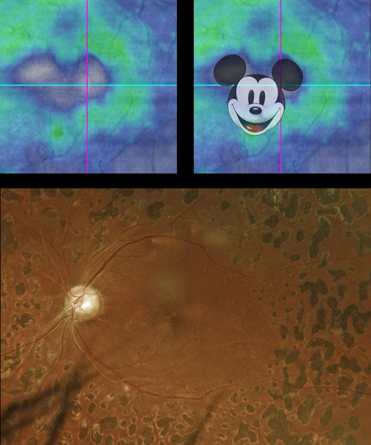
MICKEY MOUSE MACULOPATHY
Originally posted on @retina.rocks November 13, 2024
This 52YO diabetic male rodent has a history of proliferative diabetic retinopathy and full panretinal photocoagulation (PRP). Vision was stable at 20/30.
Optos color RGB imaging shows full PRP scarring. The macula is clinically ischemic with some vascular sheathing. Triton swept-source OCT shows nasal and central atrophy consistent with Mickey Mouse Maculopathy (MMM).
Created by Walt Disney and Ub Iwerks, Mickey Mouse celebrates his birthday on November 18th. This date commemorates his first appearance in the animated short film “Steamboat Willie” in 1928, famous for Mickey’s debut and for its synchronized sound.
On January 1, 2024, the famous mouse entered the public domain, allowing anyone to use this version of the character in their creative works without needing Disney’s permission or payment. However, Disney still retains the copyright on any subsequent variations of Mickey until they reach the 95-year mark.
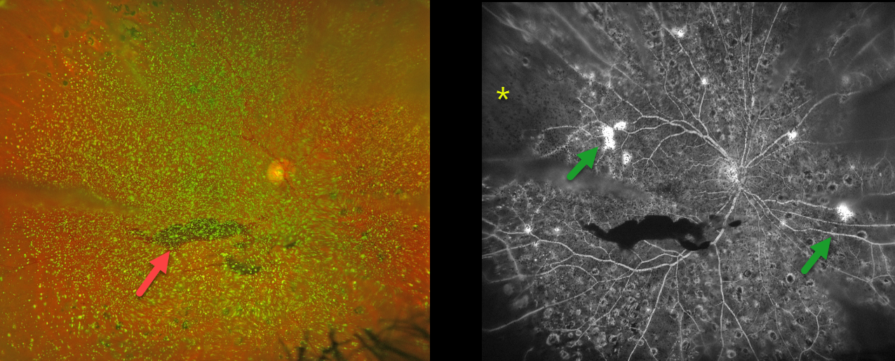
PROLIFERATIVE DIABETIC RETINOPATHY (PDR) WITH ASTEROID HYALOSIS
Originally posted on @retina.rocks October 16, 2024
This 50YO female with a history of type 2 diabetes was referred for new floaters. Several years earlier, we began panretinal photocoagulation (PRP) for proliferative diabetic retinopathy (PDR), but she had been lost to follow-up.
Optos color RG imaging shows dense asteroid hyalosis with a hazy inferior preretinal hemorrhage. Fluorescein angiography (FA) shows significant retinal ischemia, scattered retinal neovascularization, and partial PRP scarring.
Examining the fundus with dense asteroid, as in this diabetic patient, can be extremely challenging. FA and OCT are great modalities because they can visualize the retina when the clinical exam can’t. Optos FA was critical in confirming proliferative retinopathy with peripheral ischemia in our patient.
We were fortunately able to complete the PRP despite the vitreous opacities.
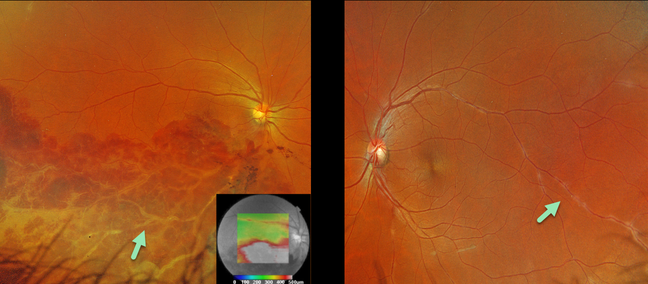
BRVO AND PERIPHLEBITIS FROM LIKELY SARCOIDOSIS
Originally posted on @retina.rocks October 8, 2024
This healthy 36YO male presented with 1 week of unilateral vision loss. Vision was 20/400 OD and 20/20 OS.
Optos color RGB imaging OD shows a major inferotemporal branch retinal vein occlusion (BRVO) with severe confluent retinal hemorrhages and a frosted appearance to the major vessels. The occlusion does not seem to arise from a more proximal arteriovenous (AV) crossing. A small area of peripheral superotemporal periphlebitis is noted OD, with more extensive periphlebitis OS. Triton swept-source OCT shows inferior macular fluid.
BRVOs nearly always arise at an AV crossing, where a thickened arterial vessel compresses the underlying vein. Sarcoidosis is the classic exception where a BRVO is not caused by an AV crossing, but by an inflammatory narrowing of the involved vein. In addition, our patients’ segmental periphlebitis (‘candle wax drippings’) is virtually pathognomonic for sarcoidosis as well.
A sarcoidosis workup was recommended, although the patient failed to show for his testing and was immediately lost to follow-up.
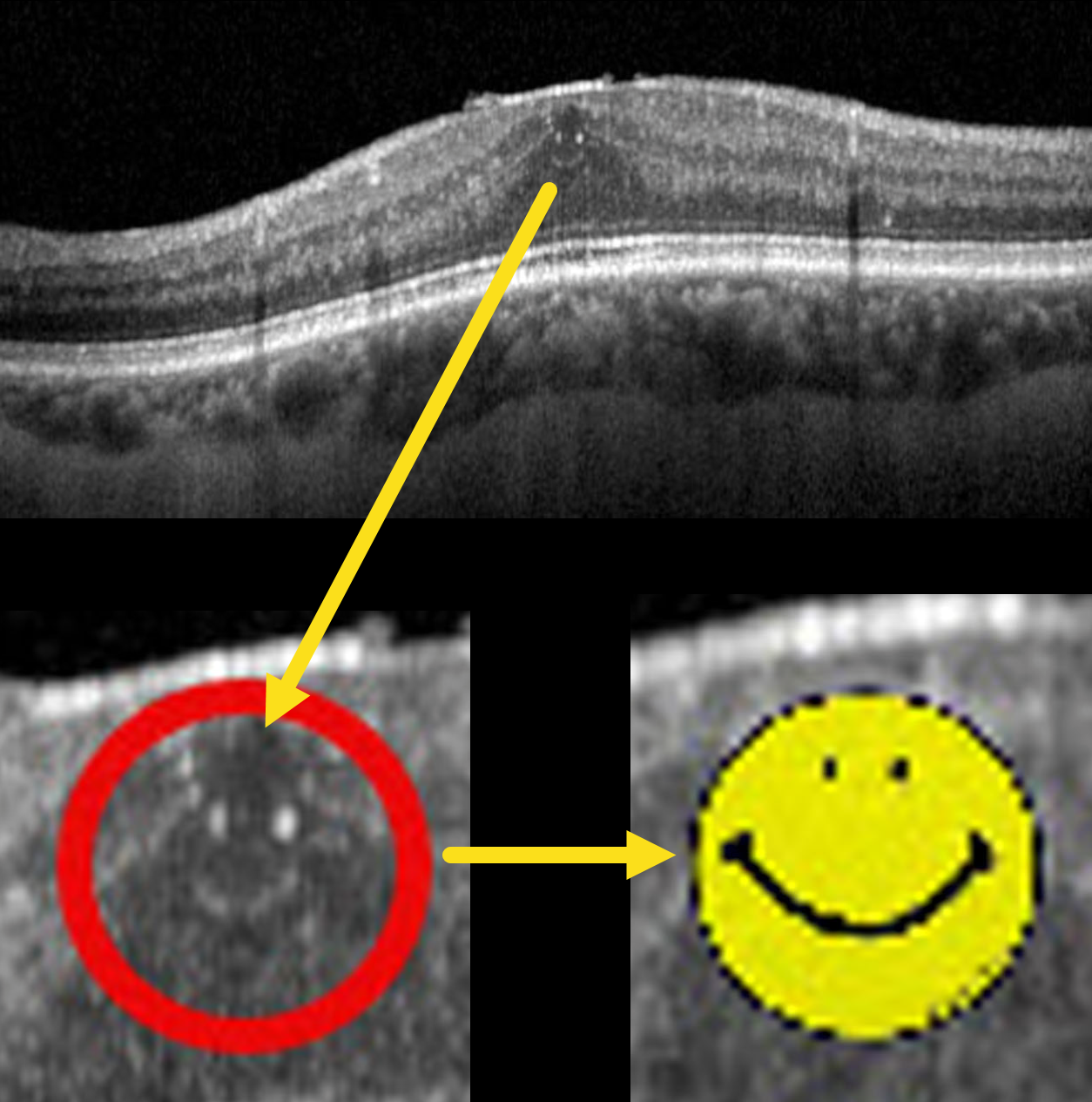
HAPPY WORLD SMILE DAY
Pier Luigi Guerin
Originally posted on @retina.rocks October 4, 2024
This 70YO male with type 2 diabetes presented with these asymptomatic OCT findings that made our day. Vision was 20/25.
World Smile Day is celebrated on the first Friday of October each year, a tradition that began in 1999. The day was created by Harvey Ball, the commercial artist who designed the iconic yellow smiley face. Ball was concerned that the over-commercialization of his symbol would dilute its original intent of spreading goodwill and cheer. To counteract this, he established World Smile Day to remind people of the power of a smile and to encourage acts of kindness around the world.
The day is marked by various activities that promote smiling and kindness. People are encouraged to perform acts of kindness and to smile more, spreading positivity and happiness. The Harvey Ball World Smile Foundation, established after Ball’s passing in 2001, continues to sponsor the day and organize events. These events range from community activities to global initiatives, all aimed at creating smiles and fostering a spirit of generosity and joy.
So, perform some random acts of kindness today and make the world a better place because you’re in it.
PROLIFERATIVE DIABETIC RETINOPATHY (PDR)
Seif Anwar
Originally posted on @retina.rocks October 2, 2024
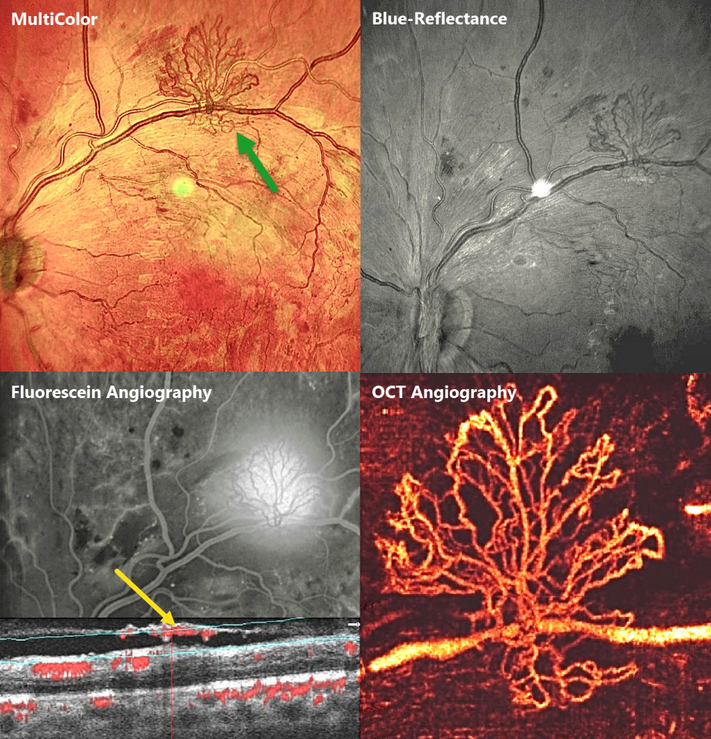
This 23YO male presented with bilateral proliferative diabetic retinopathy (PDR). Vision was 20/60 OD and 20/80 OS. Multimodal imaging of his left eye highlights an area of retinal neovascularization along the superotemporal arcade.
Which modality do you feel best images the neovascularization? Or are they all complementary?
MultiColor imaging shows the lacy active red vessels overlying the orange-red RPE and choroid. These vessels are better highlighted in the Blue-Reflectance image, since they appear black on a grey background. They profoundly leak on fluorescein angiography. En face OCT angiography (OCTA) beautifully shows the fine neovascular details. Finally, B-scan OCTA shows the neovascular vessels growing along the back surface of the partially detached posterior hyaloid.
BRVO + MYELINATED NERVE FIBER LAYER
Evgenii Chernov
Originally posted on @retina.rocks September 30, 2024
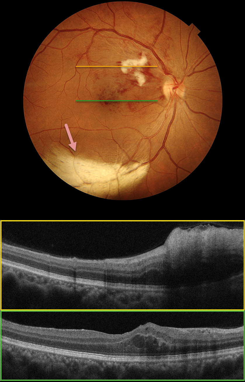
Color photography of this 48YO female’s right eye shows a major superotemporal branch retinal vein occlusion (BRVO) with foveal blood. A prominent nerve fiber layer infarct (cotton-wool spot, CWS) is present in the superonasal macula. OCT scanning shows diffuse hyperreflective inner retinal thickening through the CWS and some central foveal fluid in the outer nuclear layer. An unrelated patch of myelinated nerve fiber layer (NFL) is noted along the inferotemporal arcade. The macular edema resolved following 3 monthly intravitreal Lucentis injections (not shown).
Our patient’s eye shows two different pathologies that each involve the NFL. Myelinated NFL occurs when retinal nerve fibers develop a myelin sheath, which usually stops posterior to the lamina cribrosa. The white myelin beautifully displays the normally transparent NFL. Myelination may represent an oligodendrocytic choristoma (see Rao et al Retina 2019;39:1125-1132). Nerve fiber layer infarcts cause a more fluffy, white ischemic opacification of the NFL and are a relatively nonspecific finding in numerous disorders, including diabetes, retinal venous occlusion, hypertension, and HIV/AIDS.
PROLIFERATIVE DIABETIC RETINOPATHY (PDR)
Originally posted on @retina.rocks September 25, 2024
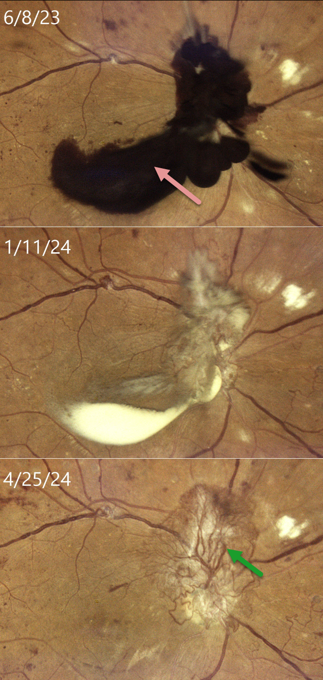
This 33YO female with type 2 diabetes was in the process of being treated with intravitreal Eylea, macular laser, and panretinal photocoagulation (PRP) for bilateral center-involved diabetic macular edema and proliferative diabetic retinopathy (PDR). She presented on 6/8/23 with recent vision loss OD and was overdue for this visit because of numerous medical issues. Vision had decreased to 20/400 OD.
Color imaging shows dark red preretinal blood obscuring the optic nerve and central macula. After several monthly Eylea injections and completion of the PRP, the preretinal blood became yellow and devitalized on 1/11/24, before finally completely absorbing, as noted at last examination on 4/25/24, with stable 20/400 vision. Despite full PRP, diffuse variably fibrosed neovascularization persists, along with venous beading. OCT scanning revealed no edema with central atrophy (not shown).
Our patient was initially in no rush for vitrectomy surgery, as vision in her fellow eye was 20/50. Conservative management with intravitreal injections and PRP was thankfully successful. This approach was also found to be safe and effective by Brar et al in a retrospective series of 22 eyes with foveal preretinal blood from PDR (Ind J Ophthalmol 2024;72:687-691).
POSTOP DIABETIC TRD WITH INNER RETINAL FOLDS
Originally posted on @retina.rocks September 5, 2024
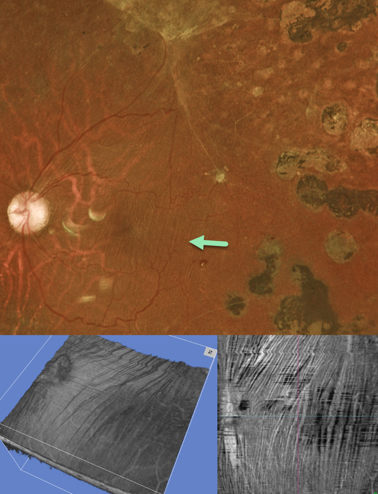
This 35YO diabetic male underwent prior vitrectomy surgery for a diabetic traction retinal detachment (TRD) five years earlier. Vision was stable at 20/40.
Optos color RG imaging shows fine macular striae extending superiorly towards the area of prior traction. Triton swept-source OCT 3D reconstruction and inner retinal en face show differing perspectives of these inner retinal folds.
Inner retinal folds are common following vitrectomy for diabetic TRD. They often fail to completely resolve, and vision can be quite good despite the persisting findings.
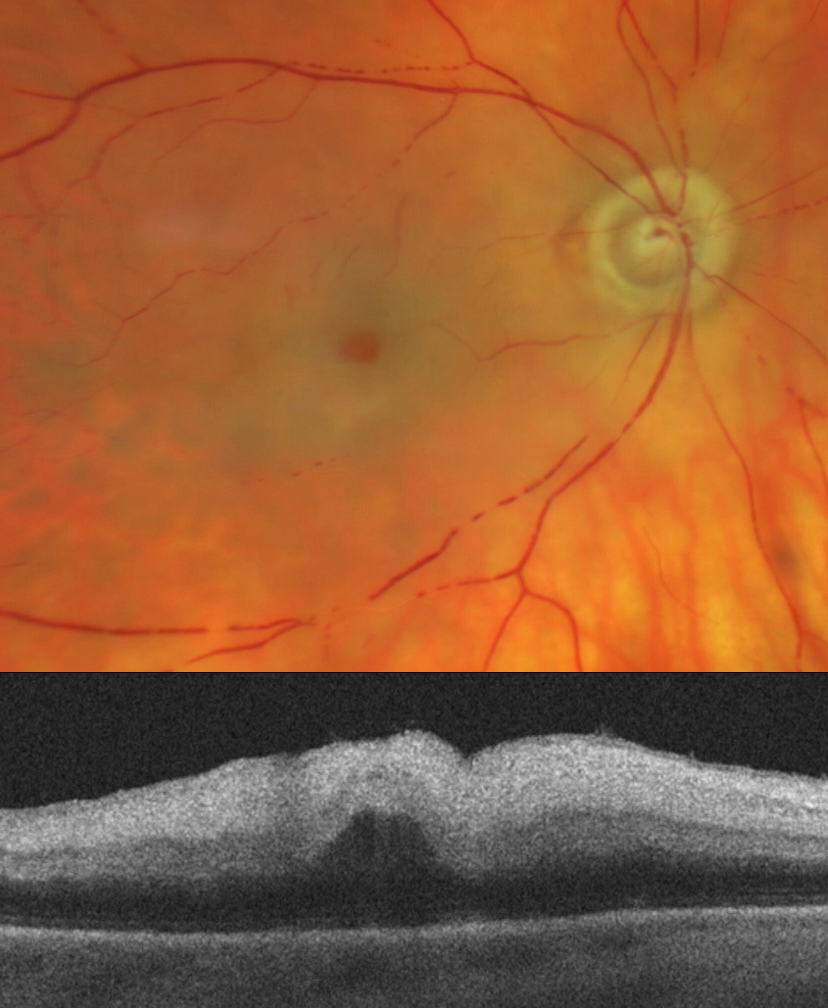
CRAO
Will Gibson
Originally posted on @retina.rocks August 29, 2024
This 81YO female presented with acute vision loss in her right eye. Vision was light perception.
Optos color RG imaging shows an acute central retinal artery occlusion (CRAO) with opaque white retina surrounding the preserved central fovea (‘cherry red spot’). A stagnant blood column is noted inferiorly with box-carring. OCT scanning shows a thickened and markedly hyperreflective inner retina. She was immediately referred to the nearest stroke center.
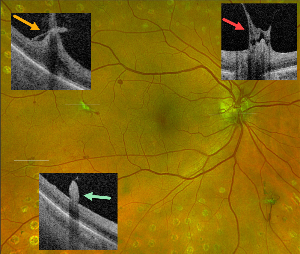
PROLIFERATIVE DIABETIC RETINOPATHY (PDR)
Originally posted on @retina.rocks August 27, 2024
This 55YO female with type 2 diabetes has involuted proliferative diabetic retinopathy (PDR) following full panretinal photocoagulation.
Optos color RG imaging shows residual disc (NVD) and peripheral retinal neovascularization (NVE). Optos Silverstone swept-source OCT through disc and temporal macular neovascularization shows active vitreous traction on the residual vessels. There is no traction on small, reddish NVE in the distal inferotemporal macula. Continued observation was recommended.
Learning Points:
An attached vitreous provides the scaffold for neovascularization to grow onto and spread. Retinal neovascularization grows from the inner retinal surface and proliferates along the outer posterior hyaloid face (Vaz-Pereira et al, Retina 2017;37:1287-1296). This tethering to a partially detached and contracting hyaloid is what causes tractional retinal detachment and vitreous hemorrhage. Thus, although an eye with a complete posterior vitreous detachment can develop small buds of neovascularization along the retinal surface (as seen in our patient’s inferotemporal NVE), the detached hyaloid is protective against the growth of large sheets of fibrovascular tissue.
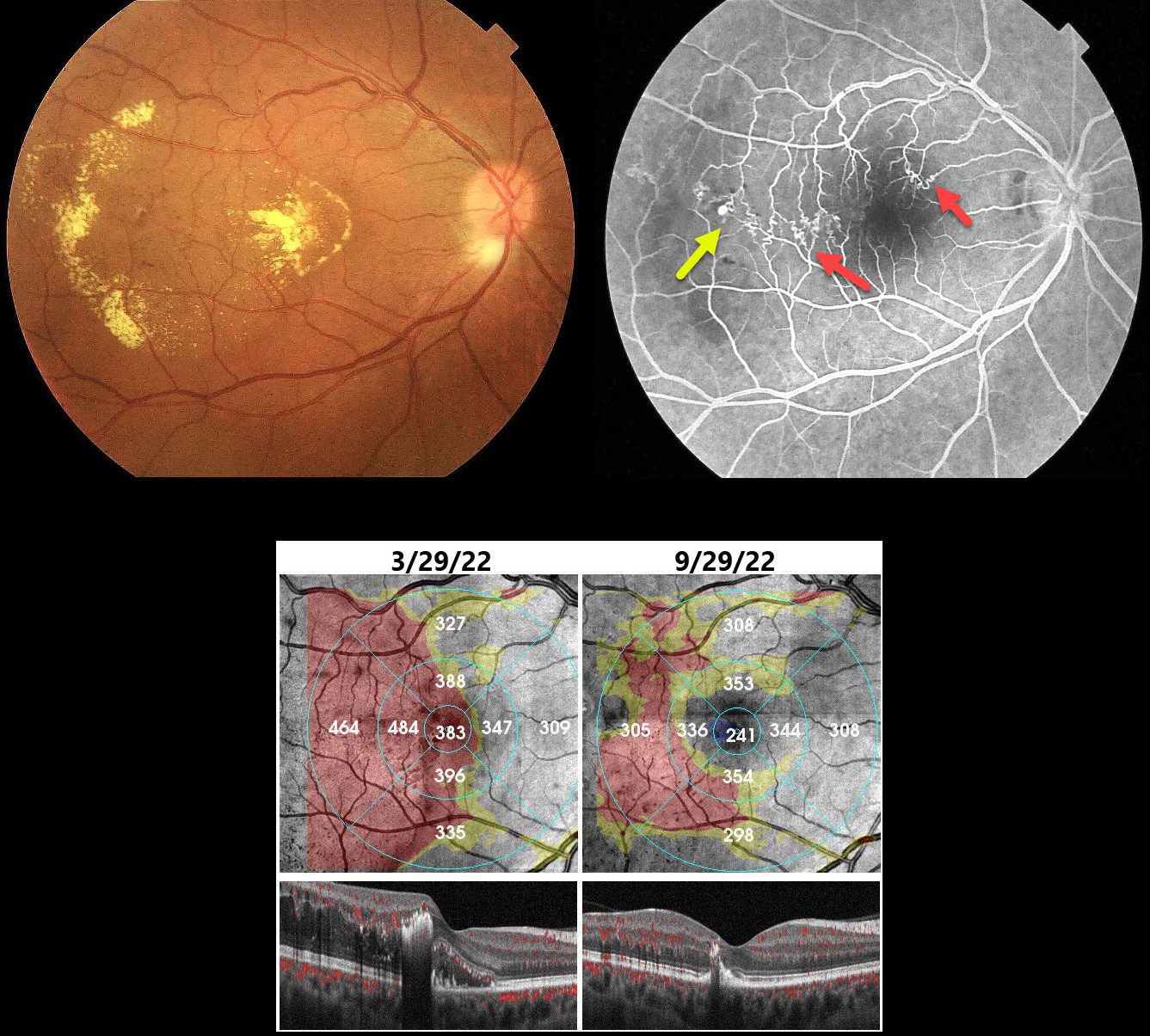
BRVO WITH RETINAL VENOUS MACROANEURYSM
Seif Anwar
Originally posted on @retina.rocks August 22, 2024
This 40YO hypertensive male presented with gradual vision loss in his right eye. Vision was 20/200 OD and 20/20 in his normal OS.
Color imaging shows central and temporal macular lipid and telangiectasia. Fundus fluorescein angiography shows a superotemporal branch retinal vein occlusion (BRVO) with superonasal and temporal collateral vessels. A retinal venous macroaneurysm (RVM) is noted distally.
Our patient was initially treated unsuccessfully with several monthly Lucentis injections. The macular edema and lipid improved following macular grid laser with focal treatment of the RVM.
Learning Points:
In 1990 Cousins et al reported that RVMs can be an additional finding in occlusive venous disease, always in association with collateral vessels or capillary or arterial macroaneurysms (AJO 1990;109:567-570). These vascular abnormalities tend to have a higher rate of intraretinal lipid (40%) but are less likely to affect central vision since they are usually outside the macula. They tend to be associated with retinal ischemia, thus increasing the risk of developing retinal neovascularization (32%).
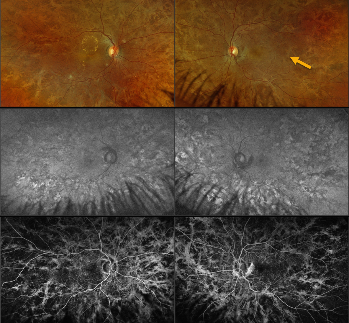
CHOROIDEREMIA CARRIER
Originally posted on @retina.rocks August 20, 2024
This 42YO female with type 2 diabetes was referred for asymptomatic diabetic macular edema (DME). There was no family history of ocular disease. Vision was 20/40 OD and 20/20 OS.
Optos color RGB imaging shows diffuse coarse subretinal hyperpigmentations throughout each posterior pole. Fundus autofluorescence (FAF) shows variable hyper- and hypo-FAF from these pigmentary changes, with fluorescein angiographic window defects. Genetic testing revealed a heterozygous pathogenic CHM mutation.
Learning Points:
Choroideremia is an X-linked recessive chorioretinal dystrophy caused by a mutation in the CHM gene located on the long arm of the X chromosome. Sons of female carriers have a 50% chance of developing choroideremia, and daughters have a 50% chance of becoming carriers. Some female carriers can develop asymptomatic pigmentary changes, as in our patient, or frank choroideremia due to irregular inactivation of the X chromosome (see Jauregui et al, AJO 2019;207:77-86).
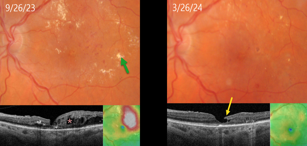
FOCAL LASER FOR DIABETIC MACULAR EDEMA (DME) WITH SUBSEQUENT DEGENERATIVE LAMELLAR MACULAR HOLE
Originally posted on @retina.rocks August 15, 2024
This 71YO female with type 2 diabetes presented diabetic macular edema (DME) and lipid exudates. Triton swept-source OCT shows central foveal thinning with surrounding cystic edema. Vision was 20/60. Thermal macular laser was applied.
Six months later, the edema completely resolved following this single treatment, with near total resolution of the macular lipid. A partial degenerative lamellar macular hole (LMH) is noted. Vision improved to 20/50.
Learning Points:
Our patient exemplifies why macular laser still has a place in any retinal surgeon’s toolbox and should be considered a viable option for treating non-central involved diabetic macular edema (NCI-DME) and even center-involved edema (CI-DME) when the leaking microaneurysms are outside the foveal avascular zone (FAZ). In our practice, macular laser remains the initial treatment for eyes with NCI-DME. When applied gently with small, low-power, and short-duration applications well outside the FAZ, patients rarely, if ever, experience scotoma. Both the treatment burden and the financial costs to society are dramatically less. And there is no risk for endophthalmitis.
Although the role of thermal laser for treating DME is currently a source of debate (see Eye 2022;36:485-486 and Eye 2022;36:483-484 for great pro-laser and anti-laser editorials) we are not sure what all the fuss is about. Old-fashioned lasers work and, in our opinion, are incredibly under-utilized.
LMH can be defined as tractional or degenerative (Govetto et al, AJO 2016;164:99-109). Our patient developed a partial degenerative LMH following likely degeneration or rupture of a foveal cyst following closure of the leaking microaneurysms. There is some debate regarding the benefits of surgery for symptomatic LMH, with some suggesting visual improvement (Morescalchi et al, Retina, 2020;40:1087-1093) and others noting little visual benefit (Mohammed and Thompson, J Vitreoretinal Diseases 2024;8:125-130). Since our patient was visually happy, observation was recommended.
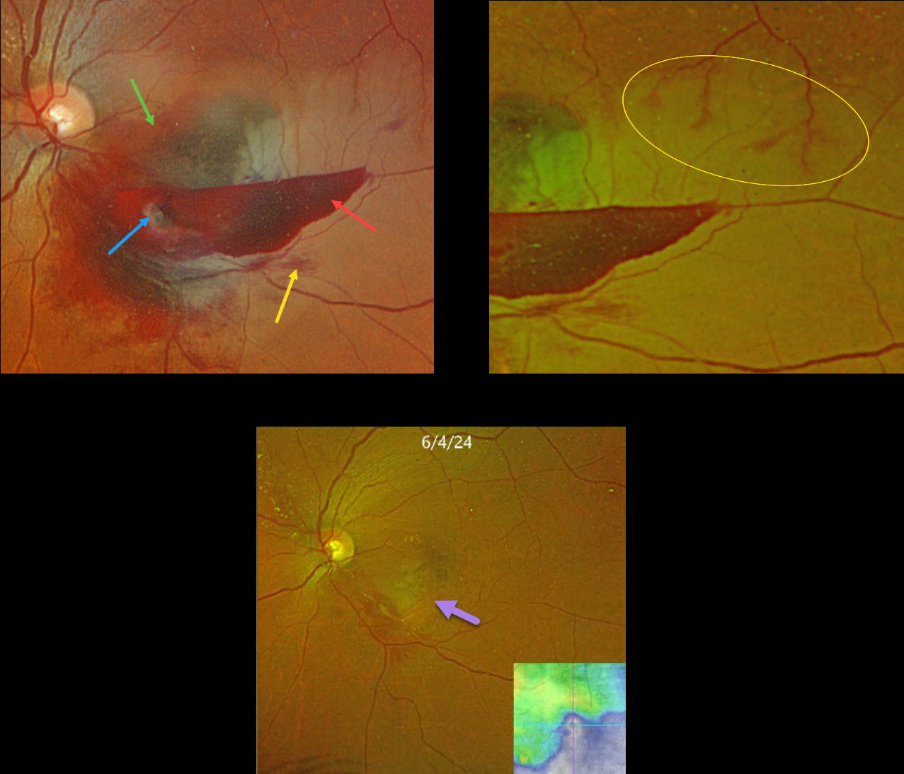
RETINAL ARTERIAL MACROANEURYSM (RAM) WITH BRAO
Originally posted on @retina.rocks August 13, 2024
This 59YO female presented with acute vision loss in her left eye. Vision was counting fingers.
Optos color RGB shows a multi-layered macular hemorrhage, including preretinal, intraretinal, and subretinal blood. The causative ruptured retinal arterial macroaneurysm (RAM) is noted within the preretinal blood inferonasally. The surrounding neurosensory retina is somewhat opaque from a secondary inferior branch retinal artery occlusion (BRAO).
Optos RG color imaging shows some periarterial sparing along the superotemporal edge of the BRAO. The perfused superior retinal arteriole, as it enters the region of the occlusion, still has some oxygen that is diffusing into the perivascular tissue.
The patient was treated with several monthly Avastin injections in hopes of more rapidly resolving the surrounding fluid to help relieve arterial compression. Although the blood and fluid resolved, secondary central and inferotemporal macular atrophy resulted from the prior BRAO. Vision was 20/400.
Learning Points:
A BRAO can be a rare complication of direct thermal laser of a RAM (Russell and Folk, AJO 1987;104:186-187), which is one reason it is almost never performed anymore. In this case, we believe the BRAO was secondary to mechanical arterial compression from the surrounding blood.
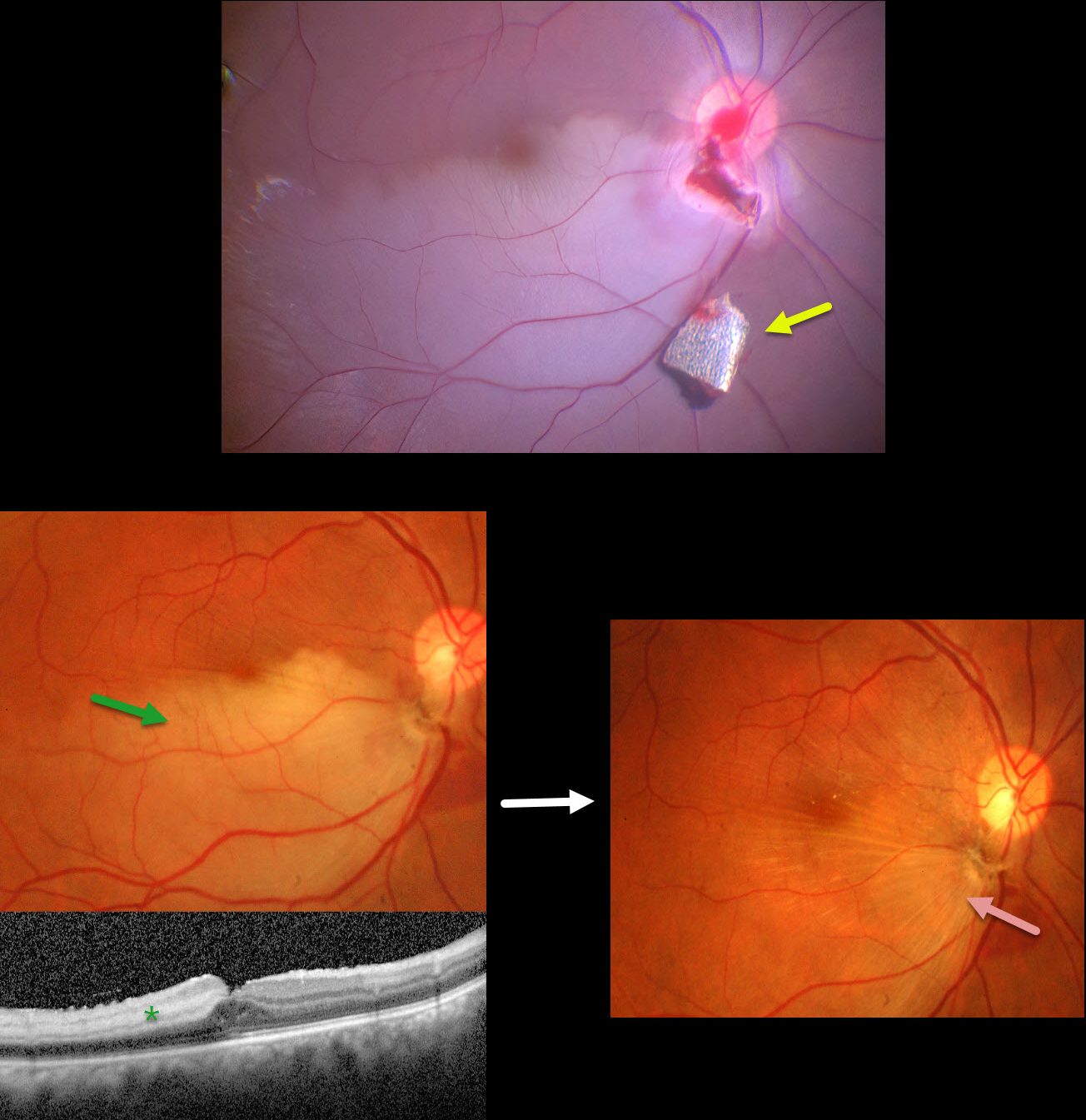
INTRAOCULAR FOREIGN BODY WITH SECONDARY BRAO
Anjana Mirajkar and Manish Nagpal
Originally posted on @retina.rocks August 5, 2024
This 34YO male presented with pain and vision loss immediately following a metal-on-metal injury at work. Vision was light perception. Slit lamp examination revealed a corneal laceration and traumatic cataract. B-scan ultrasonography and CT scanning revealed an intraocular foreign body (IOFB). The patient was immediately taken to surgery for corneal wound repair, lensectomy, and vitrectomy.
Intraoperative photography shows a metallic intraocular foreign body (IOFB) resting just inferior to the optic nerve. The impact site is just inferior to the optic nerve, with an inferotemporal branch retinal artery occlusion (BRAO) distally. The IOFB was removed with a rare-earth magnet.
Two weeks following surgery, vision was 20/120 with an aphakic correction. The inferior retina remains opaque, and the OCT shows inner retinal hyperreflectivity from the BRAO. A secondary intraocular lens implantation was performed 3 months postoperatively. Eleven months following the initial surgery, the BRAO is fully resolved, and secondary retinal striae extend outwards from the impact site. Vision remarkably improved to 20/30.
This case was published in the Indian Journal of Ophthalmology (Nagpal M et al, Ind J Ophthalmol 2018;66;146-148).
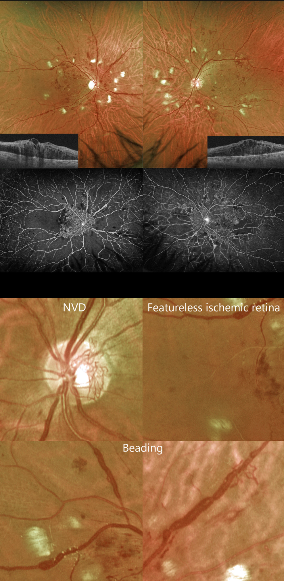
PROLIFERATIVE DIABETIC RETINOPATHY (PDR)
Originally posted on @retina.rocks August 1, 2024
This 41YO male with type 2 diabetes complained of vision loss for 5 months. He was recently hospitalized for severe systemic hypertension. Vision was counting fingers OD and 20/60 OS.
Optos color RG imaging shows bilateral venous beading and extensive cotton wool spots. Diffuse central edema is noted on Triton swept-source OCT, and severe ischemia is seen on fluorescein angiography.
But on closer inspection, the severe ischemia is evident clinically. The retina between the larger vessels is featureless, and many of the veins are sausaged and beaded. Once these clinical features are recognized, the angiographic findings can usually be predicted.
There is a relative lack of severe retinal hemorrhages and neovascularization (only disc neovascularization is present). The extensive cotton wool spots are more hypertensive than diabetic changes but indicate ischemia of the nerve fiber layer.
The patient subsequently underwent full bilateral panretinal photocoagulation and anti-VEGF therapy.
BRVO
Originally posted on @retina.rocks July 18, 2024
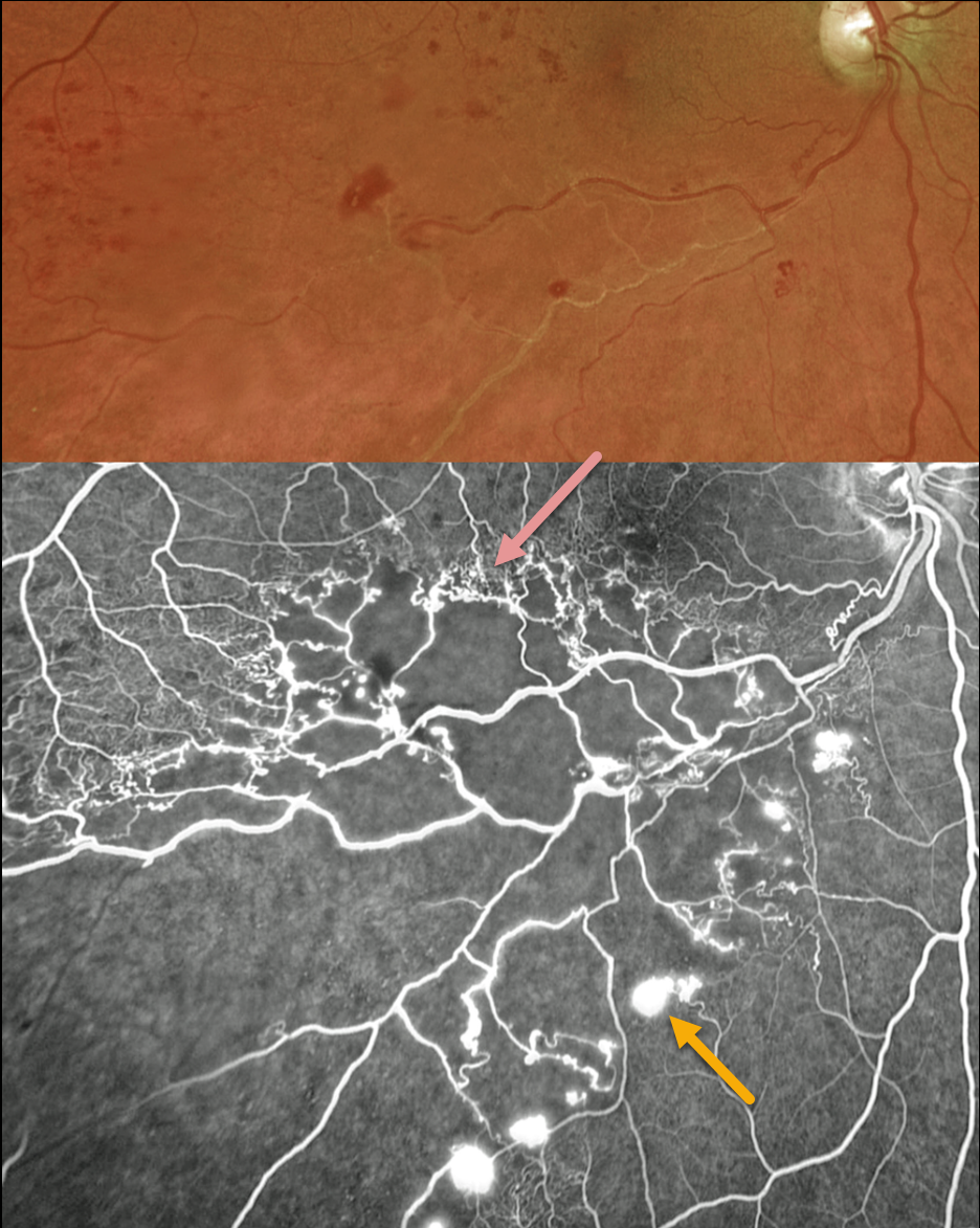
This 56YO male presented with a major inferotemporal ischemic branch retinal vein occlusion (BRVO) in his right eye and 20/40 vision. Despite the deceptively good vision, Optos ultra-widefield imaging showed marked ischemia throughout the distribution of the occlusion, which fortunately spared the central macula. A row of collateral vessels extends along the superior edge of the occlusion, with retinal neovascularization extending along its inferior extent. Leaking disc neovascularization is also present. A scatter laser throughout the area of ischemia was performed, but unfortunately, he was then lost to follow-up.
Learning Points:
In 1986, the Branch Vein Occlusion Study Group reported that scatter laser decreased the risk of vitreous hemorrhage from retinal neovascularization in BRVO, and suggested that laser should be applied once neovascularization develops (Arch Ophthalmol 1986;104:34-41). Thirty-five years later, scatter laser remains the gold standard, although some may prefer to first stabilize neovascularization with anti-VEGF therapy before laser.
Collateral vessels are dilated, pre-existing vessels that bypass an occlusion. These are most commonly seen at the posterior pole following retinal venous occlusions and are exceedingly rare with arterial occlusions. The endothelial tight junctions are intact, so they don’t leak angiographically. This helps differentiate them from neovascularization, which does leak.
CRAO
Originally posted on @retina.rocks July 16, 2024
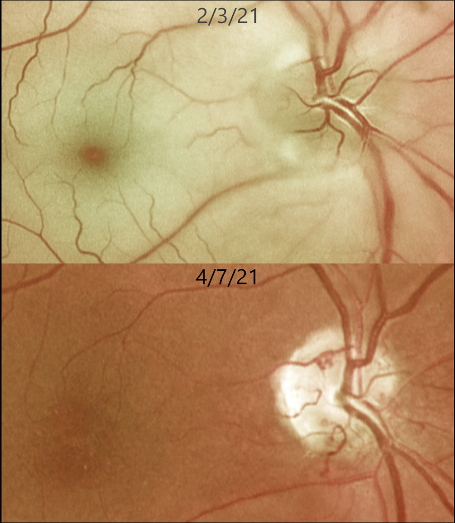
This 62YO female presented with an acute counting-fingers central retinal artery occlusion. Emergent stroke workup revealed a silent acute lacunar infarct. The retinal opacification completely resolved 2 months later. New fine collateral vessels were noted on the nerve. Vision remained at counting fingers.
Learning Points:
Collateral vessels are dilated, pre-existing vessels that bypass an occlusion. These are most commonly seen at the posterior pole following retinal venous occlusions and are exceedingly rare with arterial occlusions. The endothelial tight junctions are intact, so they don’t leak angiographically. This helps differentiate them from neovascularization, which does leak.
PROLIFERATIVE DIABETIC RETINOPATHY (PDR)
Originally posted on @retina.rocks July 15, 2024
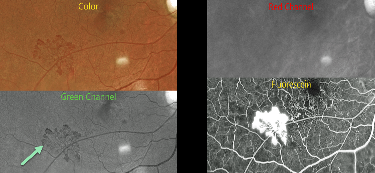
This 54YO male with proliferative diabetic retinopathy (PDR) and retinal neovascularization was evaluated with Optos multimodal imaging.
Optos color RG imaging shows active neovascularization in the inferotemporal macula.
The neovascularization is best seen in the green channel, which isolates the RPE and neurosensory retina. The blood vessels virtually disappear with the red channel, which is best for viewing the choroid. Finally, the vessels profusely leak on fluorescein angiography.
Learning Points:
Optos RG color images are generated from a red (635nm) and green (532nm) laser. The absence of a blue channel results in their greenish tint, which, at first, many clinicians find somewhat unsettling. However, much information is contained in these images once one understands how they are constructed. As shown in our patient, these images allow isolation of the neurosensory retina/RPE and choroid and often reveal more information than is visible in a true-color image.
Red neovascularization on an orange background (color image) is much more difficult to see than black neovascularization on a grey background (green channel). Recently, Optos introduced a blue channel to its California model, which, in our opinion, now offers the best of all worlds: stunning true-color images along with the ability to view each channel separately.
LOCALIZED TRACTIONAL RETINAL DETACHMENT
Ali Lamin and Ritu Chaturvedi
Originally posted on @retina.rocks July 4, 2024
This 55YO female with type 2 diabetes presented for a routine follow-up without visual complaint. Vision was 20/30 bilaterally.
MultiColor imaging of her right eye shows a localized traction retinal detachment (TRD) in the distal superotemporal macula. The TRD is much less obvious with Optos color RG imaging.
The source of the traction is a small area of retinal neovascularization that leaks on fluorescein angiography. This localized traction is also confirmed on OCT. Although vitrectomy was not recommended given the localized nature of the TRD, panretinal photocoagulation was performed.
Learning Points:
Although pretty color images that match what we see on biomicroscopy are most intuitive, newer imaging modalities that ‘distort’ the normal fundus colors often contain very clinically relevant information. Standard Optos color RG images are generated from a red (635nm) and green (532nm) laser. Although these images have a greenish tint, they essentially contain two images in one: the RPE/neurosensory retina is best captured with the green channel, and the choroid is best seen with the red channel. A newer Optos true-color RGB unit has recently been introduced, which maintains the ability to view the separate RG channels while also generating a true-to-life color image.
Heidelberg Engineering’s Multicolor imaging module similarly utilizes a blue (488nm), green (515nm), and infrared (820nm) confocal laser to generate a fundus image. The blue laser best images the vitreoretinal interface and inner retina; the green laser images the mid retina; and the infrared laser images the outer retina, RPE, and choroid (Roy et al., Surv Ophthalmology 2024;69:378-402).
Combining these 3 wavelengths into a single file produces a pseudocolor image that, like the Optos, provides the clinician with more information than a standard color photo. As seen in our patient, Multicolor imaging best illustrates both the presence and extent of TRD (Gadde et al, Indian J Ophthalmol 2022;70:465-470).
CRVO
Originally posted on @retina.rocks July 2, 2024
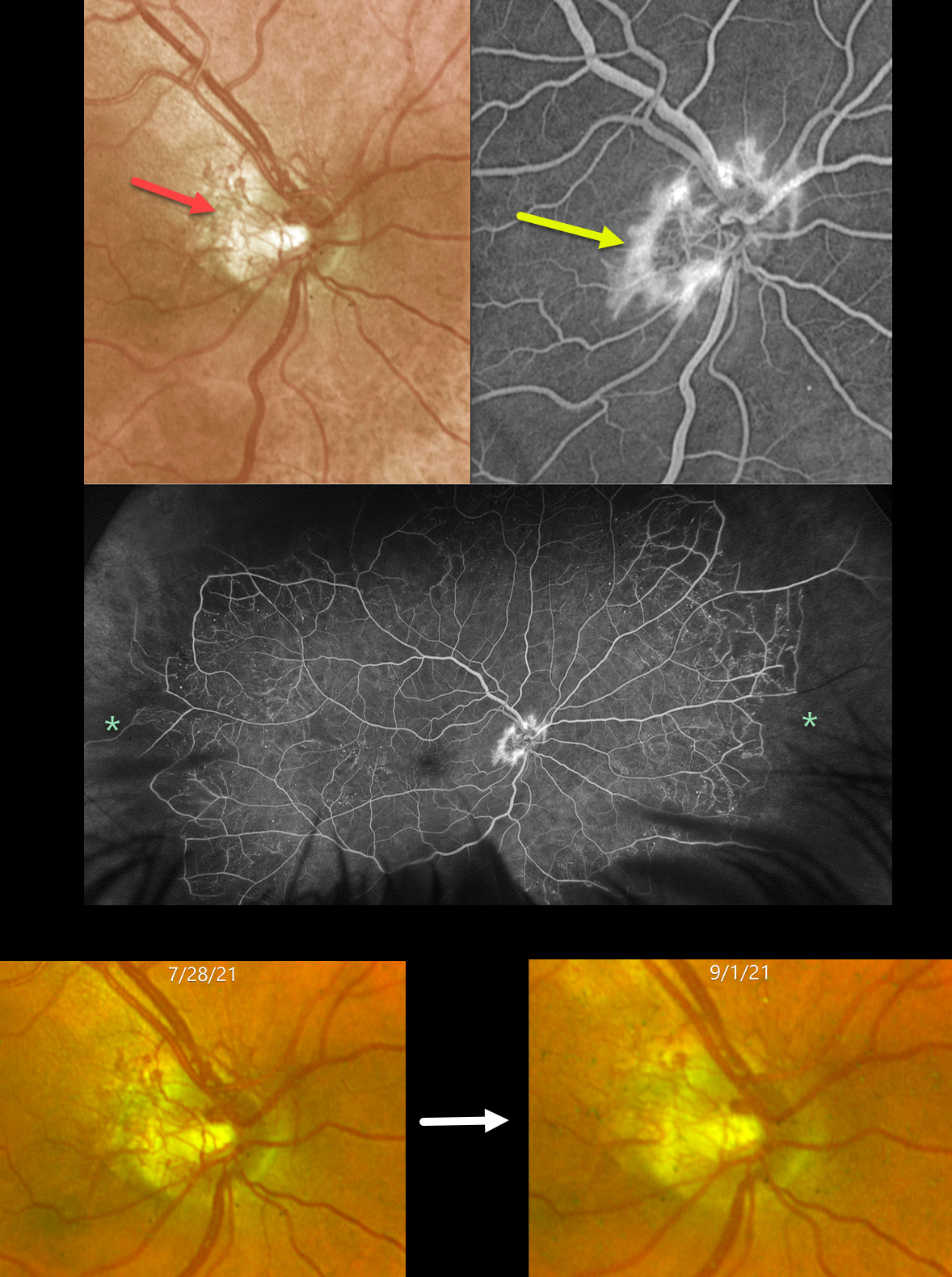
This 57YO male had a history of an old central retinal vein occlusion (CRVO) in his left eye with hand motion vision. After being lost to follow-up for 5 years, he presented on 7/28/21 with an asymptomatic CRVO with 20/25 vision in his right eye.
Optos color RG imaging shows diffuse disc neovascularization (NVD) that leaks on angiography. Ultrawidefield fluorescein angiography shows good posterior perfusion but with marked encircling peripheral ischemia. The neovascularization partially regressed 6 weeks after panretinal photocoagulation and completely regressed with subsequent laser and anti-VEGF injections (not shown).
Learning Points:
Anterior segment neovascularization is more commonly noted with central retinal vein occlusions, whereas posterior segment neovascularization is more common with branch retinal vein occlusions. Hayreh’s classic 1983 paper noted that only 5% of eyes with CRVO will develop NVD, as in our patient (Ophthalmology 1983;90:488-506).
The Central Vein Occlusion Study (CVOS) defined ischemic CRVO as having at least 10 disc areas of angiographic retinal nonperfusion as determined by standard photographic views with the Canon (60 degrees) or Topcon (45 degrees) wide-angle fundus camera (Ophthalmology 1995;102:1434-1444). Although clearly ischemic peripherally, our patient would have been classified by the CVOS as a perfused occlusion.
As with diabetic retinopathy (see Brucker, Retina 2021;41:461-463), ultrawidefield imaging and anti-VEGF injections have thrown a literal wrench into how we think about, classify, and treat retinal disorders.
CRVO + CILIORETINAL ARTERY OCCLUSION
Amit Nandan Tripathi and Vipin Rana
Originally posted on @retina.rocks June 18, 2024
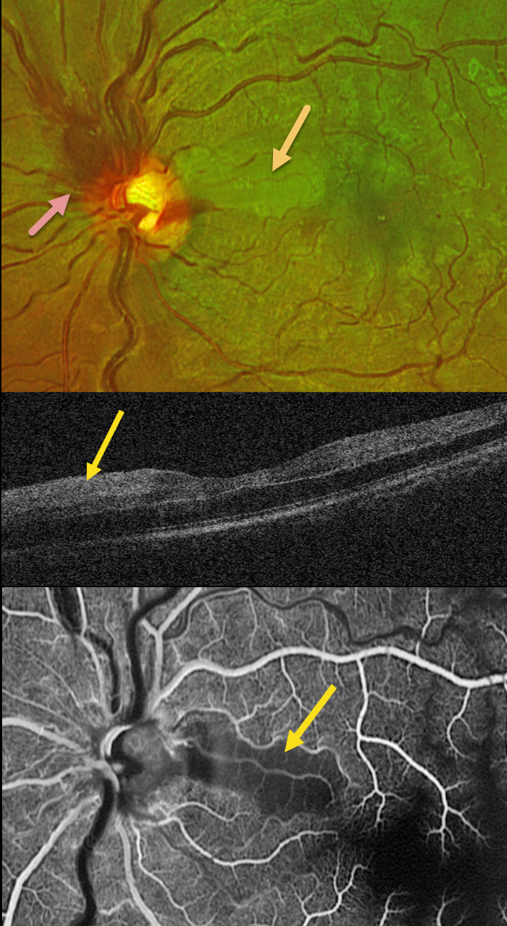
This healthy 30YO male presented with sudden vision loss in his left eye. Vision was 20/20 in his healthy right eye and counting fingers in his left eye.
Optos color RG imaging shows an acute non-ischemic central retinal vein occlusion (CRVO) with mild inner retinal hemorrhages scattered in all quadrants. The optic nerve is somewhat swollen superiorly, and there is an acute cilioretinal artery occlusion in the superior papillomacular bundle.
This area is hyperreflective on OCT. Fluorescein angiography shows a well-perfused CRVO except for a localized area of ischemia within the nasal occlusion.
Learning Points:
Some patients with CRVO will develop a secondary non-embolic cilioretinal artery occlusion caused by either compression from optic nerve edema or from a sudden rise in intraluminal retinal capillary bed pressure (Hayreh et al, Retina 2008;28:581-594). Observation was recommended, and vision improved to 20/60 five days after symptom onset.
TRACTIONAL RETINAL DETACHMENT
Originally posted on @retina.rocks June 10, 2024
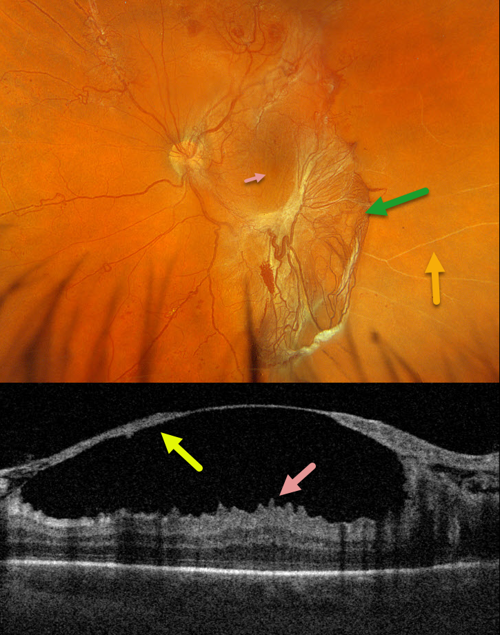
This 42YO male with type 2 diabetes presented with 1 week of vision loss in his left eye. Vision was 20/40 OD and 20/100 OS.
Optos color RGB imaging shows a large patch of variably fibrosed retinal neovascularization extending from the temporal macula into the inferior midperiphery. Vertical macular striae are drawn towards the neovascularization inferiorly. Scattered neovascularization is seen elsewhere, along with nonperfused large retinal vessels temporally.
Triton swept-source OCT through the macular center shows a thickened detached posterior hyaloid that extends from the optic nerve into the temporal macula. The macular striae noted on the Optos image correspond to inner macular folds on OCT. Vitrectomy surgery was scheduled.
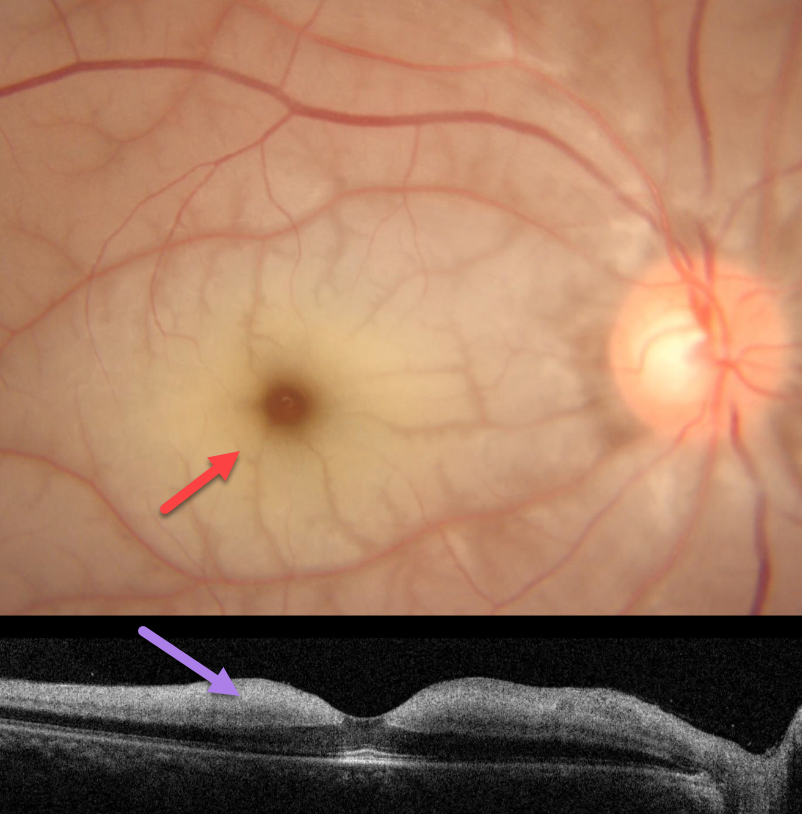
CRAO
Asma Samsudeen and Ashish Sharma
Originally posted on @retina.rocks June 6, 2024
This 41YO male presented with a 2-day history of acute vision loss in his right eye. Vision was counting fingers OD and 20/20 in his normal OS.
Fundus photography shows an acute central retinal artery occlusion (CRAO) with a pale, opaque inner retina and a cherry-red spot. The retina immediately adjacent to the radiating perifoveal arterioles is relatively transparent, creating a fern-like appearance. OCT shows a slightly thickened and markedly hyperreflective inner retina. The patient was immediately sent to the nearest stroke center.
Learning Points:
Acute CRAO often exists along a spectrum from complete occlusion with a stagnant blood column to a more partial blockage with variable patchy ischemia. Our patient still has enough active flow to supply the neurosensory retina immediately adjacent to the smaller arterioles, but not enough to supply the more distal retinal tissue.
About 90% of CRAO are embolic, with the remainder most commonly due to giant cell arteritis. The lack of a visible embolus, as in our patient, does not preclude an embolic cause since the causative embolus can either be posterior to the visible disc or have already traveled further downstream.
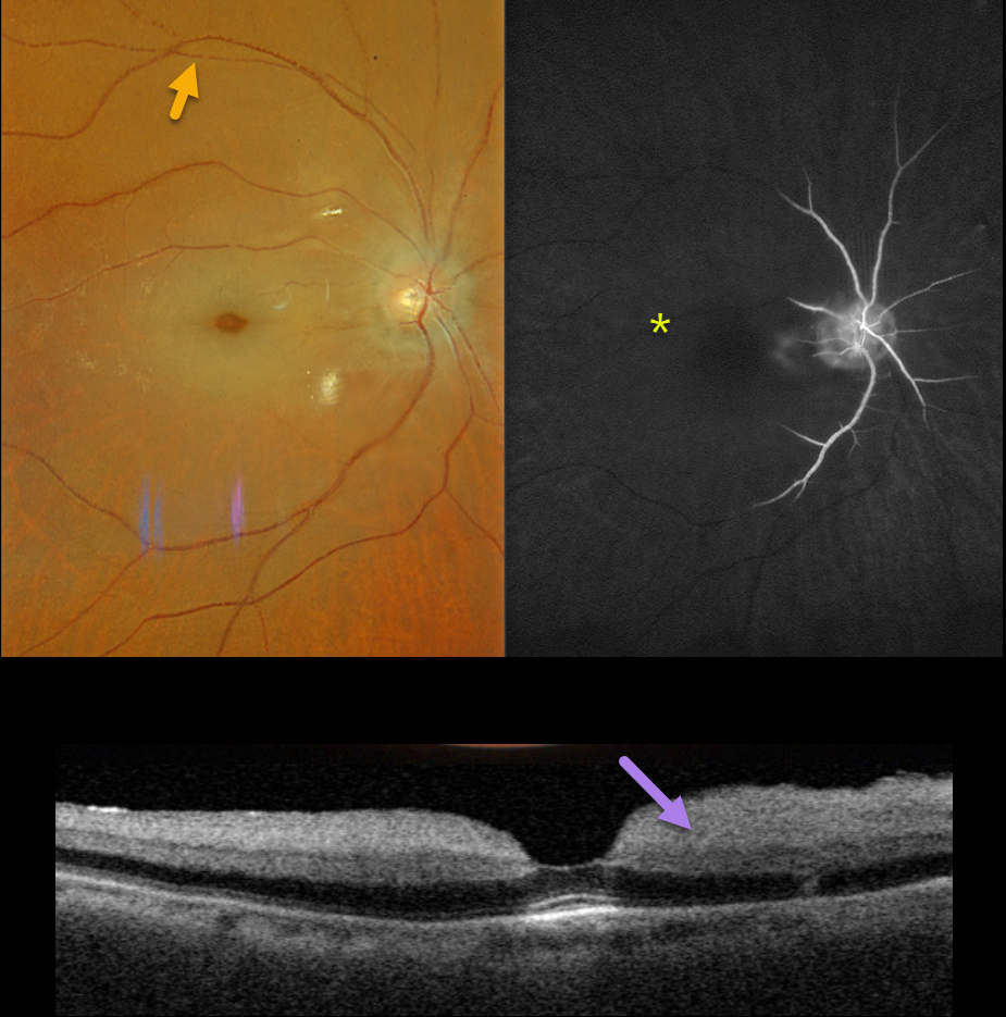
CRAO
Originally posted on @retina.rocks April 18, 2024
This 89YO male presented with 2 days of acute and severe vision loss in his right eye. Vision was no light perception.
Optos color RGB imaging shows an acute central retinal artery occlusion (CRAO) with a cherry-red spot and a stagnant blood column, best seen superiorly as box-carring of the column.
Fluorescein angiography shows profound ischemia, with no vascular filling beyond the peripapillary region.
Triton swept-source OCT shows a diffusely slightly thickened and markedly hyperreflective inner retina. He was immediately referred to the nearest stroke center.
Learning Points:
Patients with acute retinal ischemia (defined as transient monocular vision loss, acute branch retinal artery occlusion, or acute CRAO) require emergent referral to a stroke center. This is especially urgent with an acute CRAO since about 75% of patients have already developed a recent stroke.
If the patient presents immediately following the occlusion, measures to acutely lower the intraocular pressure to move the causative embolus downstream can be performed, including ocular massage, paracentesis, and breathing into a bag.
Unfortunately, most patients present outside this window (Shah et al, Ophthalmology Retina 2023;7:527-531) and even then, these measures are usually ineffective.
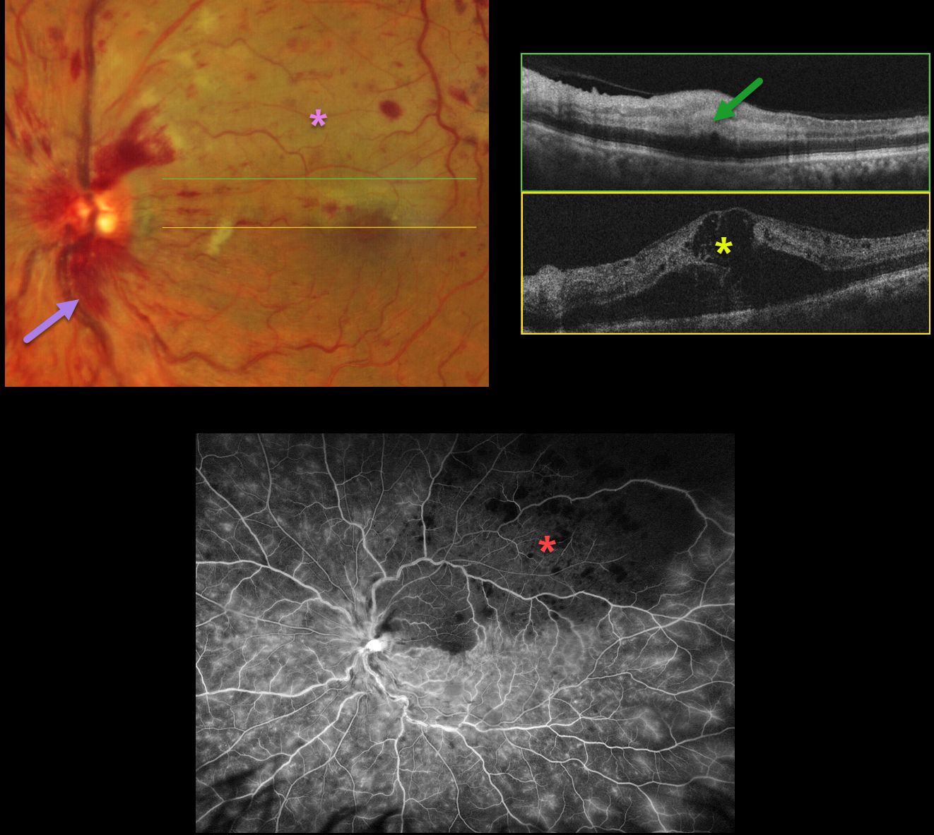
CRVO + SECONDARY BRAO
Will Gibson
Originally posted on @retina.rocks April 15, 2024
This 66YO male presented with sudden vision loss in his left eye. Vision was 20/30 in his normal right eye and counting fingers in his left eye.
Optos color RG imaging shows a central retinal vein occlusion (CRVO) with mild retinal hemorrhages in all quadrants, except for more moderate hemorrhages superotemporally. A secondary superior branch retinal artery occlusion (BRAO) bisects the fovea.
OCT scanning shows marked central edema, with hyperreflective and thickened inner retina within the BRAO. Fluorescein angiography shows a well-perfused CRVO, except for delayed arterial filling and ischemia within the BRAO.
Learning Points:
Some patients with CRVO will develop a secondary non-embolic BRAO caused by compression from optic nerve edema. However, our patient’s optic nerve did not appear that swollen. Our patient was started on anti-VEGF therapy for the severe macular edema.
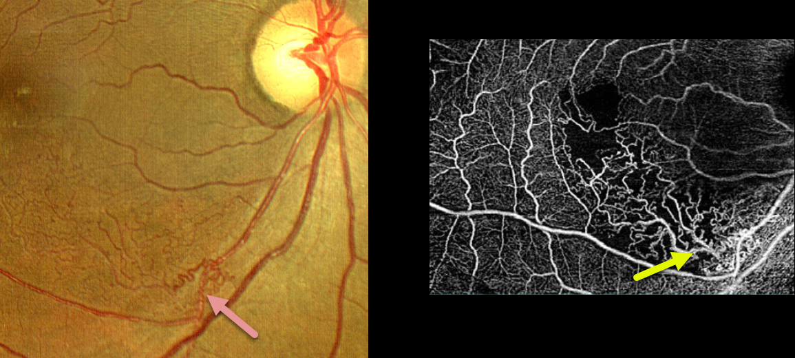
BRVO
Nilesh Kumar
Originally posted on @retina.rocks April 11, 2024
This 53YO female presented with 20/80 vision in her right eye from a macular branch retinal vein occlusion (BRVO) with macular edema. The edema resolved following a single injection of a Lucentis biosimilar. She was then lost to follow-up for 8 months, when she returned with 20/20 vision.
Extensive inferonasal macular collateral vessels are seen on fundus photo and OCTA. OCT showed no edema (not shown).
Learning Points:
Collateral vessels are dilated, pre-existing vessels that bypass an occlusion. These are most commonly seen at the posterior pole following retinal venous occlusions, but can also occur at the optic disc with optic nerve meningiomas. The endothelial tight junctions are intact, so they don’t leak on fluorescein angiography. This helps differentiate them from neovascularization, which does leak.
Collaterals most commonly form in the deep vascular plexus, which represents the primary venous drainage for the inner retina (Freund et al, JAMA Ophthalmology 2018;1262-1270).
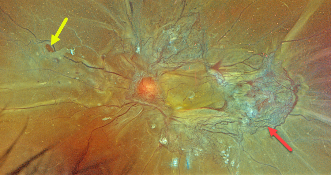
COMBINED TRACTIONAL-RHEGMATOGENOUS RETINAL DETACHMENT
Originally posted on @retina.rocks March 27, 2024
This 46YO female with type 2 diabetes presented with recent bilateral vision loss. Vision was 20/200 OD and hand motion OS.
Optos color RGB imaging of her left eye shows a combined tractional-rhegmatogenous total retinal detachment with fibrovascular tissue encircling the macula. Temporally radial outer retinal folds are dragged toward the more succulent, red neovascularization. A retinal tear is noted temporally.
Vitrectomy surgery was scheduled.
Proliferative diabetic retinopathy with disc and peripheral neovascularization was noted in her right eye (not shown). Panretinal photocoagulation was performed in her left eye and surgery was scheduled for her left eye.
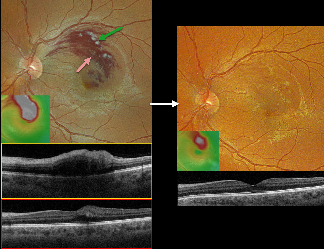
BRVO
Originally posted on @retina.rocks February 27, 2024
This healthy 33YO male presented with a 1-week history of vision loss in his left eye. Vision was 20/20 in his normal right eye and 20/60 in his left eye.
Optos color RGB imaging shows a superior macular branch retinal vein occlusion (BRVO) with relatively confluent inner retinal hemorrhages and superiorly located nerve fiber layer infarcts. Retinal fluid extends through the fovea.
Triton swept-source OCT through the superior macula shows retinal thickening and inner hyperreflectivity from the blood. Scanning through the macular center shows very shallow central edema. The macular cube shows the extent of the retinal thickening.
Following several monthly Avastin injections, vision improved to 20/30, and the macula was virtually free of fluid.
Learning Points:
Macular grid laser photocoagulation (Branch Vein Occlusion Study Group, AJO 1984;96:271-282) was the mainstay for treating persistent vision loss due to BRVO macular edema until the advent of intravitreal steroid and anti-VEGF therapy in the early-to-mid 2000s.
Although we were initially ‘WOW’ed by the near immediate and dramatic edema improvement that was never seen with thermal laser, the SCORE Study Research Group in 2009 suggested that grid laser should “remain the benchmark against which other treatments are compared” due to similar vision between intravitreal triamcinolone and laser but with fewer laser adverse events (Arch Ophthalmol 2009;127:1115-1128).
Intravitreal anti-VEGF injections have subsequently become the standard of care for initial therapy. A recent systematic review and meta-analysis showed that a treat-and-extend protocol is as effective as monthly and PRN regimens (OSLI Retina 2023;54:131-138).
Adding macular laser probably does not reduce the treatment burden or further improve vision (Ojima et al, Retina 2023;43:999-1004).
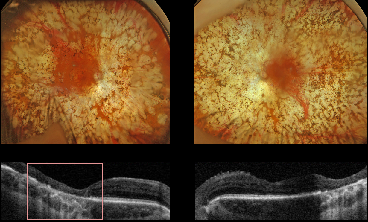
PANRETINAL PHOTOCOAGULATION
Originally posted on @retina.rocks February 18, 2024
This 53YO female with type 1 diabetes received bilateral panretinal photocoagulation (PRP) for proliferative diabetic retinopathy (PDR) about 30 years previously from an outside practice. She presented for her yearly retinopathy examination, and vision was stable at 20/200 OD and 20/30 OS.
Optos color RGB imaging shows bilateral confluent PRP scarring extending from within the arcades into the peripheries. Triton swept-source OCT of the right eye shows outer retinal atrophy extending through the foveal center from confluent macular laser scarring, with more temporal outer retinal atrophy in the left eye from confluent posterior PRP scarring.
Learning Points:
PRP should begin at least 1 disc diameter from the optic nerve and outside the major temporal arcades. Treatment is performed with a wide-angle lens or an indirect ophthalmoscope, and should be applied from the posterior retina towards the periphery to avoid accidental macular burns. The laser spots should be 200-500 microns in size at the retina, be spaced about one burn-width apart, and should extend as far anteriorly as possible.
The laser performed on this patient is wrong on so many levels, including spots extending into the macula, touching the nerve, and confluent elsewhere. The laser surgeon essentially gave this patient severe iatrogenic retinitis pigmentosa.
Although possibly appropriate before the advent of modern vitrectomy surgery and anti-VEGF therapy, this degree of laser should never be seen in today’s patients.
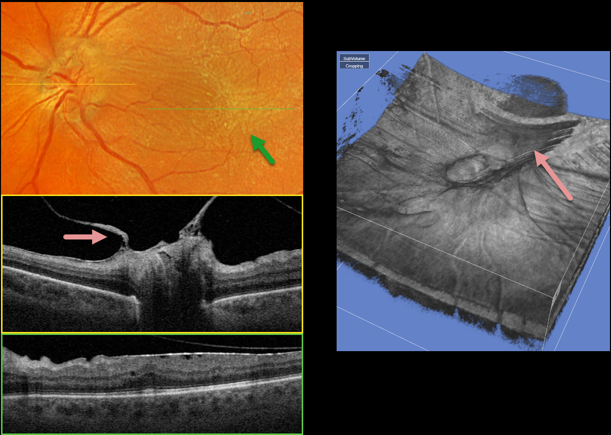
PROLIFERATIVE DIABETIC RETINOPATHY (PDR)
Originally posted on @retina.rocks January 29, 2024
This 58YO male with type 2 diabetes has long-involuted proliferative diabetic retinopathy (PDR) following full panretinal photocoagulation (PRP). He has no visual complaints, and vision is 20/50 OS.
Optos color RGB imaging shows a diffuse macular pucker with striae extending nasally towards the nerve and temporally into the distal macula.
A condensation of elevated fibrosed tissue, representing a partially detached Weiss’ ring, surrounds the optic nerve. This traction is clearly evident in Triton swept-source OCT (image 1, middle) and in the 3D reconstruction (image 2). A moderate epiretinal membrane extends throughout the macula (image 1, bottom).
Observation was recommended, and vitrectomy with membrane peeling will be performed if the patient becomes more symptomatic.
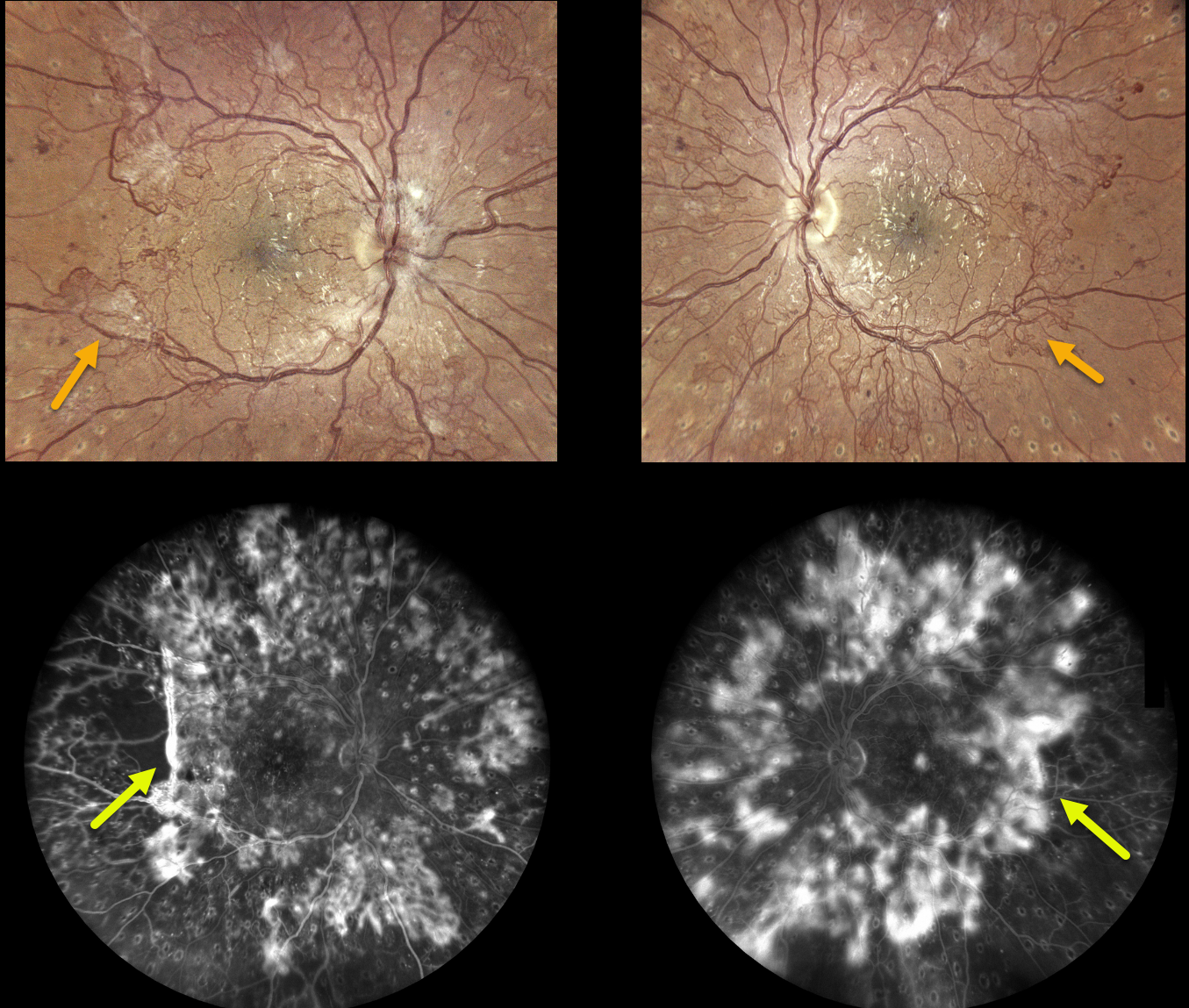
PROLIFERATIVE DIABETIC RETINOPATHY (PDR)
Originally posted on @retina.rocks January 25, 2024
This 28YO female with type 1 diabetes recently received bilateral full panretinal photocoagulation (PRP), completing treatment several months earlier.
Despite this, active neovascularization encircles the major arcades, extending into the midperipheries, clinically and angiographically. She was visually asymptomatic with vision of 20/25 OD and 20/30 OS. There was no edema or traction on OCT (not shown).
After extensive discussion with our patient, we decided against fill-in PRP or anti-VEGF therapy for fear of possibly causing contraction of the neovascularization and traction detachment. We will follow her closely and recommend vitrectomy if vitreous hemorrhage or traction develops.
What would you do?
Learning Points:
Before PRP, patients with proliferative diabetic retinopathy (PDR) routinely went blind, and one of the treatments actually included pituitary ablation (for an amazing historical perspective on diabetic laser photocoagulation, see Goldberg and Jampol, Ophthalmology 1987;94:741-746).
Following PRP, the retinal neovascularization usually morphs from active (tight small red vascular networks) into involuted (residual larger vessels, fibrosed tissue, or occasionally complete vessel disappearance) vasculature.
Once considered a “one and done” treatment, more recent studies show that patients may still need additional laser, anti-VEGF injections, or vitrectomy (see Gonzalez et al, Ophthalmology 2021;128:1448-1457).
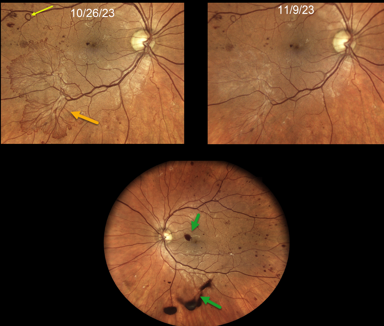
PROLIFERATIVE DIABETIC RETINOPATHY (PDR)
Originally posted on @retina.rocks January 15, 2024
This 37YO female with type 1 diabetes presented with recent floaters in her left eye. It had been over 5 years since her last eye examination. Vision was 20/30 OD and 20/50 OS.
Colour photography shows a large flat area of active retinal neovascularisation in the inferotemporal macula of the right eye. Some foveal preretinal blood and scattered small retinal neovascularisation are seen in her left eye.
Panretinal photocoagulation (PRP) was recommended for her left eye. The decision for how to treat the right eye was more complex. After a long talk with the patient, we gave an intravitreal Avastin injection in her right eye.
She failed to show for her visit one week later, but thankfully came in 2 weeks following the injection. Vision was 20/30 OD, and the neovascularisation in the right macula had nearly completely regressed. PRP was started.
Learning Points:
Intravitreal anti-VEGF is commonly used before diabetic vitrectomies to cause rapid involution of the vascularized preretinal tissue and control intraoperative bleeding during membrane dissection. These injections are given within a week of surgery, as neovascularisation can contract, increasing traction. This uncommon yet well-known phenomenon is called the ‘crunch’ syndrome (Tan et al, Survey of Ophthalmology 2021;66:926-932).
We also sometimes like to use anti-VEGF injections to stabilize these more succulent retinal neovascularizations before proceeding with PRP. In our experience, anti-VEGF therapy causes a more rapid and complete involution than we see with PRP.
Of course, the flip side is an increased risk of ‘crunch’, so these patients need to understand the possibility of early vitrectomy and be followed quite closely afterward.
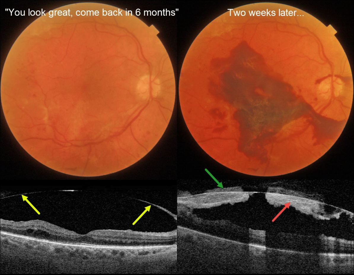
DIABETIC SUBHYALOID HEMORHAGE
Originally posted on @retina.rocks January 9, 2024
This 71YO diabetic male was examined over a year following completion of panretinal photocoagulation (PRP) for proliferative diabetic retinopathy (PDR). Vision was 20/50.
Triton color photography shows no macular edema, and swept-source OCT reveals a detached hyaloid elevated over the macula. He was told that everything looked great and to come back in 6 months. Famous last words…
Two weeks later, he presented with acute vision loss in this eye. Vision was counting fingers from a fresh macular subhyaloid hemorrhage. Both subhyaloid and prehyaloid blood are noted on Triton swept-source OCT.
An intravitreal Avastin injection was given, and vitrectomy will be recommended if the blood fails to clear.
Learning Points:
Although PRP is often a ‘one and done’ procedure, this is not always the case (Gonzalez et al, Ophthalmology 2021;128:1448-1457), and patients therefore need continued monitoring. Although our patient appeared perfect 2 weeks earlier, this case reminds us to remain humble in the presence of the retina and that even an involuted PDR requires lifelong follow-up.
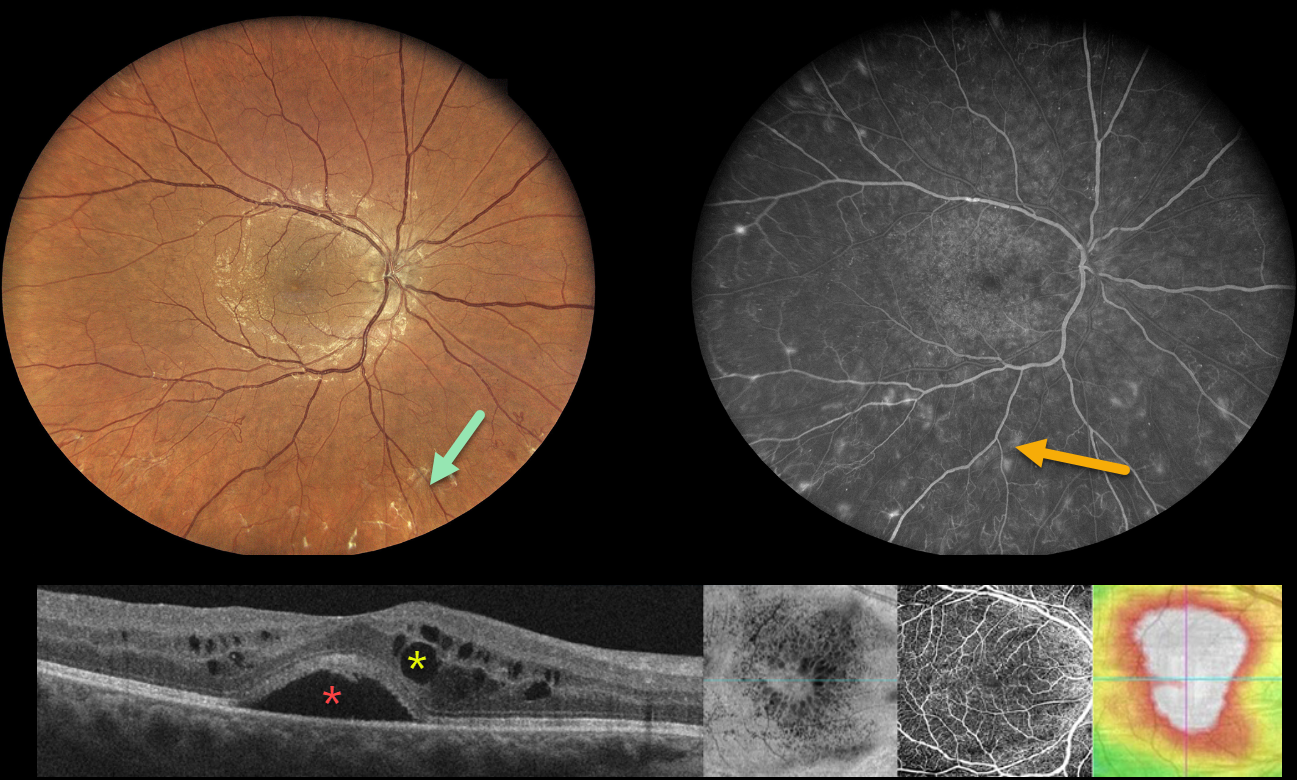
PROLIFERATIVE DIABETIC RETINOPATHY (PDR)
Originally posted on @retina.rocks January 3, 2024
This 38YO diabetic female was referred for diabetic retinal changes. Vision was 20/50 OD and 20/40 OS. Multimodal imaging shows the often subtle proliferative changes with various perspectives of her center-involved diabetic macular edema (CI-DME).
Widefield color imaging shows minimal retinal hemorrhages with a foveal cyst. Upon closer inspection, subtly fibrosed retinal neovascularization is noted, particularly inferiorly. Fluorescein angiography shows numerous buds of retinal neovascularization outside the macula.
Multiple OCT images are shown below, including a B-scan, which shows subfoveal fluid with cysts in both the inner and outer nuclear layers. En face imaging shows variably sized radiating cysts. OCT angiography is fairly normal with a preserved foveal avascular zone. Finally, a thickness map shows diffuse thickening.
Learning Points:
These presentations can be very humbling for us clinicians and remind us of the importance of peripheral retinal examination and ultra-widefield imaging with fluorescein angiography in screening for proliferative disease. Widefield OCT angiography may also prove to be an equally effective yet less invasive screening tool (Al-Khersan et al., AJO 2021;224:292-300).
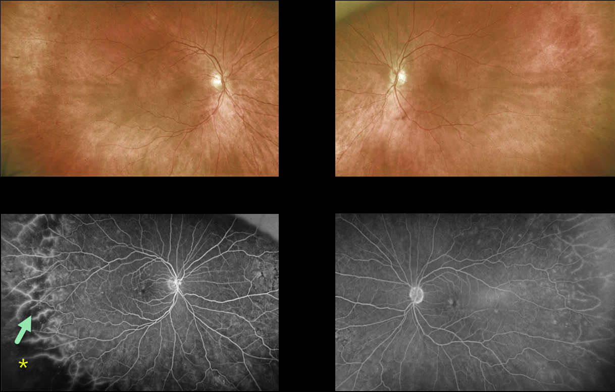
NON-PROLIFERATIVE DIABETIC RETINOPATHY (NPDR)
Originally posted on @retina.rocks November 20, 2023
This 69YO female with a history of type 2 diabetes was referred for asymptomatic retinal changes. Vision was 20/25 OU.
Optos color RG imaging shows virtually normal posterior poles with temporal peripheral retinal hemorrhages.
Fluorescein angiography shows temporal ischemia with diffuse vascular staining.
Learning Points:
The DRCR Retina Network’s Protocol AA found that ultrawidefield (UWF) imaging, compared to the standard ETDRS 7-field images, increases the frequency of diabetic retinopathy identification by nearly two fold (JAMA Ophthalmology 2019;137:65-73).
UWF fluorescein angiography further increases the sensitivity for assessing retinal ischemia, with about 70% of nonperfusion located outside the posterior pole (Retina 2022;42:1302-1310).
Finally, eyes with angiographically predominantly peripheral lesions, as in our patient, are at a higher 4-year risk of worsening disease, independent of the baseline diabetic retinopathy severity score (JAMA Ophthalmology 2022;140:946-954).
UWF fundus imaging has transformed the field of retina. In our practice, we are constantly humbled by the superiority of UWF imaging in finding peripheral pathology compared to our clinical examination. And increasingly, this technology is becoming the gold standard for identifying pathology and guiding our treatment decisions.
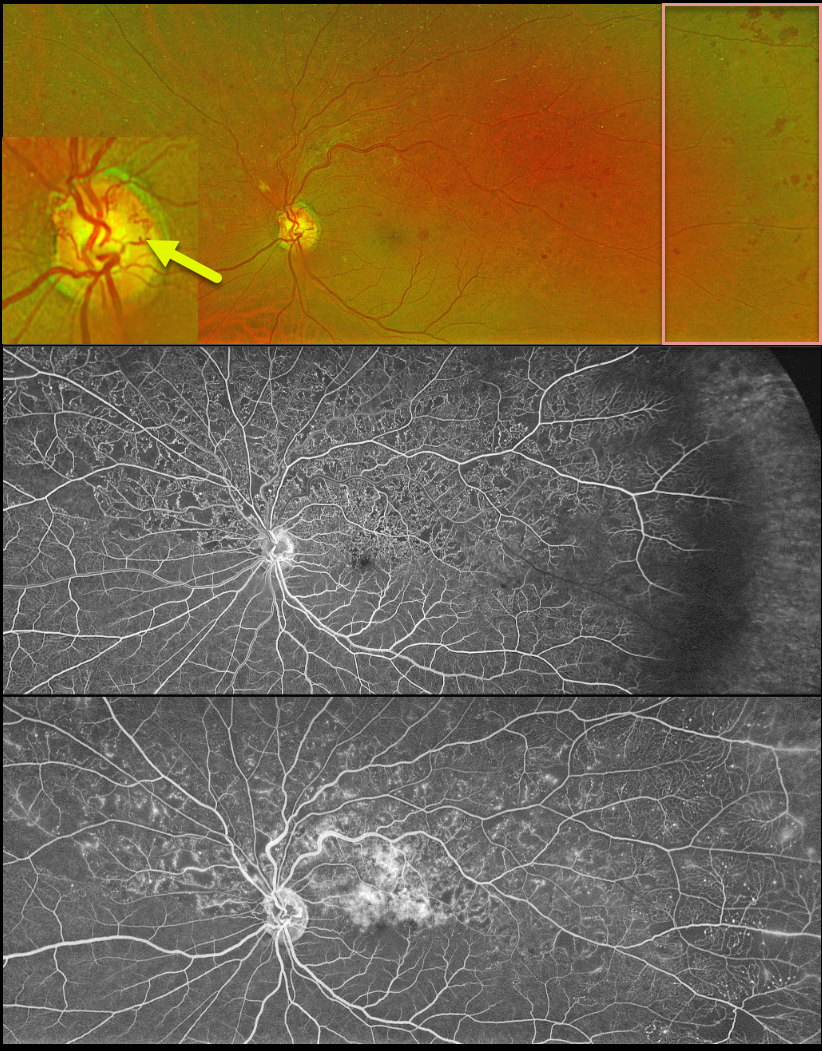
HRVO
Originally posted on @retina.rocks November 8, 2023
This 63YO male was referred for relatively asymptomatic retinal findings in his left eye. Vision was 20/60.
Optos color RG imaging shows a superior hemiretinal vein occlusion (HRVO) with mild hemorrhages, mostly in the temporal periphery. The occlusion is well-perfused without retinal neovascularization on fluorescein angiography. Prominent collateral vessels are noted on the optic disc superiorly. Observation was recommended.
Learning Points:
Branch retinal vein occlusions are virtually always caused by a retinal artery compressing a retinal vein within its common adventitial sheath. Some people are born with an anomalous two-trunked central retinal vein that independently drains the superior and inferior retinal hemispheres.
HRVO, similar to central retinal vein occlusion, is caused by compression of the retinal vein posterior to the lamina cribrosa (see Hayreh, Arch Ophthalmol 1980;98:1600-1609).
Collateral vessels are dilated, pre-existing vessels that bypass an occlusion. These are seen in the posterior pole most commonly following retinal venous occlusions, but can also occur on the optic disc with optic nerve tumors. The endothelial tight junctions are intact, so they don’t leak on angiography. This helps differentiate them from neovascularization, which does leak.
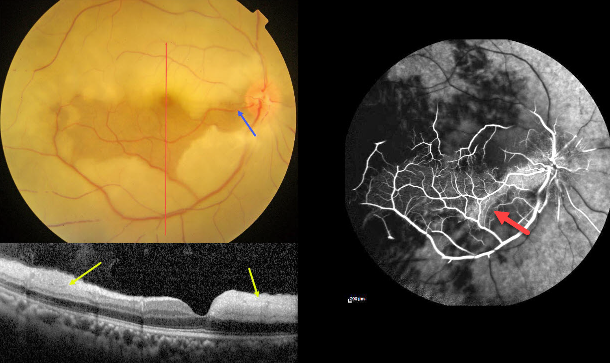
CRAO WITH CILIORETINAL ARTERY SPARING
Anjana Mirajkar and Manish Nagpal
Originally posted on @retina.rocks November 2, 2023
This 45YO male presented with acute vision loss in his right eye 4-5 days earlier. Vision was 20/80 in his right eye and 20/20 in his normal left eye.
Color photography shows an acute central retinal artery occlusion (CRAO) with sparing of the central and inferior macula due to a large cilioretinal artery.
The vertical OCT B-scan shows opaque hyperreflective paracentral inner retina with normal central and inferior foveal layers. Fluorescein angiography confirms preserved central and inferior macular arterial flow.
Workup elsewhere included a normal brain MRI and cardiac 2D Echo. Carotid Doppler showed a plaque in the right internal carotid artery.
Learning Points:
The inner two-thirds of the neurosensory retina is supplied by the central retinal artery, with the choroid supplying the RPE and photoreceptor layer. Acute CRAO, therefore, presents with a white edematous inner retina that spares the fovea.
Cilioretinal arteries, which arise from the posterior choroidal circulation, are present in up to 50% of individuals, and bilateral in about 25% (Schneider et al, Acta Ophthalmologica 2021;99:e310-e318). Our patient was quite fortunate to have a large cilioretinal vessel that relatively preserved central vision.
Patients with acute retinal ischemia (defined as transient monocular vision loss, acute BRAO, or acute CRAO) need to be emergently referred to a stroke center. This is especially urgent with an acute CRAO since about 75% of patients have already developed a recent stroke.
If the patient presents immediately after occlusion, measures to acutely lower intraocular pressure to move the causative embolus downstream can be performed, including ocular massage, paracentesis, and breathing into a bag. Unfortunately, most patients present outside this window (Shah et al, Ophthalmology Retina 2023;7:527-531), and even then, these measures are usually ineffective.
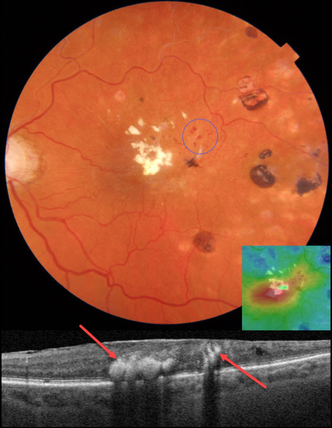
BRVO WITH RETINAL VENOUS MACROANEURYSM
Originally posted on @retina.rocks October 2, 2023
This 83YO female presented with recent vision loss in her left eye. There was a prior history of a branch retinal vein occlusion (BRVO), for which she received scatter laser elsewhere 8 years earlier. Vision was 20/60 OS.
Color photography shows superotemporal macular telangiectasia with likely retinal venous macroaneurysms (RVMs). Fairly dense lipid is noted in the fovea along with temporal laser scarring.
On OCT scanning, the lipid is markedly hyperreflective with posterior shadowing. There is central macular edema on the thickness map. Focal thermal laser was recommended to the RVMs.
Learning Points:
In 1990, Cousins et al reported that RVMs can be an additional finding in occlusive venous disease, always in association with collateral vessels or capillary or arterial macroaneurysms (AJO 1990;109:567-570).
These vascular abnormalities tend to have a higher rate of intraretinal lipid (40%) but are less likely to affect central vision since they are usually outside the macula. They tend to have a higher rate of developing retinal neovascularization (32% of eyes).
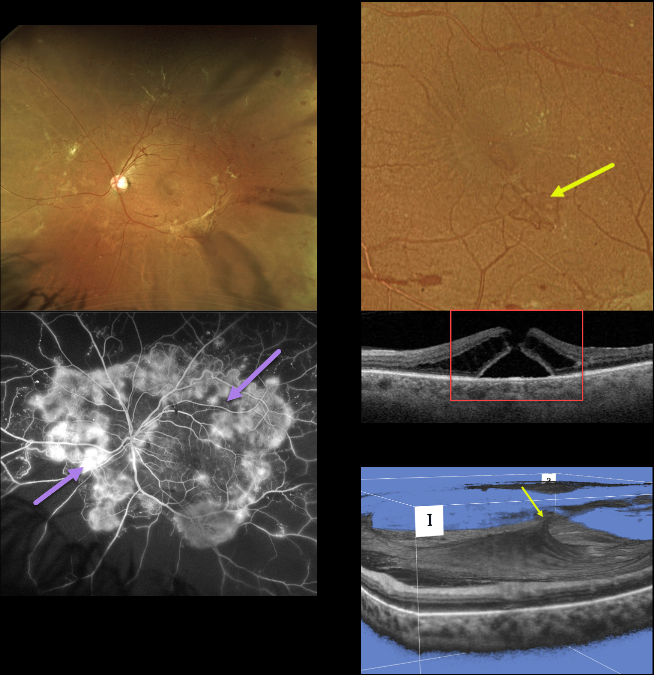
MACULAR HOLE DUE TO DIABETIC TRACTION
Originally posted on @retina.rocks September 21, 2023
This 49YO female with type 2 diabetes presented with a several-year history of blurred vision in her left eye. Vision was 20/40 OD and 20/60 OS.
Optos RG imaging shows variably fibrosed retinal neovascularization encircling the posterior pole. Fluorescein angiography shows active leakage from these vessels.
An area of small neovascularization is present in the inferior temporal macula, which caused a tractional full-thickness macular hole. A 3D OCT view best shows this diabetic traction.
Vitrectomy surgery was scheduled for her left eye. Primary vitrectomy will be performed for her right eye as well once the left eye has stabilized.
Learning Points:
In 1988, Dr. Gass described his concept for the pathogenesis of idiopathic macular holes (Arch Ophthalmol 1988;106:629-639). His classification system, from stage 1 for an impending macular hole through stage 4 for a fully developed macular hole with complete posterior vitreous detachment, was based on his concept of localized, mostly tangential traction caused by the shrinking foveal vitreous cortex. This classic paper heralded modern macular hole surgery for a previously untreatable condition.
The advent of OCT technology, however, has shown that macular hole development is more often due to focal vitreomacular traction, as our case beautifully illustrates.
The current classification system, described by the International Vitreomacular Traction Study Group, emphasizes the characteristics that determine treatment choice and visual prognosis (Ophthalmology 2013;120:2611-2619). This includes size (small <= 250 microns, medium >250 microns and <= 400 microns, large >400 microns), presence or absence of VMT, and cause (primary from VMT or secondary).
Our patient emphasizes why this new classification system was implemented, since it provides information that guides our treatment. Our patient’s hole is secondary to diabetic traction, which obviously requires a very different vitrectomy procedure compared to a primary hole.
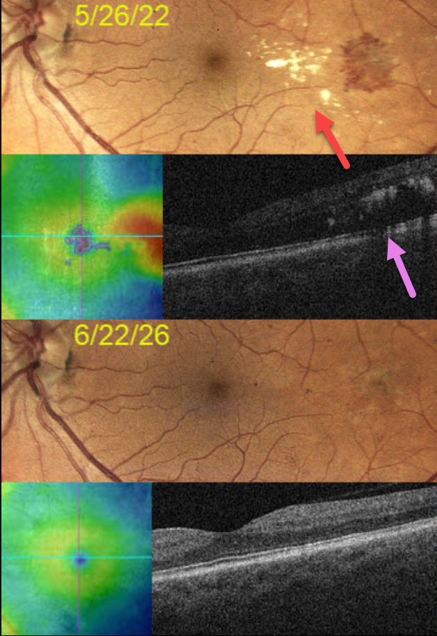
DIABETIC MACULAR EDEMA (DME)
Originally posted on @retina.rocks August 29, 2023
This 57YO man presented on 5/26/22 with asymptomatic diabetic macular edema (DME) in his left eye. Vision was 20/20.
Extensive lipid exudates with associated macular thickening were noted in the temporal macula. A gentle thermal laser was applied to the leaking vessels.
One year later, vision remains at 20/20, with total resolution of all lipid and DME.
Learning Points:
Beginning in the 1970s, macular laser was the gold standard for treating DME. In the early to mid-2000s, a perfect storm brewed with the advent of OCT technology and intravitreal injections. Unlike lasers, which take many months to work and rarely significantly improve vision, intravitreal injections cause a rapid and dramatic improvement in vision, making them the mainstay for treating DME ever since.
Clinically significant macular edema (CSME) is now an outdated term, as we are more concerned with whether the edema is central (central involved, CI-DME) or non-central (NCI-DME). Eyes with CI-DME are usually best treated with injections for superior visual outcomes.
However, our patient exemplifies why macular laser still has a place in any retinal surgeon’s toolbox, and should be considered a viable option for treating NCI-DME and even CI-DME when the leaking microaneurysms are outside the foveal avascular zone (FAZ).
In our practice, macular laser remains the initial treatment for eyes with NCI-DME. When applied gently with small, low-power, and short-duration applications well outside the FAZ, patients rarely, if ever, experience scotoma. Both the treatment burden and the financial costs to society are dramatically less. And there is no risk for endophthalmitis.
Although the role of thermal laser for treating DME is currently a source of debate (see Eye 2022;36:485-486 and Eye 2022;36:483-484 for great pro-laser and anti- laser editorials) we are not sure what all the fuss is about. Old-fashioned lasers work and, in our opinion, are incredibly under-utilized.
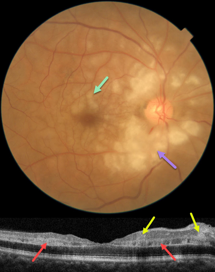
CRAO
Asma Samsudeen and Ashish Sharma
Originally posted on @retina.rocks August 10, 2023
This 60YO male presented with an abrupt, painless loss of vision in his right eye 1 week earlier. There was no prior ocular history or recent history of trauma. Vision was 20/30 in his right eye, and 20/20 in his normal left eye.
Fundus photography shows patches of mostly inner-peripapillary ischemia, with deeper ischemia throughout the macula. This is shown on OCT as patches of inner and mid-retinal hyperreflectivity. Fluorescein angiography showed a markedly delayed transit time (not shown).
We immediately referred him to his cardiologist for an emergency stroke protocol evaluation.
Learning Points:
Purtscher retinopathy was first described by Otmar Purtscher in 1910. Although originally described in a man who fell from a tree with cranial trauma, these white patches of retinal ischemia and hemorrhages are found in numerous other etiologies, including pancreatitis, chest trauma, and collagen vascular diseases.
The findings are called Purtscher retinopathy when due to trauma, and Purtscher-like retinopathy when seen from other causes.
Our patient’s fundus appearance most resembles Purtscher-like retinopathy, with patches of inner- and middle-retinal ischemia. However, given the acute symptoms and delayed angiographic filling time, we believe our patient most likely has an incomplete central retinal artery occlusion (CRAO).
See Mangla et al for the OCT findings in CRAO of varying severities (Int J Retina Vitreous 2023;9;37).
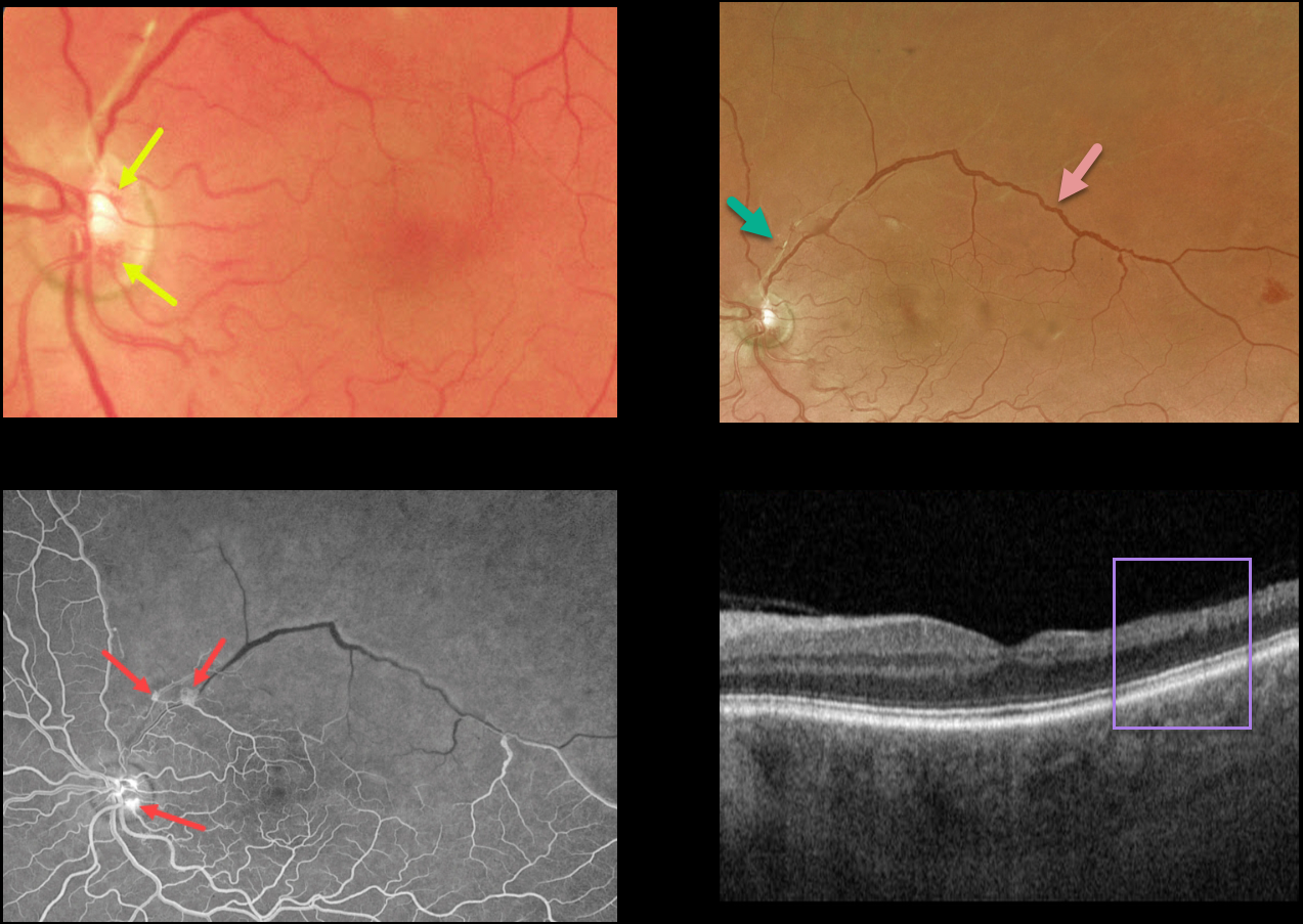
BRVO
Originally posted on @retina.rocks August 1, 2023
This 55YO male presented with relatively asymptomatic inferior field loss in his left eye.
Color imaging of the left nerve and macula shows a few small areas of disc neovascularization (NVD) and a fibrosed arteriole superiorly. Optos color RG imaging shows an irregularly dilated superotemporal major retinal vein and a featureless superior retina.
Fluorescein angiography shows severe retinal ischemia throughout the entire distribution of the branch retinal vein occlusion (BRVO) with leaking NVD and neovascularization elsewhere.
OCT scanning shows temporal inner retinal atrophy with the inner retinal layers replaced by a single hyperreflective band.
Scatter laser photocoagulation was recommended.
Learning Points:
In 1986, the Branch Vein Occlusion Study Group reported that scatter laser decreased the risk of vitreous hemorrhage from retinal neovascularization in BRVO, and suggested that laser should be applied once neovascularization develops (Arch Ophthalmol 1986;104:34-41).
Decades later, scatter laser remains the gold standard, although some may prefer to first stabilize neovascularization with anti-VEGF therapy prior to laser.
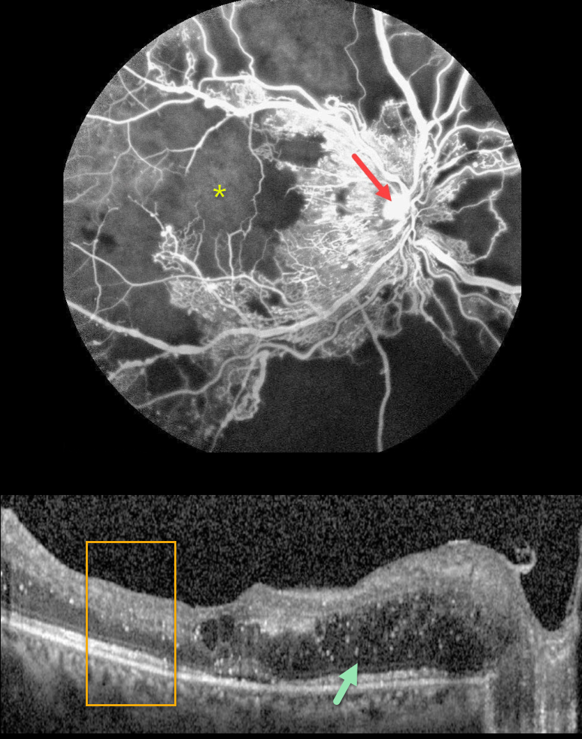
PROLIFERATIVE DIABETIC RETINOPATHY (PDR)
Sehrish Momin and Haroon Tayyab
Originally posted on @retina.rocks July 31, 2023
This 27YO female with a history of uncontrolled type 1 diabetes presented with bilateral vision loss. Vision was 20/40 OD and 20/200 OS.
Fluorescein angiography shows severe capillary loss that extends through the central macula. The remaining vessels are telangiectatic, especially bordering the areas of frank loss, and an area of leaking disc neovascularization is noted.
OCT scanning shows mostly nasal edema of the outer nuclear layer with numerous suspended hyperreflective particles (hyperreflective foci, HRF), along with more central cysts and trace subretinal fluid. The inner retinal layers have lost some of their normal hyper- and hypo-reflective structures (disorganization of the retinal inner layers, DRIL). Similar findings were noted in her left eye (images not shown).
Monthly anti-VEGF therapy was recommended although the patient was subsequently lost to follow-up.
Learning Points:
Anti-VEGF therapy has become the standard treatment for central-involved diabetic macular edema (CI-DME), with the vast majority of eyes showing improvements in macular thickness and vision.
Although we expect our patient’s edema to improve if she returns for treatment, we are more guarded regarding her final acuity since the capillary loss is permanent. Eyes with CI-DME and baseline central DRIL have a worse visual prognosis (Sun et al, JAMA Ophthalmol 2014;123:1309-1316).
HRF are small dot-like retinal opacities found in some eyes with diabetic macular edema. Although their pathogenesis is debated, they likely represent extravasated lipoproteins that can later become clinically evident lipid exudates (Ganne et al, Indian J Ophthalmol 2021;69:3208-3217).
Although HRF numbers decrease with treatment, it is unclear whether they serve as a biomarker for predicting visual outcome (Huang et al, Ophthalmology Retina 2022;6:814-827).
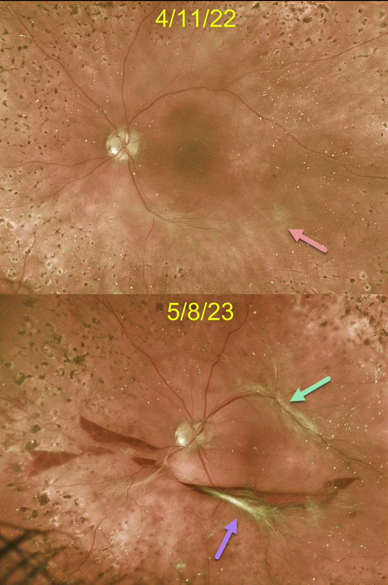
TRACTIONAL RETINAL DETACHMENT
Originally posted on @retina.rocks July 17, 2023
This 45YO female with type 1 diabetes responded beautifully to full panretinal photocoagulation (PRP) for her proliferative diabetic retinopathy (PDR). When examined 6 weeks following completion of her PRP, faint fibrotic neovascularization was noted along the inferotemporal arcade. Vision was 20/400 from residual central edema that was being treated with ongoing anti-VEGF therapy.
Unfortunately, she was lost to follow-up for over a year. Although vision remained at 20/400, increased traction was noted inferiorly, with new traction superiorly, resulting in an extrafoveal traction detachment. Symptomatic vitreous hemorrhage was also seen. Vitrectomy surgery was scheduled.
Learning Points:
Although PRP is often a ‘1 and done’ procedure, this is not always the case (Gonzalez et al, Ophthalmology 2021;128:1448-1457), and patients therefore need continued monitoring.
Although our patient showed involution of her PDR following PRP, increased fibrosis and traction still developed about a year later.
These findings would likely have been caught earlier if she had not been lost to follow-up, which is unfortunately quite common among patients with diabetes (Obeid et al., Ophthalmology 2018;125:1386-1392; Gao et al., Ophthalmology Retina 2019;3:230-236; Green et al, AJO 2020;216:18-27, etc.).
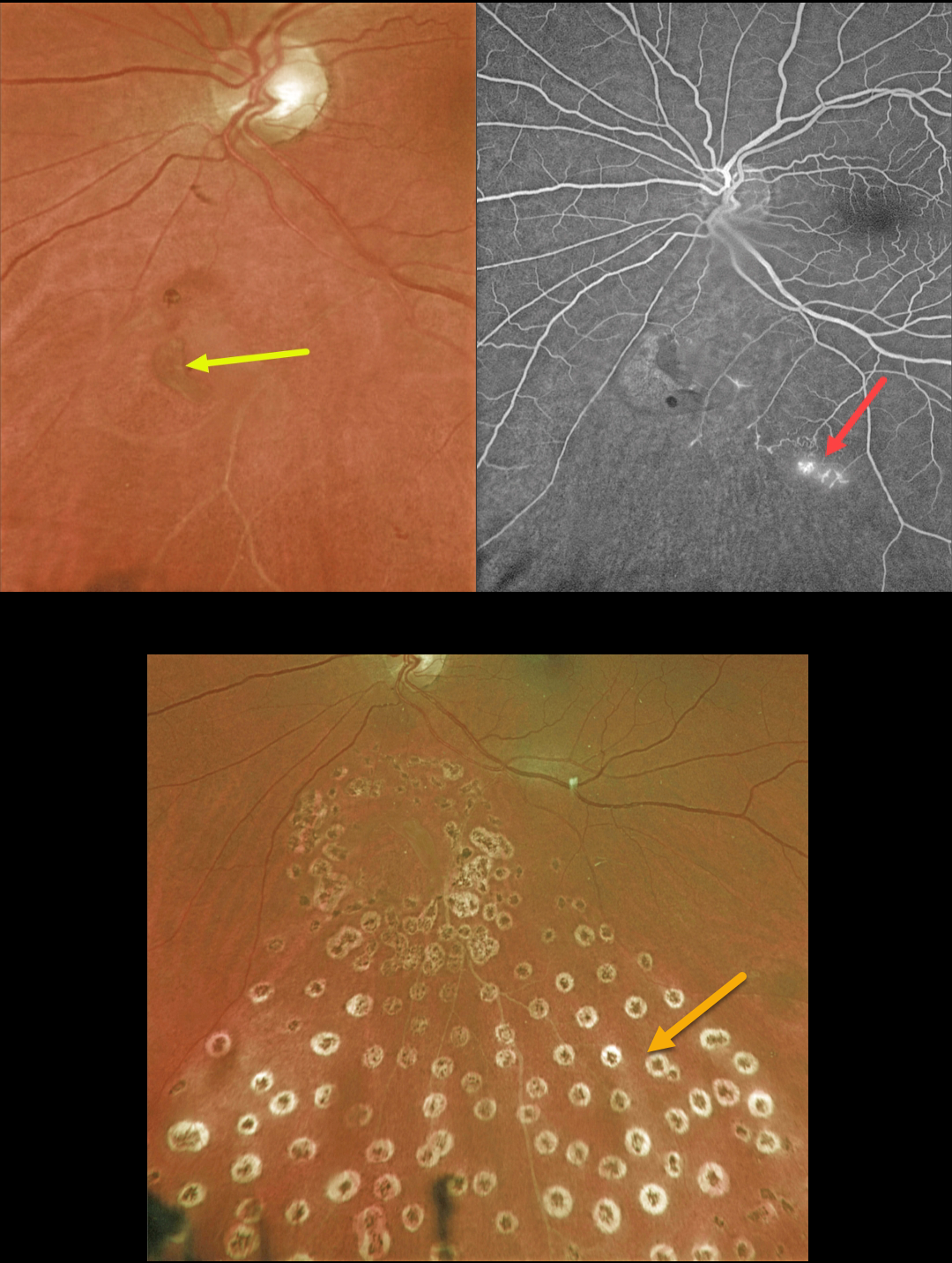
RETINAL TEAR + BRVO
Originally posted on @retina.rocks June 5, 2023
This 63YO female presented with symptomatic flashes and floaters in her left eye.
Optos color imaging shows a retinal tear just inferior to the nerve. An ischemic branch retinal vein occlusion (BRVO) is noted inferiorly. A posterior vitreous detachment (PVD) was noted clinically.
Fluorescein angiography confirmed the inferior ischemic BRVO with some small areas of retinal neovascularization. The tear appeared to be completely independent of the venous occlusion.
The retinal tear was surrounded by a few nearly confluent rows of laser photocoagulation burns, and inferior scatter laser was applied into the distribution of the BRVO. Fundus photography, a few months later, shows the laser scarring.
Learning Points:
Retinal tears are virtually always seen in the retinal periphery and are caused by an adherent vitreous pulling the retina anteriorly towards the vitreous base, most commonly during an acute PVD.
Our patient’s tear is unusual due to its quite posterior location and the flap occurring along its posterior edge. This implies that the vitreous was tugging towards the optic nerve. The optic nerve is usually the last site for the vitreous to release from during an acute PVD (Johnson, AJO 2010;149:371-382).
We hypothesize that the vitreous was completely detached except at the optic nerve and just inferiorly, and the tear was created from anterior to posterior traction when the vitreous separated from the nerve.
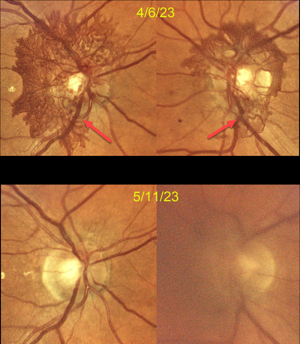
PROLIFERATIVE DIABETIC RETINOPATHY (PDR)
Mattie Adams
Originally posted on @retina.rocks May 30, 2023
This 59YO female was referred for proliferative diabetic retinopathy (PDR). She noted floaters in her right eye for several months. There was a 20-year history of type 2 diabetes, which was currently insulin-dependent. Vision was 20/25 bilaterally.
Color photography dated 4/6/23 shows florid bilateral disc neovascularization (NVD) secondary to previously undiagnosed PDR. Bilateral Avastin injections were given that day. She returned two weeks later with several days of blurred vision OS. Vision was 20/60 in this eye due to a new moderate vitreous hemorrhage.
She returned on 5/11/23, subjectively better, with 20/25 OD and 20/30 OS. The vitreous hemorrhage in her left eye was much improved, and the NVD had dramatically resolved in each eye. Panretinal photocoagulation (PRP) will be started bilaterally.
Learning Points:
PRP has been around since the 1960s and still remains the mainstay for treating proliferative disease. Although the majority of treatment-naive retinal neovascularizations will respond to PRP, in our experience, this type of succulent NVD rarely regresses significantly with laser alone.
As in this case, we prefer to first stabilize the neovascularization with anti-VEGF therapy, and then add PRP as a more permanent solution to prevent reproliferation.
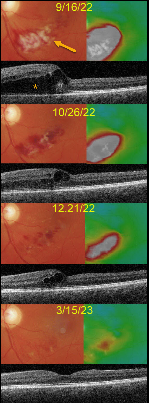
BRVO
Originally posted on @retina.rocks May 8, 2023
This 50YO male presented on 9/16/22 with 20/50 vision in his left eye from a macular branch retinal vein occlusion with edema.
Although we recommended anti-VEGF injections, he reported that a similar episode improved without treatment about 10 years earlier. He therefore asked if we could initially refrain from treatment.
The occlusion and edema slowly and spontaneously regressed. When last examined on 3/15/23, vision was 20/20 and just a small amount of non-central macular edema remained.
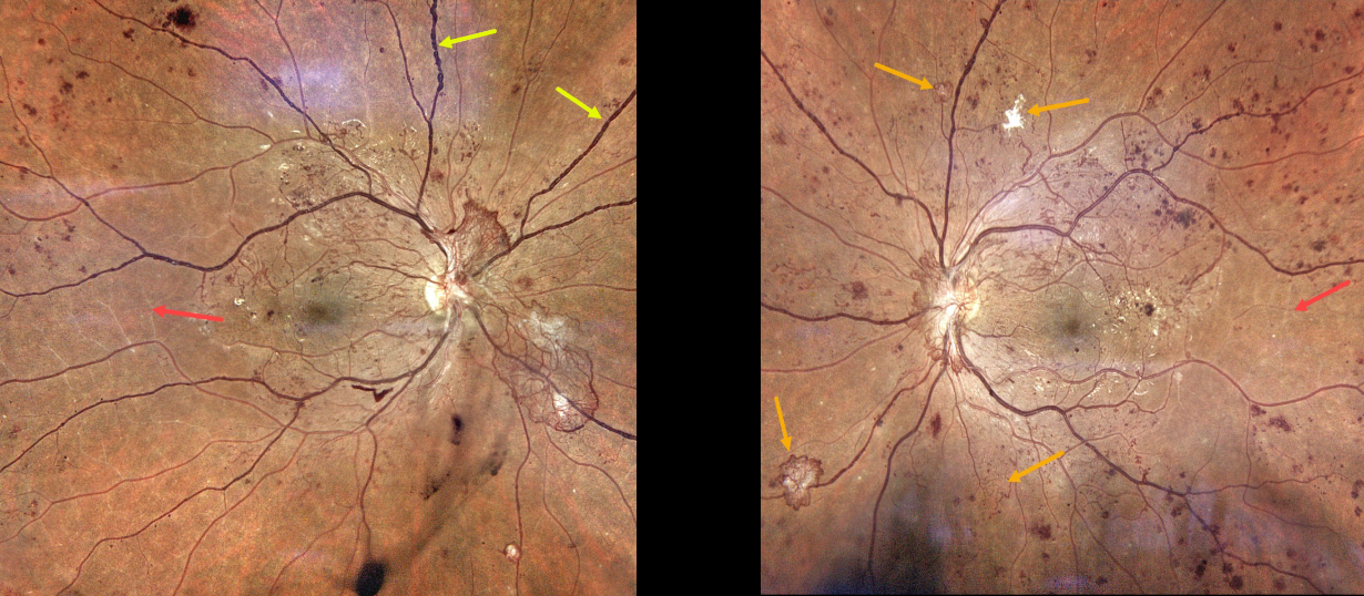
PROLIFERATIVE DIABETIC RETINOPATHY (PDR)
Originally posted on @retina.rocks March 16, 2023
This 28YO male with a history of type 1 diabetes was referred for asymptomatic retinal findings. He noticed some recent floaters in his right eye. Vision was 20/50 OU.
Color photography shows severe bilateral proliferative diabetic retinopathy. The right eye has a large area of active disc neovascularization, venous beading, and white vessels temporally indicating severe ischemia. There is some mild inferior vitreous blood.
The left eye has a smaller area of succulent NVD along with scattered smaller areas of variably active and fibrosed peripheral neovascularization. Clinical ischemic retina is also noted. Panretinal photocoagulation will be started bilaterally.
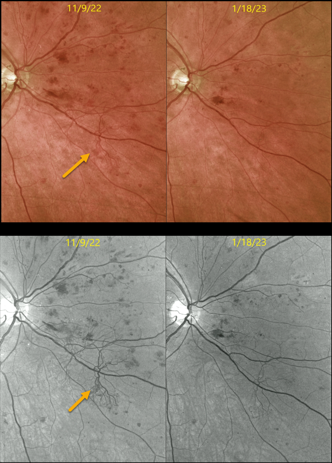
PROLIFERATIVE DIABETIC RETINOPATHY (PDR)
Originally posted on @retina.rocks March 1, 2023
This 56YO male with type 2 diabetes presented on 11/9/22 with 20/30 vision OD with both central-involved diabetic macular edema (CI-DME) and proliferative diabetic retinopathy. Optos color and green-channel imaging show a large patch of retinal neovascularization.
Two monthly intravitreal injections of Avastin were administered for the CI-DME. On 1/18/23, vision improved to 20/25, and the edema completely resolved. The retinal neovascularization dramatically regressed. Macular laser for the injection treatment burden will be performed, followed by panretinal photocoagulation (PRP).
Learning Points:
The DRCR.net’s Protocol S explicitly compared PRP to Lucentis for the primary treatment of proliferative diabetic retinopathy (JAMA 2015;314(20):2137-2146). At 2 years, vision was equivalent between both treatment groups, and both treatments were equally effective in controlling neovascularization.
However, Lucentis-treated eyes experienced less peripheral visual field loss, developed macular edema less often, and needed fewer vitrectomies.
A secondary analysis of Protocol S suggested that Lucentis monotherapy was cost-effective for PDR+vision-impairing CI-DME but not for PDR without CI-DME (JAMA Ophthalmology 2019;137:1424-1432).
The role of intravitreal therapy vs PRP as the primary management for proliferative disease is intriguing, but in our practice, we find it difficult to justify, given the well-tolerated, long-lasting effects of laser therapy, contrasted with the risk of poor patient follow-up and the expense and treatment burden of anti-VEGF therapy.
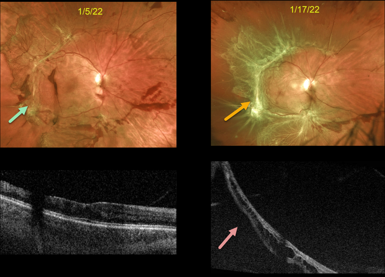
CRUNCH SYNDROME
Originally posted on @retina.rocks February 22, 2023
This 37YO female presented with a symptomatic diabetic traction retinal detachment (TRD) extending from the peripheral macula towards the mid-peripheries. Vision was 20/60. An intravitreal Avastin injection was given in preparation for vitrectomy within the week.
However, due to work-related reasons, the patient delayed the surgery and returned 2 weeks later, complaining of several days of increasing distortion and vision loss. Vision was 20/200. There was marked fibrosis of all neovascularization, and the macula was now detached. Vitrectomy was scheduled within the next several days.
Learning Points:
Intravitreal anti-VEGF is commonly used before diabetic vitrectomies to cause rapid involution of the vascularized preretinal tissue and control intraoperative bleeding during membrane dissection. These injections are given within a week of surgery since the neovascularization can contract with increased traction. This uncommon but known phenomenon is called the ‘crunch’ syndrome.
See Tan et al for a recent crunch review (Survey of Ophthalmology 2021;66:926-932).
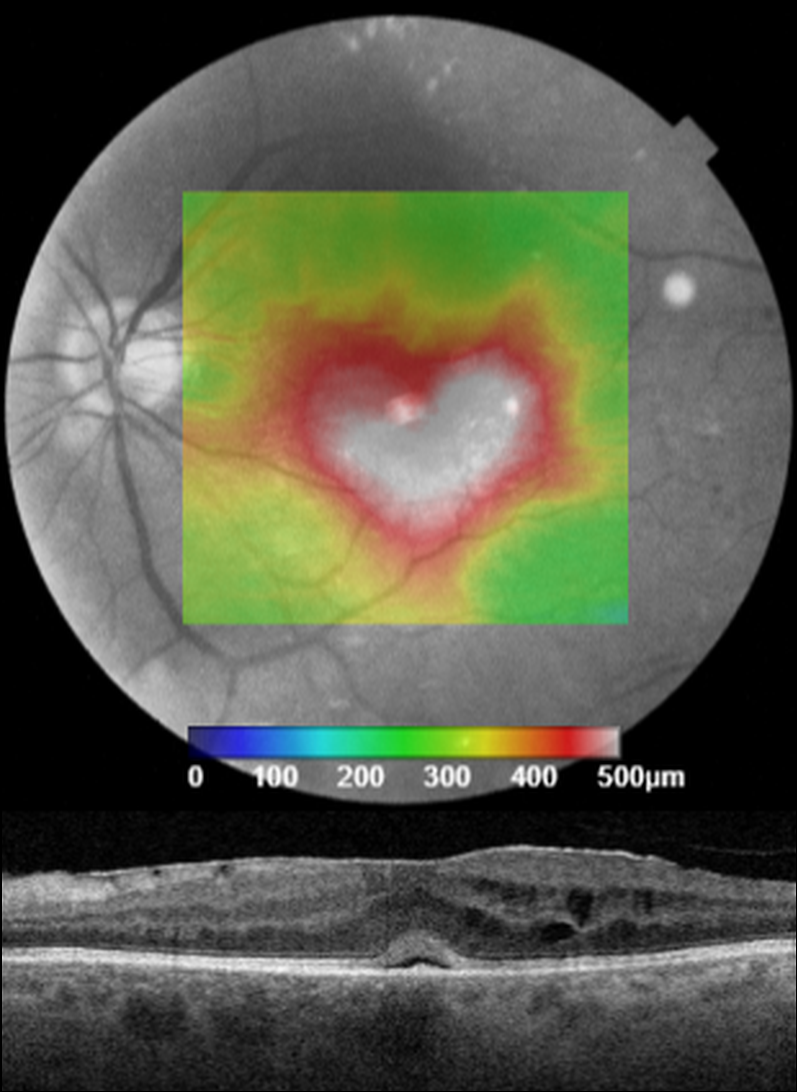
VALENTINE MACULAR EDEMA
Originally posted on @retina.rocks February 14, 2023
This patient presented with 20/200 vision from center-involved diabetic macular edema.
Unfortunately, we don’t have any further clinical information for this patient, but we still wish everyone a ‘heart-felt’ Happy Valentine’s Day!
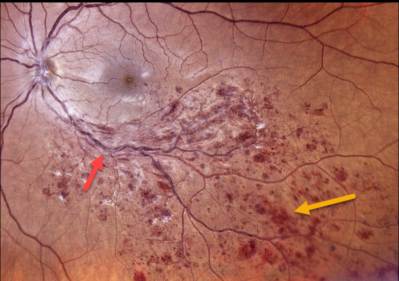
BRVO
Originally posted on @retina.rocks February 2, 2023
This 42YO female was referred for a completely asymptomatic branch retinal vein occlusion (BRVO) in her left eye. Vision was 20/20 due to a lack of macular edema or ischemia.
Color photography shows classic findings, including a quadrant of dilated tortuous veins with mostly inner retinal hemorrhages.
We will initially follow her every 3-4 months and recommend treatment if macular edema or retinal neovascularization develops.
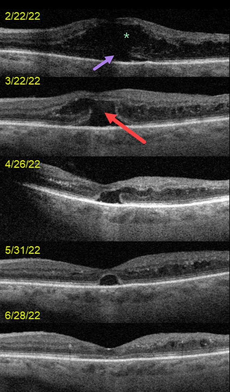
DIABETIC MACULAR EDEMA (DME)
Originally posted on @retina.rocks January 26, 2023
This 72YO female presented on 2/22/22 with counting-finger vision in her left eye.
OCT scanning at the initial visit shows severe center-involved diabetic macular edema (CI-DME) with shallow subretinal fluid. Anti-VEGF therapy was started.
One month later, vision remained at counting fingers. The retinal thickness had improved, but a new outer macular hole was noted.
Following 3 additional monthly injections, the edema continued to resolve with spontaneous closure of the outer macular hole. Vision was 20/70 at the 6/28/22 visit.
Learning Points:
Traditional primary full-thickness macular holes are caused by vitreomacular traction. Outer macular holes, however, have a much broader range of etiologies.
In our practice, we most commonly see these lesions in optic pit maculopathy and paraproteinemia (see Mansour et al, Ophthalmology 2014;121:1925-1932).
Our patient’s outer macular hole was likely caused by inner retinal exudation that extended through the outer retina. The return of central vision and closure of the defect, with relatively intact outer macular architecture, likely indicates that this was an outer macular dehiscence rather than a true loss of tissue. See Kumawat et al for a great review of atypical macular holes (Retina 2019;39:1236-1264).
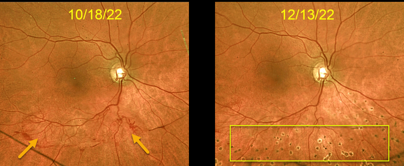
BRVO
Originally posted on @retina.rocks January 25, 2023
This 57YO female was referred for an asymptomatic branch retinal vein occlusion (BRVO) in her right eye. Vision was 20/30.
Optos color imaging shows a normal right macula, with large active retinal neovascularization extending along and distal to the inferotemporal arcade.
Six weeks after the inferior scatter laser, the neovascularization regressed dramatically and virtually completely.
Learning Points:
The results of the Branch Vein Occlusion Study Group (Arch Ophthalmology 1986;104:34-41) still remain the gold standard for treating BRVO-related retinal neovascularization.
Once neovascularization develops, scatter laser into the involved quadrant significantly reduces the risk for vitreous hemorrhage.
Although anti-VEGF therapy can initially be given to rapidly stabilize the neovascularization, laser is usually all that’s needed, as illustrated by our patient.
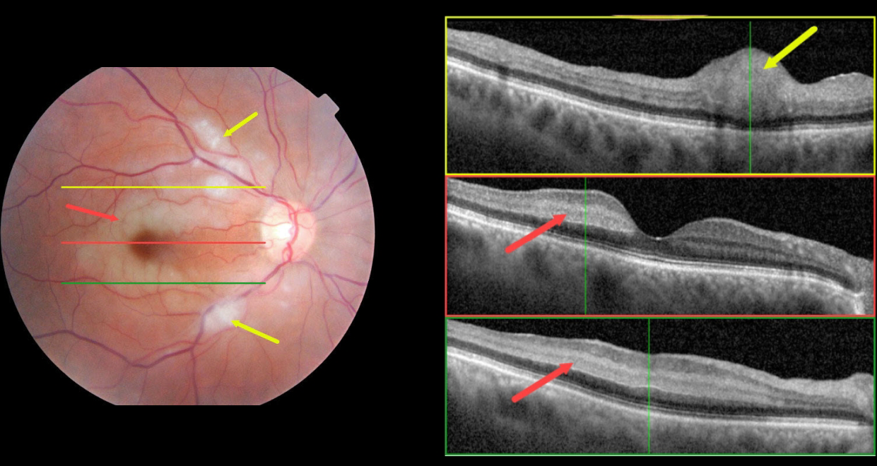
CRAO
Ryan Kern
Originally posted on @retina.rocks January 10, 2023
This 70 YO female presented with acute vision loss in her right eye starting 1 day earlier. Vision was 20/70 OD and 20/25 in her normal left eye.
Color fundus photography shows an incomplete central retinal artery occlusion (CRAO) with focal areas of acute inner retinal infarcts along with paracentral areas of deeper retinal opacification.
OCT scanning through the superior macula shows a focal area of retinal opacification and thickening that extends from the outer plexiform layer to the internal limiting membrane. Scans of the central and inferior macula show hyperreflectivity involving the middle retinal layers (outer plexiform, inner nuclear, and inner plexiform).
Blood pressure was 183/110. Emergent stroke evaluation was fortunately negative. Her blood pressure was stabilized, and baby aspirin was started. Visual acuity improved to 20/30 one month later.
This case was submitted by Ryan Kern.
Learning Points:
Paracentral acute macular maculopathy (PAMM) is a phenotype caused by acute mid-retinal ischemia due to obstruction of the deep (DCP) and intermediate (ICP) capillary plexi. PAMM is associated with an increasing constellation of conditions, including retinal vein and artery occlusions.
It is unclear if patients with a pure PAMM phenotype require an immediate stroke protocol referral. In our practice, we have seen several elderly patients who developed a complete CRAO within days of presenting with PAMM, so we do send these patients for emergent evaluation. We do not believe that typical PAMM in younger patients or found as a secondary finding in other ocular conditions requires a stroke workup.
Our patient’s fundus appearance has more of a Purtscher ’s-like appearance with focal areas of cotton wool spot-like inner retinal ischemia (although on OCT these involved the middle retinal layers as well) and mid-retinal ischemia.
However, given her age and history, acute onset, and unilateral presentation, her diagnosis is most consistent with an incomplete CRAO.
For a great recent PAMM review, see Scharf et al, Progress in Retinal and Eye Research 2021;81;100884.
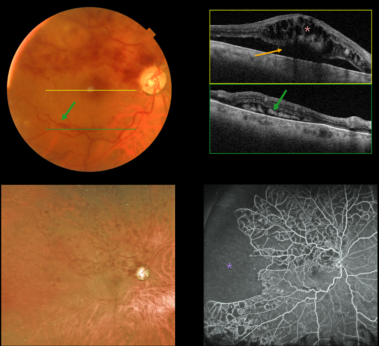
BRVO
Originally posted on @retina.rocks January 9, 2023
This 79YO male presented with recent vision loss of 20/400 in his right eye from a fresh branch retinal vein occlusion (BRVO). Layered subretinal blood is noted in the inferior macula.
Swept-source OCT through the macular center shows marked cystoid macular edema with underlying subretinal fluid. OCT through the inferior macular subretinal blood shows subretinal hyperreflectivity. Optos fluorescein angiography shows marked peripheral ischemia without neovascularization.
Learning Points:
Layered preretinal blood is a common occurrence with proliferative retinopathies, including proliferative diabetic retinopathy and BRVO. The retinal neovascularization grows from the retinal surface onto the back of the posterior hyaloid, and blood accumulates in the subhyaloid space.
The appearance of layered inferior macula blood in our patient is a ‘fake-out’ because the blood was neither neovascular nor preretinal. Blood, likely due to hydrostatic pressure, was forced into the subretinal space, and the subretinal fluid allowed it to settle inferiorly.
The blood and fluid completely resolved following several monthly intravitreal anti-VEGF injections.
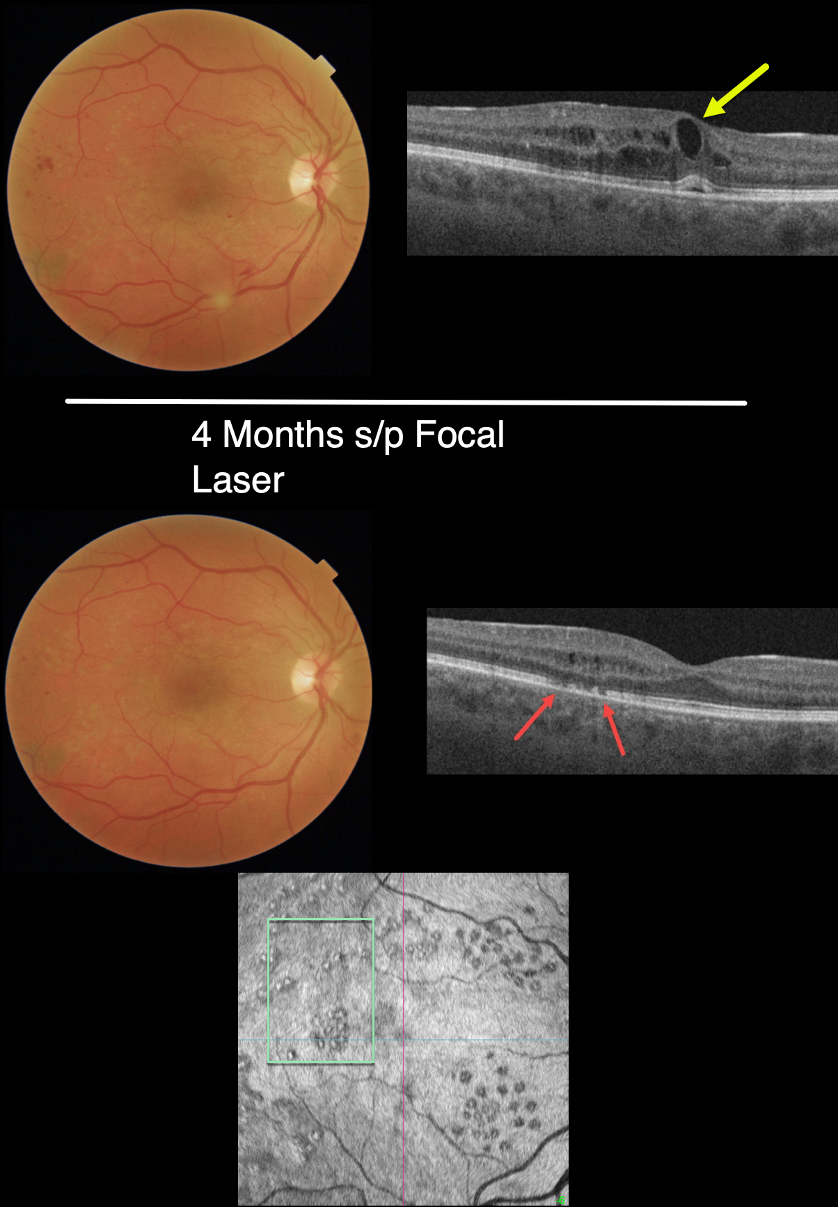
FOCAL LASER FOR DIABETIC MACULAR EDEMA (DME)
Originally posted on @retina.rocks December 29, 2022
This 32YO female with type 1 diabetes was examined with no visual symptoms, and vision was 20/30 OD and 20/20 OS. Macular photography shows a foveal cyst and mild peripheral macular hemorrhages, and OCT scanning shows central and temporal cystic edema with trace subfoveal fluid.
Due to the lack of symptoms and good vision, we were hesitant to start anti-VEGF therapy, but due to concern for progressive, increased edema and vision loss, we decided to treat with macular laser.
Four months later, the edema virtually completely resolved. The laser scars appear as temporal defects in the ellipsoid zone, although they are barely visible funduscopically. The laser scars are more visible with outer retinal en face imaging.
Learning Points:
In their landmark 1985 paper (Arch Ophthalmology 1985;103:1796-1806), the Early Treatment Diabetic Retinopathy Study (ETDRS) introduced the term clinically significant diabetic macular edema (CSME). Broadly speaking, eyes with CSME, as determined by retinal thickening found on contact lens examination, have the threshold amount of macular edema that benefits from laser photocoagulation.
Macular laser remained the gold standard for diabetic macular edema (DME) for about 15 to 20 years. In the early to mid-2000s, a perfect storm brewed with the advent of OCT technology and intravitreal injections. Unlike lasers, which take many months to work and rarely significantly improve vision, intravitreal injections show a rapid and dramatic visual improvement, making them the mainstay for treating DME ever since.
CSME has become an antiquated term since we are now more concerned with whether the edema is central (central involved, CI-DME) or non-central (NCI-DME). Eyes with CI-DME are best treated with injections due to superior visual results.
However, our patient exemplifies why macular laser still has a place in any retinal surgeon’s toolbox, and should be considered a viable option for treating NCI-DME and even CI-DME when the leaking microaneurysms are outside the foveal avascular zone (FAZ). In our practice, macular laser remains the initial treatment for eyes with NCI-DME. When applied gently with small, low-power and short-duration applications well outside the FAZ, patients rarely, if ever, experience scotoma. Both the treatment burden and the financial costs to society are dramatically less. And there is no risk for endophthalmitis.
Although the role of thermal laser for treating DME is currently a source of debate (see Eye 2022;36:485-486 and Eye 2022;36:483-484 for great pro-laser and anti- laser editorials) we are not sure what all the fuss is about. Old-fashioned laser works and, in our opinion, is incredibly under-utilized.
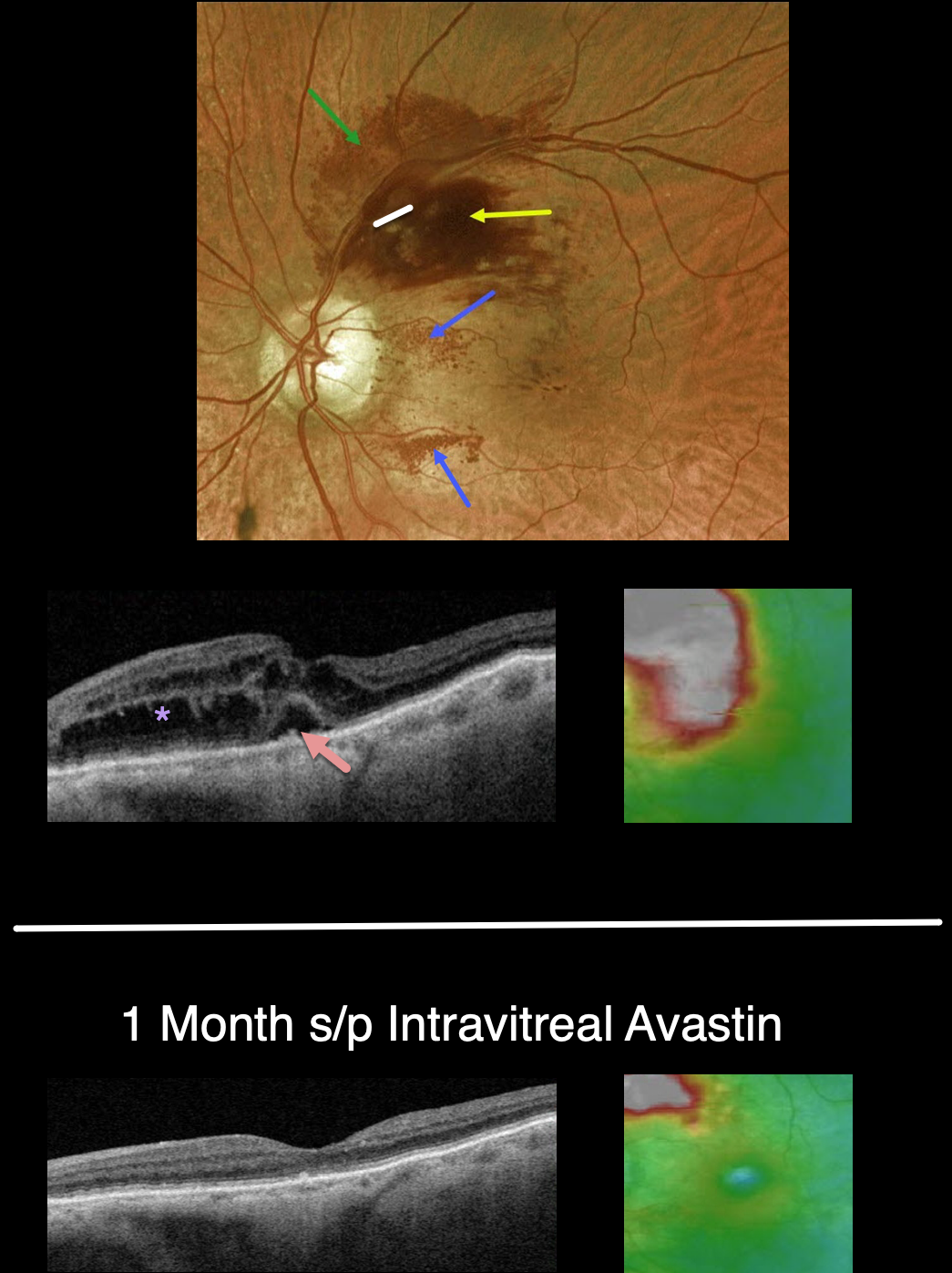
BRVO
Originally posted on @retina.rocks December 28, 2022
This 88YO female presented with recent vision loss in her right eye. Vision was 20/50.
Optos color imaging shows an unusual appearing superotemporal branch retinal vein occlusion (BRVO). Confluent inner retinal hemorrhages are noted in the superonasal macula, with milder deep retinal blood more superiorly. Unusual dots of intraretinal blood are also noted nasally and inferonasally.
Swept-source OCT shows moderately severe central and superonasal macular edema with some foveal subretinal fluid.
An intravitreal Avastin injection was given. One month later, vision improved to 20/30. The retinal hemorrhages dramatically improved. OCT scanning shows complete resolution of all central fluid, with some residual distal superonasal edema.
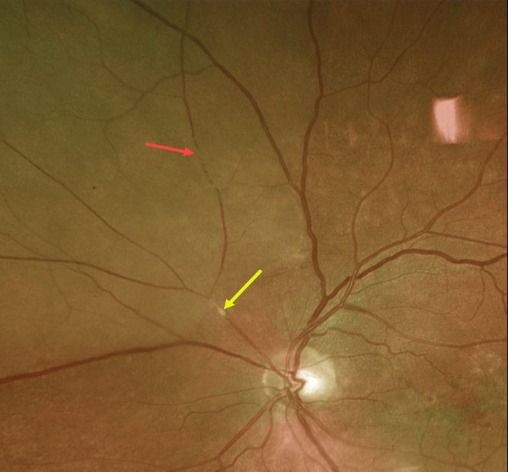
BRAO
Originally posted on @retina.rocks December 26, 2022
This 73YO male presented with acute inferotemporal visual field loss.
Optos imaging shows an acute extramacular branch retinal artery occlusion (BRAO) caused by a likely platelet-fibrin embolus just superonasal to the optic nerve. The distal retina is somewhat opaque, and the blood column is stagnant superiorly.
This patient had acute retinal ischemia (defined as acute BRAO, central retinal artery occlusion or transient monocular vision loss), and was immediately referred to the nearest stroke center (Biousse et al, Ophthalmology 2018;125:1597-1607).
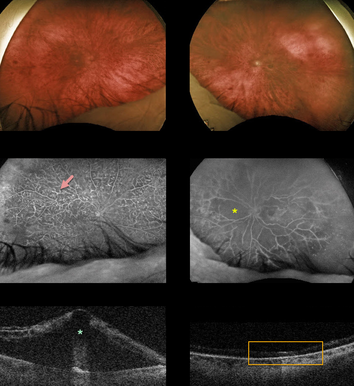
CRVO
Originally posted on @retina.rocks December 22, 2022
This 78YO male presented with counting finger vision in his right eye. He had a prior CRVO in his left eye, causing hand motion vision.
Optos color imaging shows mild to moderate bilateral scattered retinal hemorrhages. Fluorescein angiography shows fairly good perfusion with leaking vessels OD with severe diffuse ischemia OS.
Swept-source OCT shows severe central macular edema OD with an atrophic left macula.
Anti-VEGF therapy was started for the edema OD.
Learning Points:
Prior to the introduction of intravitreal injections in the early 2000s, there were essentially no treatments for CRVO-related macular edema.
Anti-VEGF injections in general are now the initial treatment of choice, with about 50-60% of eyes gaining at least 3 lines of vision. Intravitreal steroids, usually Ozurdex, are more often used as secondary treatment.
Macular grid laser photocoagulation is not performed since it was shown to have no visual benefit in the Central Vein Occlusion Study Group (see Ophthalmology 1995;102;1425-1433).
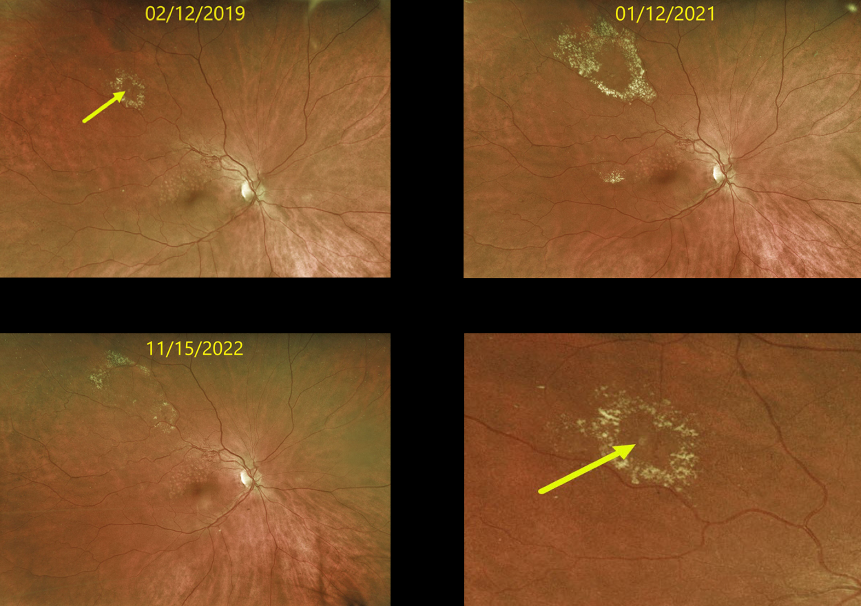
RETINAL VENOUS MACROANEURYSM
Originally posted on @retina.rocks December 15, 2022
This 63 YO female originally presented in 2019 with a history of a major superotemporal branch retinal vein occlusion (BRVO). Successful macular laser was performed for macular edema.
When initially examined, a midperipheral retinal venous macroaneurysm (RVM) with some surrounding lipid was noted. This RVM has been observed due to its distant location from the fovea. The series of photos over several years shows the exudation to wax and wane. At the last examination on 11/15/22, there was almost complete lipid resolution. Vision remains at 20/25.
Learning Points:
In 1990 Cousins et al reported that RVMs can be an additional finding in occlusive venous disease, always in association with collateral vessels or capillary or arterial macroaneurysms (AJO 1990;109;567-1700).
These vascular abnormalities tend to have a higher rate of intraretinal lipid (40%) but are less likely to affect central vision since they are usually outside the macula.
They tend to have a higher association with retinal ischemia, thus being at a higher rate of developing retinal neovascularization (32%). The lesions will usually involute, as was the case in our patient.
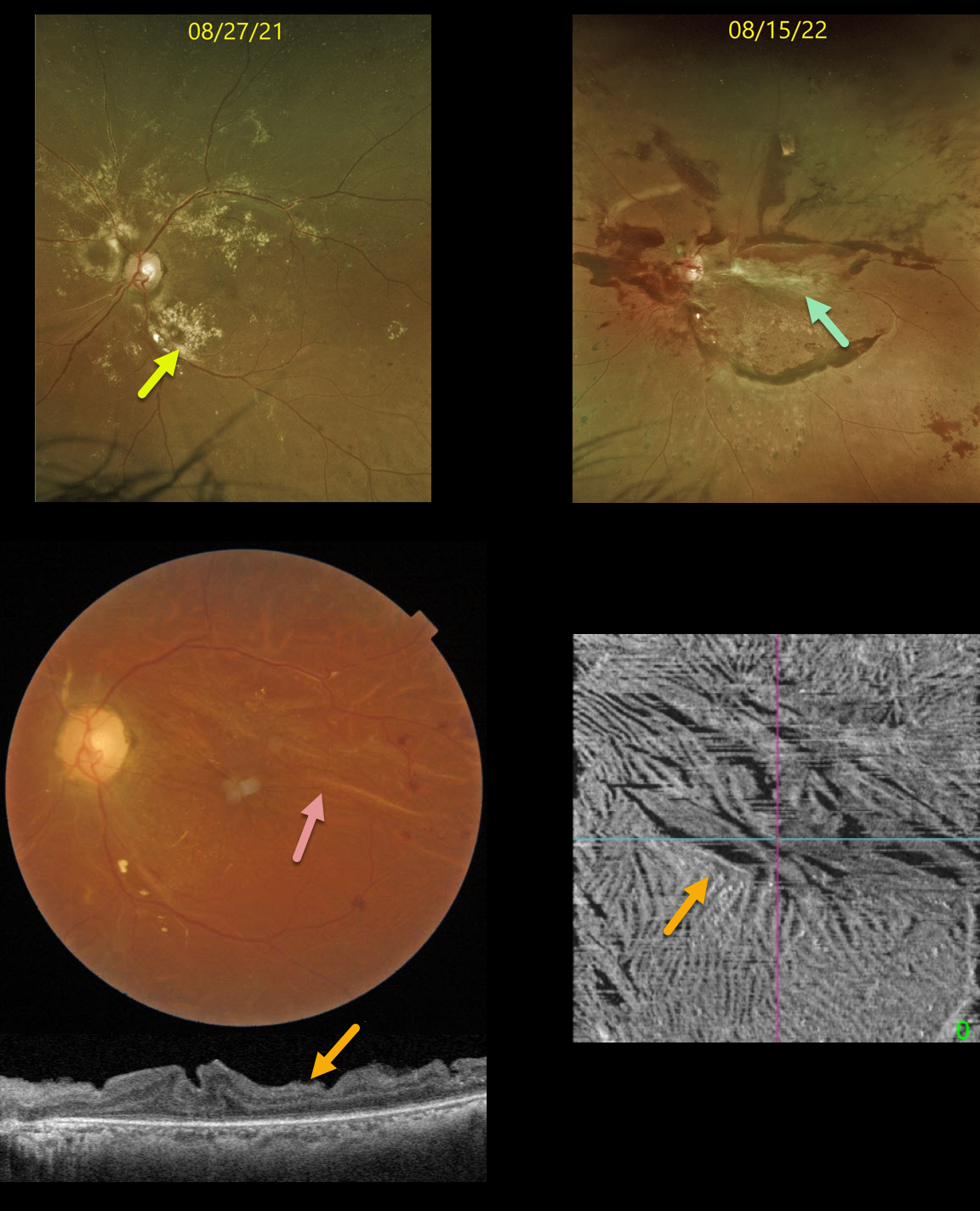
TRACTIONAL RETINAL DETACHMENT
Originally posted on @retina.rocks December 12, 2022
This 38YO diabetic female was initially seen on 8/27/21 with extensive dry macular lipid in her left eye and proliferative diabetic retinopathy. Vision was 20/40. Panretinal photocoagulation was recommended.
Unfortunately, she was lost to follow-up until she returned on 8/15/22 with 20/200 vision from a traction retinal detachment. The lipid spontaneously absorbed.
Vitrectomy surgery was performed, and three weeks later, the retina was completely attached with 20/70 vision.
Color imaging of the macula shows variable retinal folds centrally and superiorly.
OCT B-scan and en face of the inner retina variably show the dramatic inner retinal folds caused by the prior traction. We expect these to improve over time, but will probably never be completely resolved.
Learning Points:
Inner retinal folds, although usually less dramatic than in our patient, are fairly common following vitrectomy for diabetic TRD. They often fail to completely resolve, and vision can be quite good despite the OCT findings.
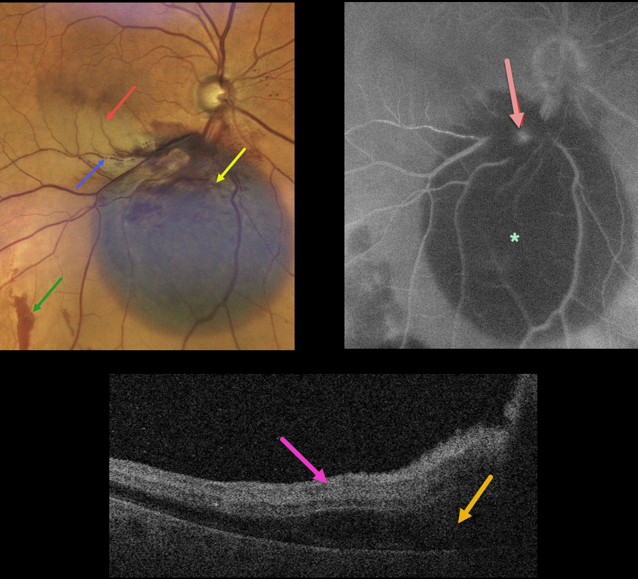
RETINAL ARTERIAL MACROANEURYSM WITH SECONDARY BRAO
Originally posted on @retina.rocks December 8, 2022
This 79YO female presented with acute inferior paracentral vision loss in her right eye. Vision was 20/50.
A large blister of dark subretinal blood was noted just inferior to the optic nerve. A few small areas of more distal red subretinal blood were seen. An inferior macular branch retinal artery occlusion likely accounted for her symptoms. The blood column is stagnant within the inferior portion of the acute occlusion.
OCT scanning through the BRAO shows hyperreflective inner retina with subretinal fluid.
Fluorescein angiography shows blockage from the subretinal blood, along with a leaking retinal arterial macroaneurysm (RAM).
Learning Points:
A BRAO can be a rare complication from direct thermal laser of a RAM (Russell and Folk, AJO 1987;104:186-187), which is one reason why this is almost never performed anymore. In this case, we believe the BRAO was secondary to mechanical arterial compression from the surrounding blood.
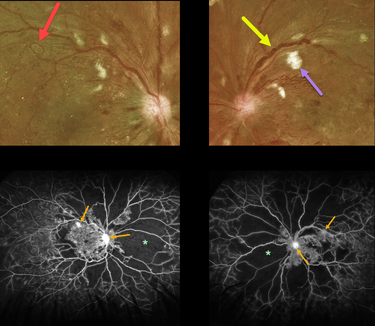
PROLIFERATIVE DIABETIC RETINOPATHY (PDR)
Originally posted on @retina.rocks December 6, 2022
This 33YO diabetic male presented with vision of 20/30 OD and 20/100 OS.
Optos color imaging shows bilateral severe posterior pole retinal hemorrhages and nerve fiber layer infarcts. Florid disc neovascularization is noted bilaterally. Marked venous beading is noted in each eye, and a large venous loop is noted in his right eye. The peripheral retina is also markedly ischemic.
Fluorescein angiography shows marked severe ischemia. The venous beading is much more global angiographically, with extensive venous staining of the irregularly beaded veins. Despite the ischemia, just a few areas of neovascularization are noted in each eye.
Vision is good OD since the foveal avascular zone is relatively normal, and vision is poor OS due to foveal ischemia.
Panretinal photocoagulation was recommended for each eye.
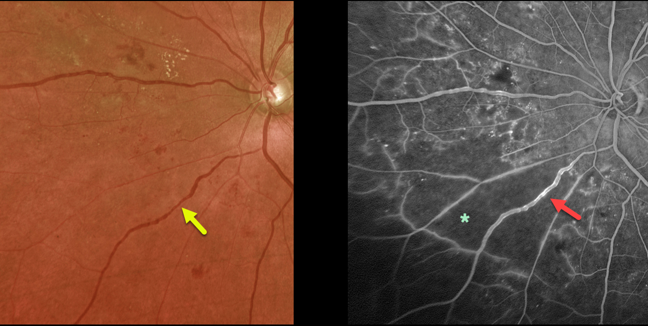
NON-PROLIFERATIVE DIABETIC RETINOPATHY (NPDR)
Originally posted on @retina.rocks December 5, 2022
This 64YO diabetic female presented with asymptomatic severe nonproliferative diabetic retinopathy (NPDR) in her left eye. Vision was 20/40.
Optos color imaging shows venous beading inferonasally. Fluorescein angiography shows severe retinal ischemia with venous staining.
Our patient’s ischemia was localized just to one quadrant with good perfusion elsewhere, so we are following her closely.
Learning Points:
Severe NPDR is defined as severe (at least 20) retinal hemorrhages in all quadrants, beading in at least 2 quadrants, or intraretinal microvascular abnormalities in 1 quadrant. These eyes are at significant risk for progressing to proliferative disease, so we will often treat with panretinal photocoagulation before neovascularization develops.
In our experience, eyes with significant venous beading or venous loops more often than not already have proliferative disease.
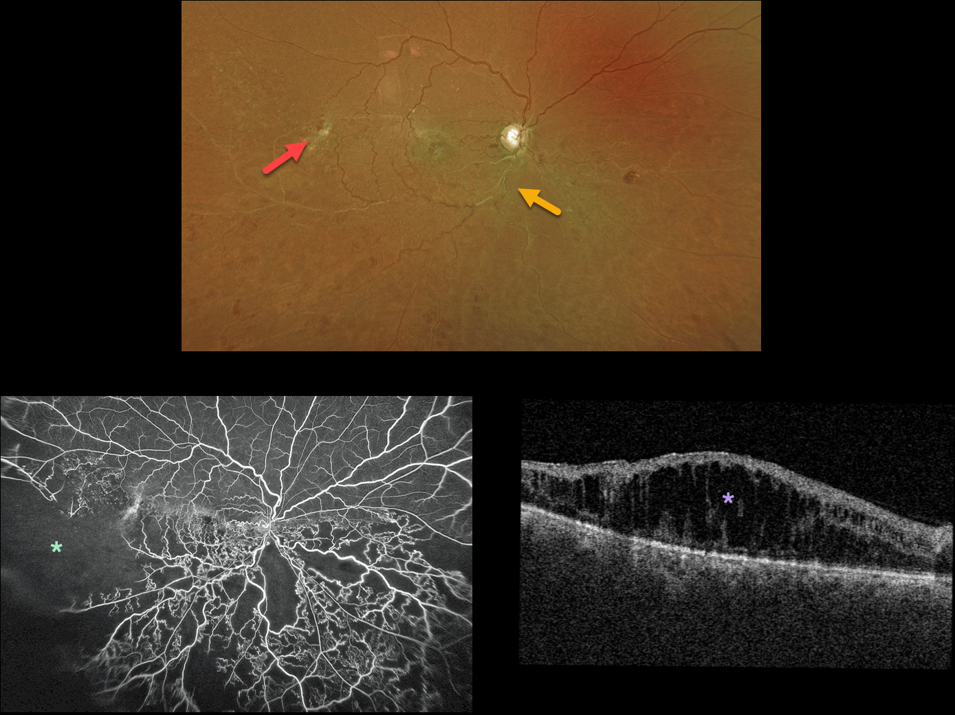
HRVO
Originally posted on @retina.rocks December 1, 2022
This 60YO male presented with hand motion vision in his right eye from an inferior hemiretinal vein occlusion (HRVO).
Optos color imaging shows white and sheathed retinal vessels inferiorly and extensive temporal collateral vessels.
Fluorescein angiography confirms the marked clinical ischemia. There was no neovascularization. OCT scanning shows marked diffuse macular edema.
The patient refused anti-VEGF therapy, and actually got up and walked out of the exam room before we could finish our discussion regarding why treatment was needed!
Learning Points:
Branch retinal vein occlusions are virtually always caused by a retinal artery compressing a retinal vein within its common adventitial sheath. Some people are born with an anomalous two-trunked central retinal vein that independently drains the superior and inferior retinal hemispheres.
HRVO, similar to central retinal vein occlusion, is caused by compression of the retinal vein posterior to the lamina cribrosa (see Hayreh, Arch Ophthalmol 1980;98:1600-1609).
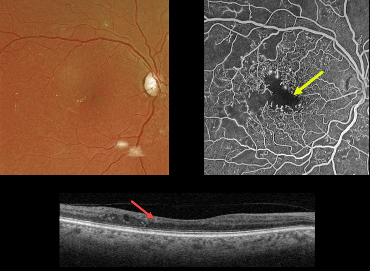
DIABETIC MACULAR ISCHEMIA
Originally posted on @retina.rocks November 23, 2022
This 67YO male has a history of type 2 diabetes. Vision is 20/40.
Optos color imaging is fairly unremarkable, with some mild temporal foveal lipid. However, fundus fluorescein angiography shows foveal ischemia with an enlarged, irregular foveal avascular zone along with more peripheral superotemporal retinal ischemia.
Swept-source OCT shows a thinned, relatively featureless inner retina, also known as disorganization of inner retinal layers (DRIL).
Observation was recommended. The ischemic retina and DRIL will not improve with anti-VEGF therapy.
Learning Points:
DRIL is seen in various retinal vascular disorders, including diabetic macular edema and retinal vein occlusions. It is generally associated with worse vision and increased anti-VEGF treatment burden (see Babiuch et al, JAMA Ophthalmol 2019;137:38-46).
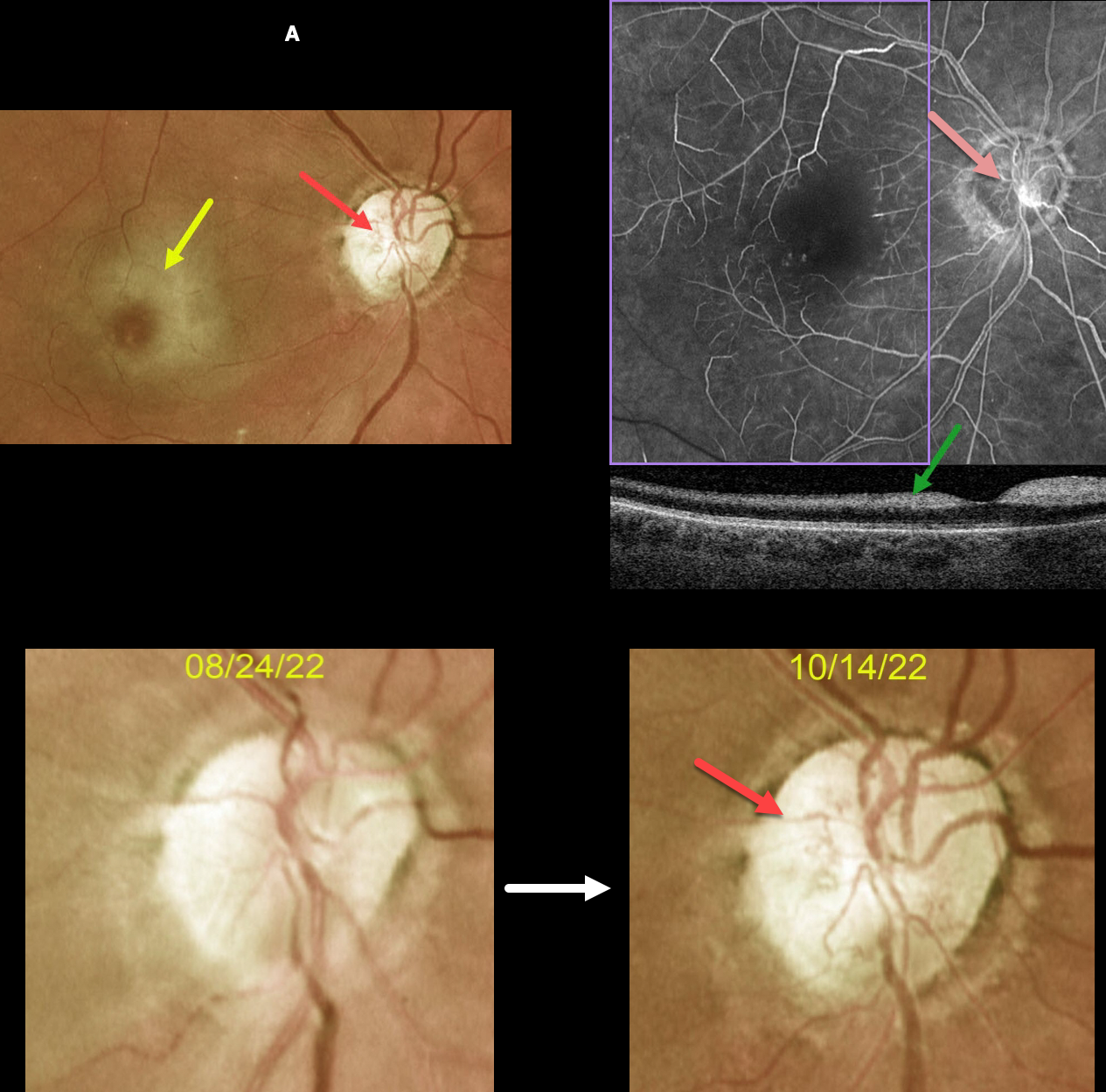
CRAO
Originally posted on @retina.rocks November 21, 2022
This 63YOF initially presented with an acute central retinal artery occlusion in her right eye. Emergency stroke referral revealed two prior strokes with a new asymptomatic stroke.
She returned two months later. There was subjective visual improvement, although vision remained at hand motion. Although the retinal opacification in general was improving, the perifoveal retina was more opaque with a pronounced cherry red spot.
New fine vessels were noted on the nerve. These vessels did not leak on fundus fluorescein angiography. Marked angiographic ischemia is also noted. OCT scanning shows increased reflectivity from the ischemic inner retina.
Learning Points:
Collateral vessels are dilated, pre-existing vessels that bypass an occlusion. These are seen in the posterior pole most commonly following retinal venous occlusions and are exceedingly rare with arterial occlusions.
The endothelial tight junctions are intact, so they don’t leak angiographically. This helps differentiate them from neovascularization, which does leak.
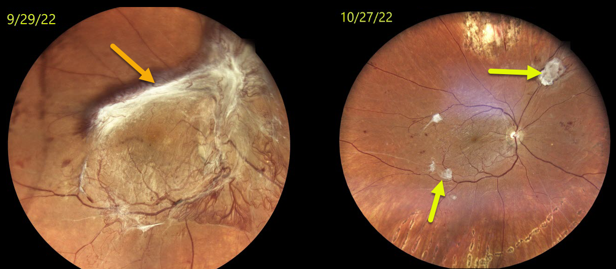
TRACTIONAL RETINAL DETACHMENT
Originally posted on @retina.rocks November 18, 2022
This 27YO female with type 1 diabetes presented with counting fingers vision OD after being lost to follow-up with a new tractional retinal detachment (TRD). Vision and retinal findings were unchanged OS.
Vitrectomy surgery was performed one week later.
Three weeks postop, vision improved to 20/40, and the retina was completely attached.
Residual fibrosed islands of neovascularization are seen in the distal temporal macula and superonasal midperiphery, representing the neovascular ingrowth sites onto the posterior hyaloid that created the TRD.
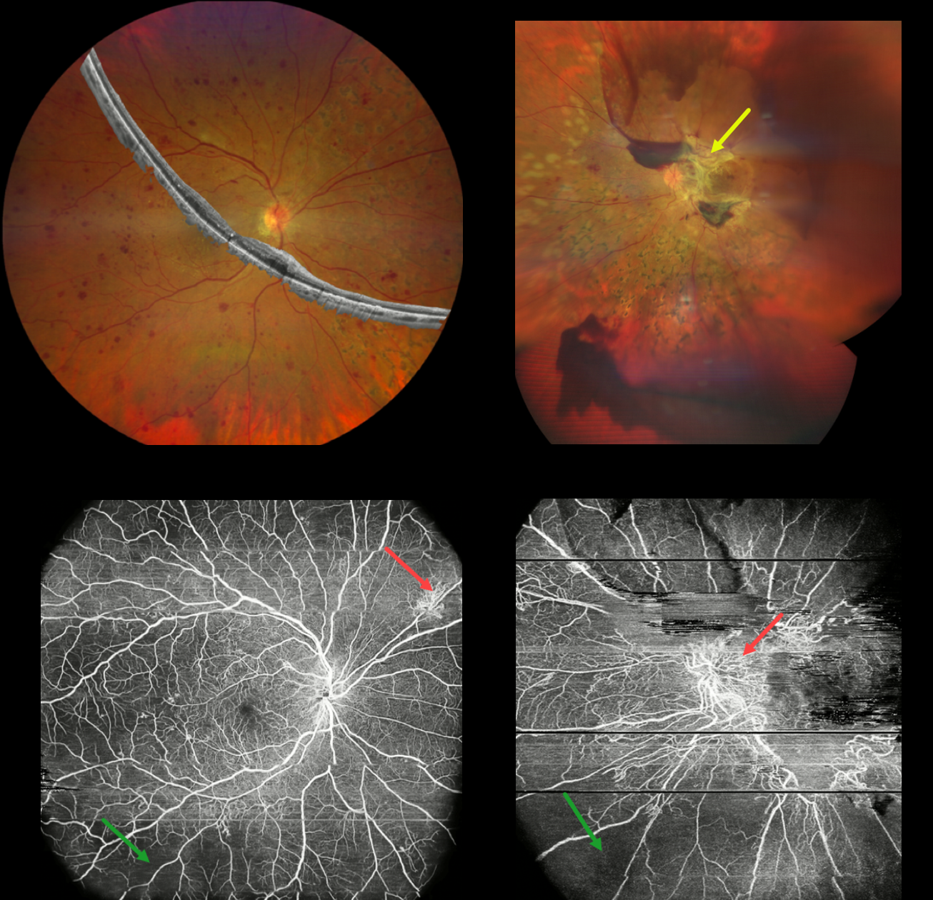
PROLIFERATIVE DIABETIC RETINOPATHY (PDR)
Veronika Matello and Barbara Parolini
Originally posted on @retina.rocks November 14, 2022
This 43YO female has a 16-year history of type 1 diabetes, and presented with 3 years of progressive vision loss in her left eye. Vision was 20/20 OD and 20/400 OS.
Composite widefield color imaging and OCT of her right eye show moderate retinal hemorrhages, nasal panretinal photocoagulation scarring, and no edema. Widefield OCT angiography shows peripheral ischemia with superonasal midperipheral neovascularization.
Multimodal imaging of her left eye shows fibrotic neovascularization around the nerve and macula. OCT angiography shows the neovascularization and peripheral retinal ischemia.
Learning Points:
Ultra-widefield imaging with fluorescein angiography is becoming increasingly important in screening for proliferative disease, especially since about 70% of angiographic nonperfusion lies outside the posterior pole (Silva et al, DRCR Protocol AA, Retina 2022;42:1302-1310). Widefield OCT angiography may also prove to be an equally effective yet less invasive screening tool (Al-Khersan et al., Am J Ophthalmol. 2021;224:292-300).
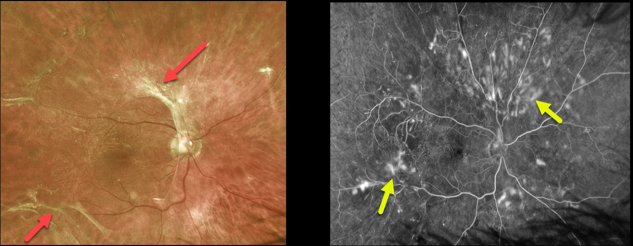
PROLIFERATIVE DIABETIC RETINOPATHY (PDR)
Originally posted on @retina.rocks November 3, 2022
This 58YO male presented with asymptomatic retinal changes in his right eye. Vision was 20/25 OD.
The left eye was phthisical. He had a history of type 2 diabetes.
Optos color imaging shows fibrosed retinal neovascularization scattered around the posterior pole. Fluorescein angiography shows scattered retinal neovascularization.
We were concerned that panretinal photocoagulation could further contract the neovascular tissue, possibly increasing traction and leading to vision loss. We were also hesitant to recommend primary vitrectomy since he is monocular, has excellent vision, and minimal macular traction.
We are therefore following him closely with observation and will likely recommend primary vitrectomy for increased traction. What would you recommend?
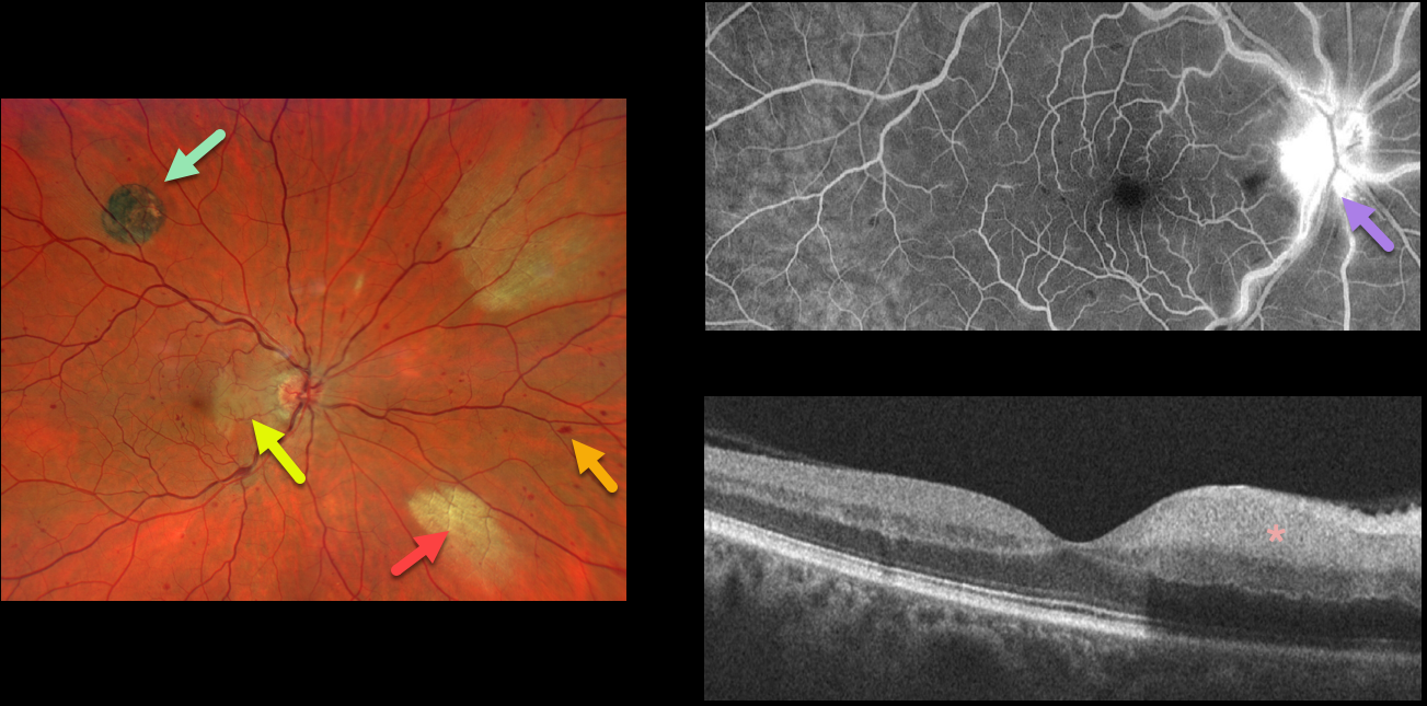
CRVO + CHRPE + MYELINATED NERVE FIBER LAYER
Will Gibson
Originally posted on @retina.rocks October 27, 2022
This 52YO male presented with acute visual symptoms in his right eye. Vision was 20/20.
Optos imaging shows a non-ischemic central retinal vein occlusion (CRVO) with mild scattered retinal hemorrhages in all quadrants. An acute nasal macular branch retinal artery occlusion (BRAO) is noted, along with a small round patch of congenital hypertrophy of the RPE (CHRPE) superotemporally, and two patches of myelinated nerve fiber layer (NFL) nasally.
Fluorescein angiography shows a well-perfused central retinal vein occlusion (CRVO) with late optic nerve leakage. OCT scanning shows hyperreflectivity of the inner nasal retina.
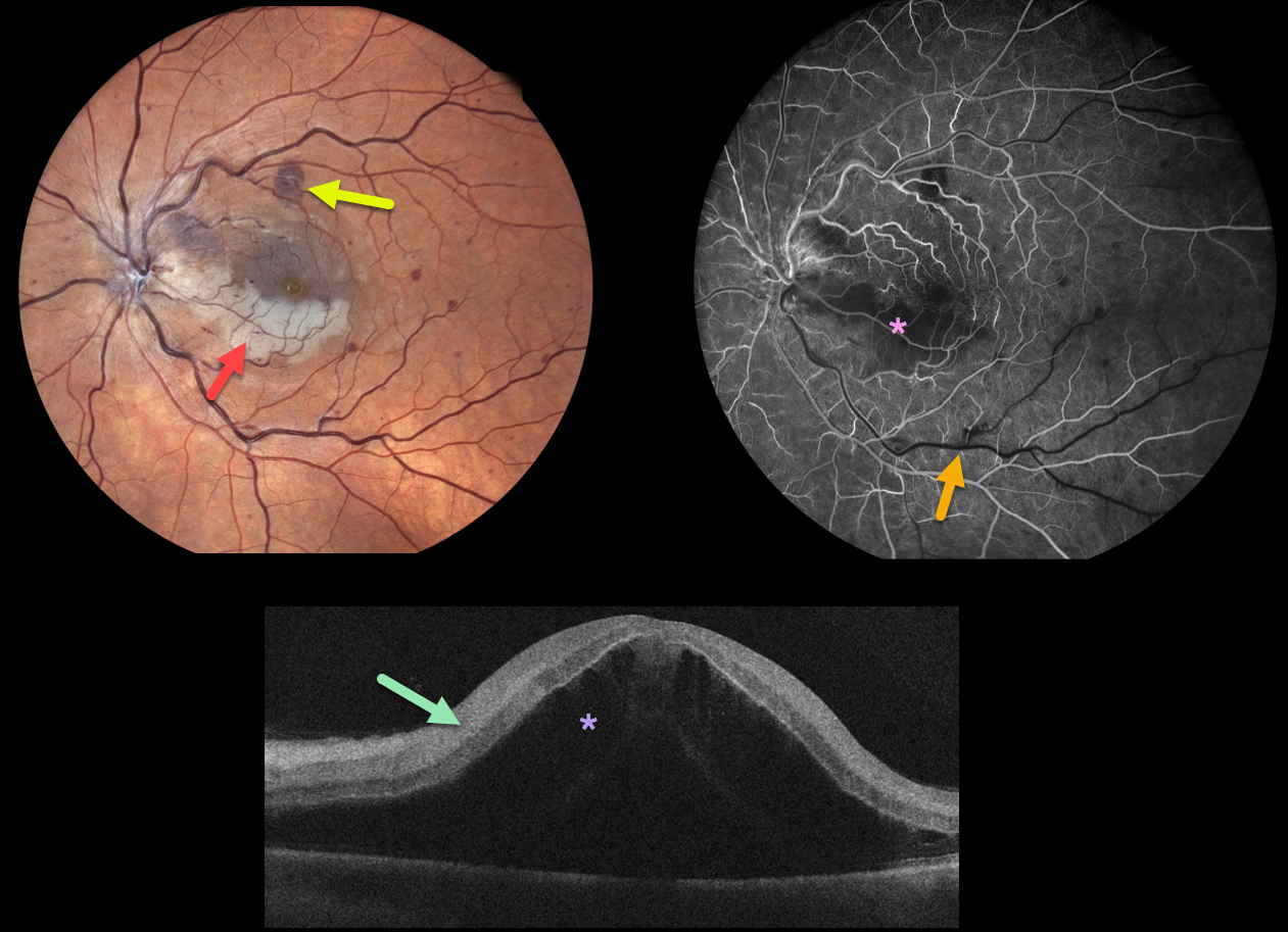
CRVO WITH SECONDARY BRAO
Originally posted on @retina.rocks October 18, 2022
This 49YO male woke with counting finger vision in his left eye. Fundus photography shows a fresh central retinal vein occlusion (CRVO) with mild retinal hemorrhages. An acute inferior macular branch retinal arterial occlusion (BRAO) is also noted.
OCT B-scan shows marked inner retinal hyperreflectivity and severe macula edema.
Fluorescein angiography in the venous laminar phase shows inferior macular hypofluorescence from combined blockage from the ischemic white retina, along with a filling defect from the BRAO. The retinal perfusion elsewhere is good.
Some patients with CRVO will develop a secondary non-embolic BRAO caused by compression from optic nerve edema. However, our patient’s optic nerve did not appear that swollen. Anti-VEGF therapy was started for the severe macular edema.
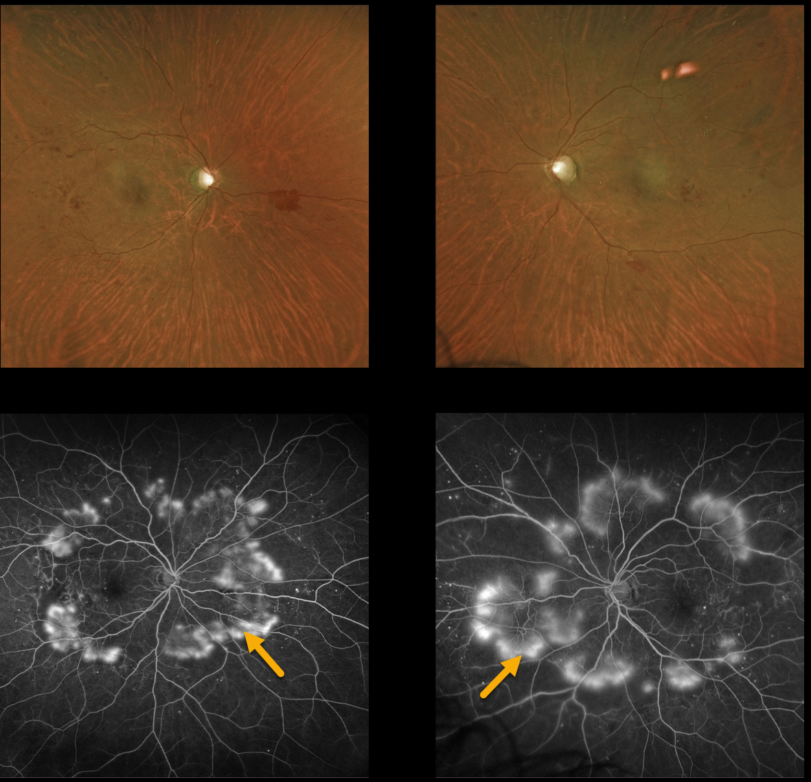
PROLIFERATIVE DIABETIC RETINOPATHY (PDR)
Originally posted on @retina.rocks October 12, 2022
This 51YO female with type 2 diabetes presented with severe peripheral proliferative diabetic retinopathy (PDR).
Despite having extensive angiographic neovascularization encircling each posterior pole, these findings are remarkably less obvious on color imaging.
Learning Points:
These types of presentations are very humbling to us clinicians and remind us of the importance of peripheral retinal examination and ultra-widefield imaging with fluorescein angiography in screening for proliferative disease.
Widefield OCT angiography may also prove to be an equally effective yet less invasive screening tool (Al-Khersan et al., Am J Ophthalmol 2021;224:292-300).
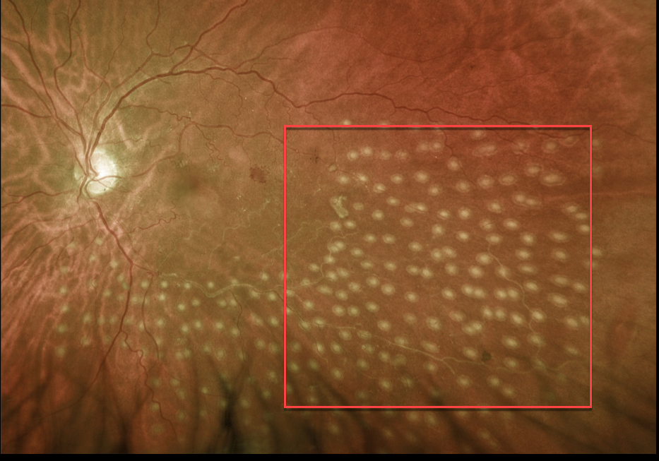
BRVO
Originally posted on @retina.rocks October 5, 2022
This 64YO female presented with one month of symptomatic vitreous floaters and 20/70 vision in her left eye.
Clinical examination and Optos photography with fluorescein angiography (not shown) confirmed an inferotemporal ischemic branch retinal vein occlusion (BRVO) with severe ischemia and scattered areas of retinal neovascularization.
She was scheduled to return for scatter laser photocoagulation, but was lost to follow-up. She presented 6 months later with one day of severe vision loss OS, now at 20/200. A significant vitreous hemorrhage precluded laser, so two monthly intravitreal Avastin injections were given.
Following this, the hemorrhage decreased enough to allow for scatter laser into the involved quadrant.
Learning Points:
In 1986, the Branch Vein Occlusion Study Group reported that scatter laser decreased the risk of vitreous hemorrhage from retinal neovascularization in BRVO, and suggested that laser should be applied once neovascularization develops (Arch Ophthalmol 1986;104:34-41).
Decades later, scatter laser remains the gold standard, although some may prefer to first stabilize neovascularization with anti-VEGF therapy before laser.
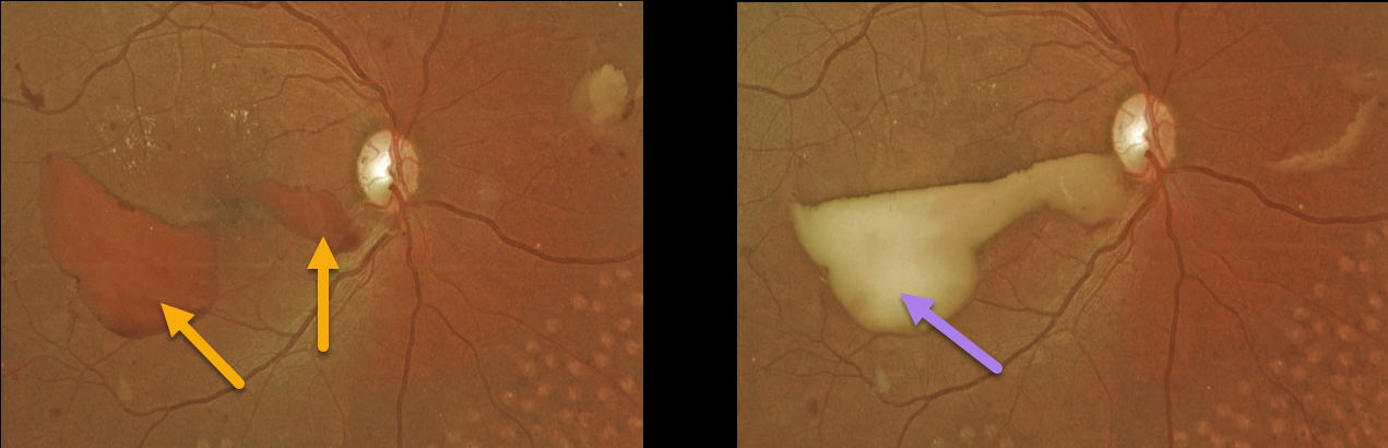
PROLIFERATIVE DIABETIC RETINOPATHY (PDR)
Originally posted on @retina.rocks August 17, 2022
This 37YO female presented with recent vision loss in her right eye. She had a history of prior laser treatment elsewhere several years ago. Vision was 20/30 OD and 20/25 OS.
Red preretinal blood was seen in the inferotemporal right macula with partial PRP scarring. One month following a single Avastin injection, the devitalized preretinal blood was yellowing. Bilateral PRP is planned.
Learning Points:
PRP has been around since the 1960s and still remains the mainstay for treating proliferative disease. Often considered a “one and done”, patients can still develop break-through hemorrhage or progressive neovascular growth and traction.
Intravitreal anti-VEGF injections are often helpful, but some will require vitrectomy.
In our patient’s case, the new preretinal blood was most likely due to her having been incompletely treated rather than a PRP failure.
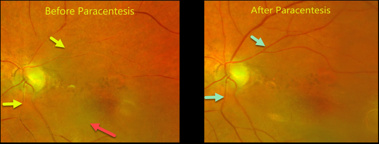
CRAO
Originally posted on @retina.rocks August 10, 2022
This 94YO female had a prior central retinal artery occlusion (CRAO) with vision that returned back to a baseline of 20/100. There was a history of multiple prior strokes, and she was not a surgical candidate.
Six months later, she came to see us emergently after waking with severe vision loss in her left eye. Vision was light perception.
A fresh CRAO was noted with a stagnant blood column and early inner retinal opacification. Blood flow was immediately restored following paracentesis.
The patient’s daughter reported sadly that the nursing home had made an error and not given her mom anticoagulants for months.
Learning Points:
Measures to relieve the occlusion by moving the causative embolus downstream include vasodilation by increasing the carbon dioxide concentration (rebreathing into a paper bag, carbogen) or rapid IOP lowering (paracentesis, ocular massage). Interestingly, a recent meta-analysis suggested that eyes treated with these measures had worse visual outcomes and recovery rates (Schrag et al, JAMA Neurology 2015;72:1148-1154).
Our patient hasn’t returned yet for us to determine if the paracentesis truly helped.
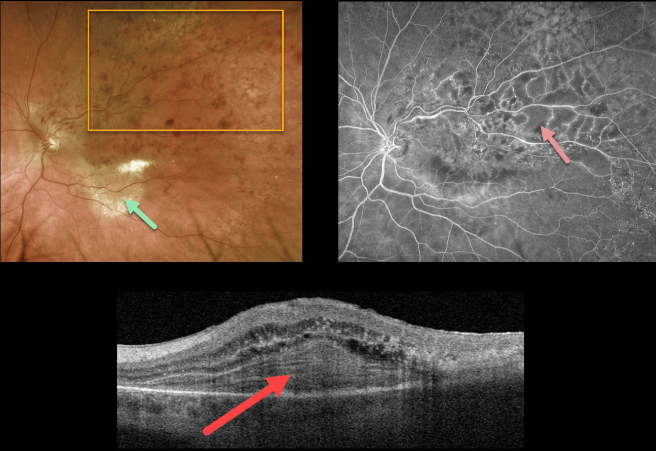
BRVO
Originally posted on @retina.rocks August 8, 2022
This 82YO female presented with a major superotemporal branch retinal vein occlusion (BRVO) and hand motion vision. She reported decreased vision for over a year.
Optos photography shows a major superotemporal BRVO. A plaque of subretinal lipid is seen in the inferotemporal macula. Swept-source OCT through this lipid shows an unusual laminated, subretinal, hyperreflective mound. Fluorescein angiography shows some macular leakage and some areas of more peripheral ischemia.
Although the patient has a poor prognosis due to the chronicity of the disease, monthly intravitreal Avastin for the central edema was initiated in hopes that it may improve her vision.
Learning Points:
Lipid exudates are rarely seen in BRVO, which may be due to a physiologic defect in the inner blood-retinal barrier (the endothelial cell tight junctions) that are too small to allow larger lipoproteins to escape into the extracellular space.
When present, lipid is often associated with a retinal venous macroaneurysm (Cousins et al, AJO 1990;109:567-170), which was not visualized in our patient.
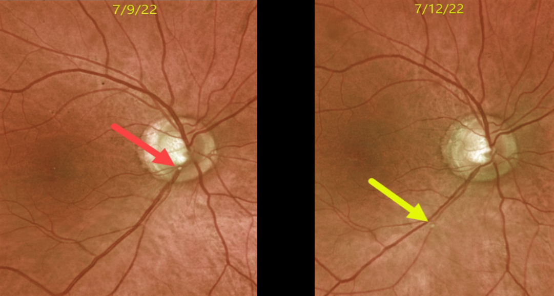
RETINAL EMBOLUS
Originally posted on @retina.rocks July 22, 2022
This 72YO male presented with acute superior field loss from an acute branch retinal artery occlusion (BRAO) in his right eye.
The causative calcific embolus is seen originally along the inferior optic nerve margin. Three days later, the plaque moved further downstream.
This patient had acute retinal ischemia (defined as acute BRAO, central retinal artery occlusion, or transient monocular vision loss), and was immediately referred to the nearest stroke center, which is the standard of care.
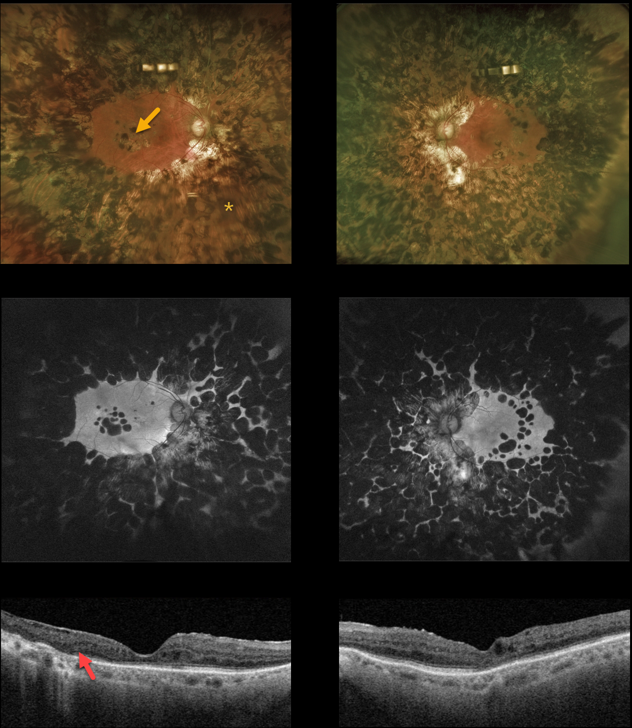
PANRETINAL PHOTOCOAGULATION
Originally posted on @retina.rocks July 7, 2022
This 80YO diabetic female gave a history 0f prior retinal lasers elsewhere about 20 years earlier. Vision was 20/30 OD and 20/80 OS.
Optos color and fundus autofluorescent imaging show heavy bilateral panretinal photocoagulation (PRP) and macular laser scarring. Swept source OCT shows variable disorganization of the retinal layers and outer retinal atrophy. Visual fields (not shown) are severely constricted due to the ‘iatrogenic’ retinitis pigmentosa caused by the posterior and confluent laser scarring.
Learning Points:
Although possibly appropriate before the advent of modern vitrectomy surgery and anti-VEGF therapy, this degree of laser should never be seen in today’s patients. Laser should begin well outside the arcades and away from the optic nerve, extending as anteriorly as possible with the 200 to 500 micron-sized retinal burns spaced about one burn-width apart.
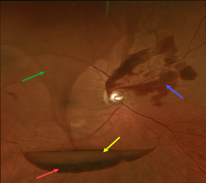
PROLIFERATIVE DIABETIC RETINOPATHY (PDR)
Originally posted on @retina.rocks June 30, 2022
The preretinal blood in this 70YO male with proliferative diabetic retinopathy (PDR) has multiple layers.
The red arrow highlights subhyaloid blood, while the yellow arrow shows the yellowing, devitalized subhyaloid blood just above. The blue arrow shows subhyaloid blood emanating from disc neovascularization. Last but not least, the twister of blood within the vitreous gives the appearance of a tornado literally shaking up all of this blood.
Panretinal photocoagulation was started.
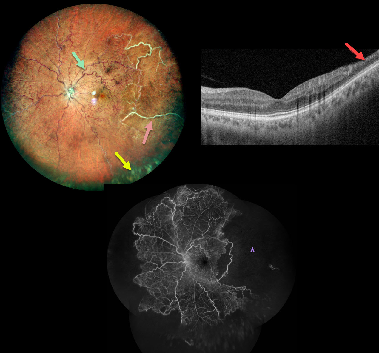
ISCHEMIC CRVO
Nivesh Gupta
Originally posted on @retina.rocks June 28, 2022
This healthy 34YO male presented with this relatively asymptomatic central retinal vein occlusion (CRVO). Vision was 20/20 OU.
Color imaging shows large temporal nonperfused retinal vessels with old inferior yellow vitreous hemorrhage. The retinal vessels are diffusely dilated and tortuous. OCT B-scan shows a remarkably normal central macula with some distal inner retinal thinning. Fluorescein angiography confirms widespread peripheral nonperfusion.
Anti-VEGF therapy, followed by panretinal photocoagulation, was administered.
Learning Points:
The Central Vein Occlusion Study (CVOS) defined ischemic CRVO as having at least 10 disc areas of angiographic retinal nonperfusion as determined by standard photographic views with the Canon (60 degrees) or Topcon (45 degrees) wide-angle fundus cameras (Ophthalmology 1995;102:1434-1444).
Although clearly ischemic, this patient might have been classified by the CVOS as a perfused occlusion, depending on the extent of peripheral ischemia imaged. As with diabetic retinopathy (see Brucker, Retina 2021;41:461-463), ultrawidefield imaging and anti-VEGF injections have thrown a literal wrench into how we think about, classify, and treat retinal disorders.
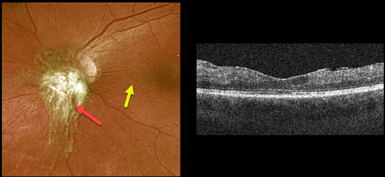
PROLIFERATIVE DIABETIC RETINOPATHY (PDR)
Originally posted on @retina.rocks June 16, 2022
This 26YO female presented with bilateral disc and peripheral retinal neovascularization from proliferative diabetic retinopathy. Vision was 20/50 OD and 20/200 OS.
Panretinal photocoagulation was started in each eye. She was then lost to follow-up for a year, when she returned with a somewhat unusual elevated patch of fibrosed disc neovascularization in her left eye. Some mild nasal macular traction is noted in this image and on OCT scanning.
Some peripheral vitreous hemorrhage was also present in her left eye (not shown). Vision was 20/50 in her stable OD and counting fingers OS.
The PRP was completed in each eye. The fibrosed NVD OS will not resolve and hopefully won’t cause further macular traction.
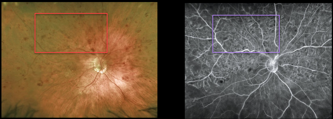
HRVO
Originally posted on @retina.rocks June 14, 2022
This 88YO female presented with counting fingers vision in her right eye from a symptomatic superior hemiretinal vein occlusion (HRVO).
Optos imaging shows moderate superior hemispheric inner retinal hemorrhages, and fluorescein angiography shows a well-perfused occlusion. OCT scanning showed moderate central macular edema (not shown). Anti-VEGF therapy was started.
Learning Points:
Branch retinal vein occlusions are virtually always caused by a retinal artery compressing a retinal vein within its common adventitial sheath. Some people are born with an anomalous two-trunked central retinal vein that independently drains the superior and inferior retinal hemispheres.
HRVO, similar to central retinal vein occlusion, is caused by compression of the retinal vein posterior to the lamina cribrosa (see Hayreh, Arch Ophthalmol 1980;98:1600-1609).
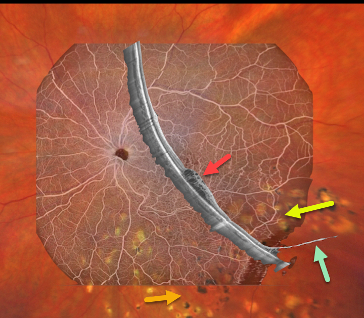
BRVO WITH DEGENERATIVE RETINOSCHISIS
Barbara Parolini and Veronika Matello
Originally posted on @retina.rocks June 13, 2022
This 67YO female has a history of branch retinal vein occlusion (BRVO) in her left eye. Asymptomatic degenerative retinoschisis was also present bilaterally. Vision was 20/33. She had received prior scatter laser and intravitreal injections about 3 years earlier.
Fortunately, all findings remained stable despite loss to follow-up due to the COVID pandemic.
Clarus 500 wide field imaging shows mild inferotemporal tortuosity with collateral vessels within the involved quadrant. Prior scatter laser scarring for secondary retinal neovascularization, along with prior barrier laser around the posterior edge of the schisis cavity, is noted.
Canon Xephilio S1 OCT angiography highlights the collateral vessels and more peripheral retinal nonperfusion. Xephilio B-scan shows some inferotemporal macular edema, and just catches the more distal inferotemporal schisis.
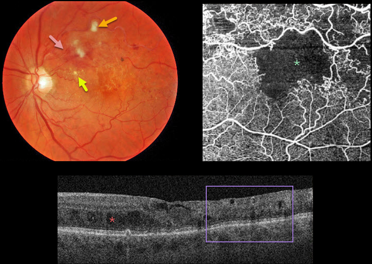
BRVO WITH MACULAR ISCHEMIA
Originally posted on @retina.rocks June 10, 2022
This 79YO male presented with a major superotemporal branch retinal vein occlusion (BRVO) and counting fingers vision.
Fundus photography shows superior macular hemorrhages with inner retinal ischemia (cotton-wool spots) and some nasal lipid. The vessels, especially around the superotemporal fovea, are nonperfused.
The clinically suspected foveal ischemia is confirmed on OCT angiography. Spectral-domain OCT shows disorganization of the inner retinal layers (DRIL), particularly within the clinically ischemic temporal macula, along with scattered inner and outer retinal cysts.
Learning Points:
DRIL is seen in various retinal vascular disorders, including diabetic macular edema and retinal vein occlusions. It is generally associated with worse vision and increased anti-VEGF treatment burden (see Babiuch et al, JAMA Ophthalmol 2019;137:38-46).
Since the patient has no central foveal thickening or retinal neovascularization, we are observing for now. Anti-VEGF therapy will not restore the foveal circulation.
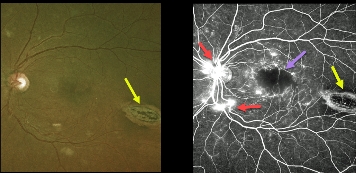
DIABETIC MACULAR ISCHEMIA WITH TORPEDO MACULOPATHY
Originally posted on @retina.rocks June 9, 2022
This 45YO female with type 2 diabetes presented with bilateral proliferative diabetic retinopathy (PDR). Vision in her left eye was 20/200 due to severe foveal ischemia, which is best seen on fluorescein angiography. Leaking disc and peripheral neovascularization are also seen angiographically. The macula was also edematous on OCT scanning (not shown).
An incidental finding completely unrelated to her diabetic retinopathy was an oval-shaped chorioretinal lesion in the distal inferotemporal macula known as torpedo maculopathy.
Although anti-VEGF therapy was started in her left eye for the macula edema, we doubt vision will significantly improve due to the foveal ischemia.
Learning Points:
Gass originally described torpedo maculopathy as a solitary hypopigmented nevus of the RPE (Arch Ophthalmology 1992;1358-1359). These benign, unilateral lesions are teardrop-shaped and almost always located in the horizontal meridian of the temporal macula.
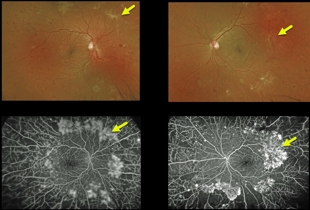
PROLIFERATIVE DIABETIC RETINOPATHY (PDR)
Originally posted on @retina.rocks May 27, 2022
This 29YO male with type 1 diabetes presented with severe peripheral proliferative diabetic retinopathy (PDR). Despite having extensive peripheral ischemia and neovascularization, the posterior poles were remarkably almost normal.
Learning Points:
These types of presentations are very humbling for us clinicians and remind us of the importance of peripheral retinal examination and ultra-widefield imaging with fluorescein angiography in screening for proliferative disease.
Widefield OCT angiography may also prove to be an equally effective yet less invasive screening tool (Al-Khersan et al., Am J Ophthalmol 2021;224:292-300).
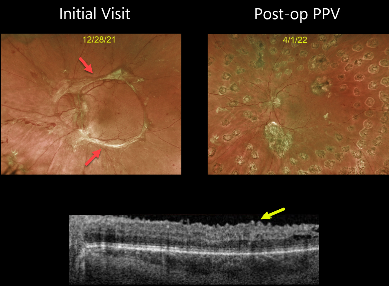
TRACTIONAL RETINAL DETACHMENT
Originally posted on @retina.rocks May 20, 2022
This 32YO female with type 1 diabetes presented with an 18-month history of vision loss in her left eye. Vision was counting fingers.
Optos imaging beautifully captures the pre-operative photos of the tractional retinal detachment, caused primarily by fibrosed retinal neovascularization extending in a circular pattern from the nerve along the arcades.
The second image shows the patient four months following vitrectomy. Swept-source OCT shows an irregular inner retinal contour, which is common following these vitrectomies.
Vision improved to 20/50. She is currently undergoing panretinal photocoagulation in her contralateral eye.
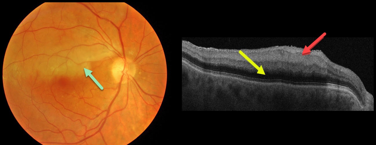
BRAO
Originally posted on @retina.rocks April 29, 2022
This 80YO male presented with an acute branch retinal artery occlusion (BRAO) and 20/25 vision. Triton color imaging shows an opaque retina in the superior macula.
Swept-source OCT shows a thickened, hyperreflective inner retina with posterior shadowing due to decreased light transmittance through the opacified inner retinal layers. There are no emboli.
This patient had acute retinal ischemia (defined as acute BRAO, central retinal artery occlusion or transient monocular vision loss), and was immediately referred to the nearest stroke center.
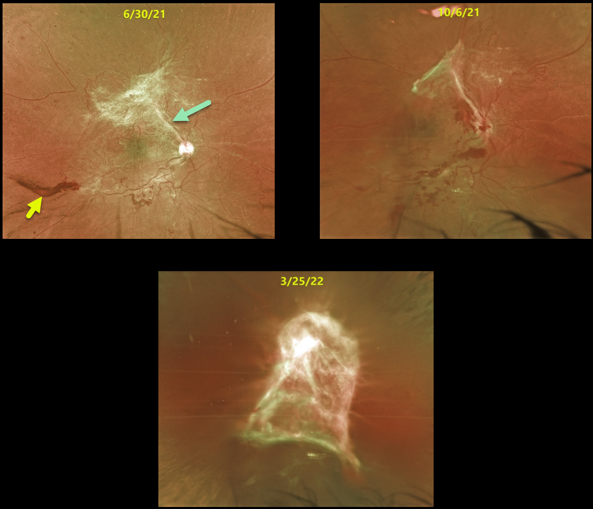
TRACTIONAL RETINAL DETACHMENT
Originally posted on @retina.rocks April 14, 2022
This 34YO female presented with hand motion vision from a progressive diabetic tractional retinal detachment with subhyaloid hemorrhage.
She was repeatedly noncompliant with appointment attendance and was lost to follow-up on numerous occasions.
The images show the progressive fibrosis and traction that developed over the course of a year. She will now require vitrectomy in hopes of recovering some vision in this eye.
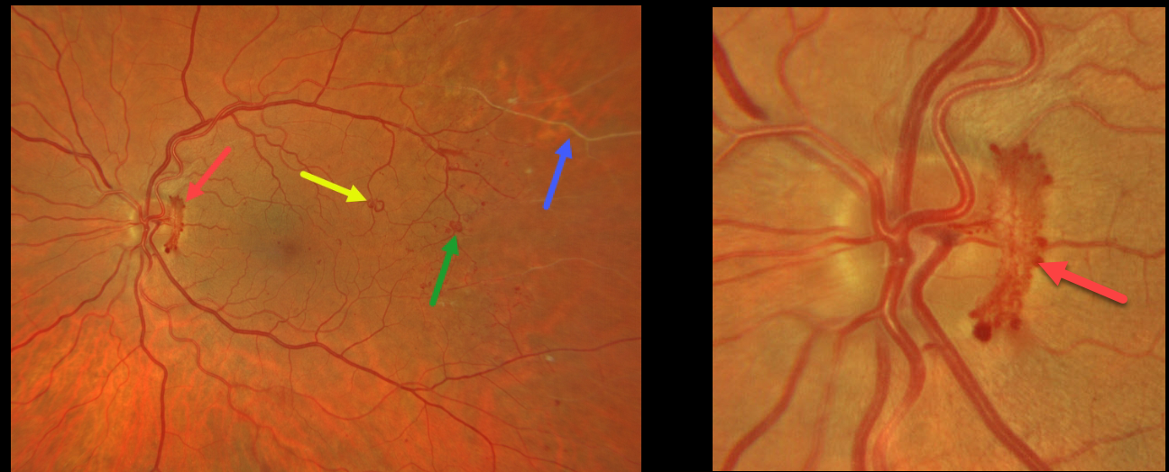
PROLIFERATIVE DIABETIC RETINOPATHY (PDR)
Originally posted on @retina.rocks April 8, 2022
This patient presented with some beautifully imaged findings for proliferative diabetic retinopathy (PDR).
A wide-angle photograph shows active disc neovascularization (NVD), a venous loop, a few small patches of distal macular peripheral neovascularization (NVE), and distal retinal ischemia. A magnified image of the nerve shows the NVD.
Although venous loops are not generally included in the clinical classification of diabetic retinopathy, they are universally seen in severely ischemic eyes.
This patient is currently undergoing panretinal photocoagulation.
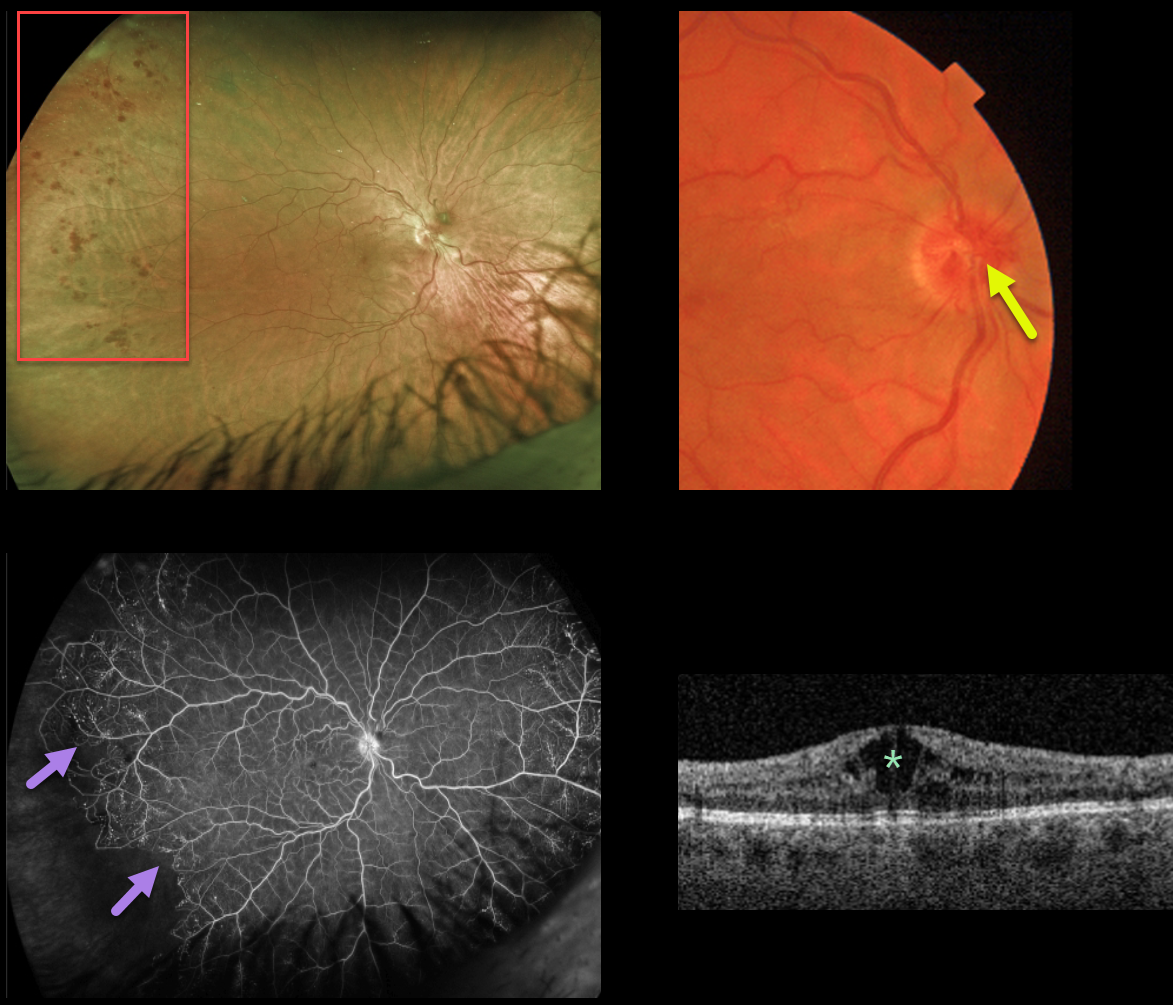
CRVO
Originally posted on @retina.rocks April 5, 2022
This 40YO female presented with 20/70 vision OD from a chronic central retinal vein occlusion (CRVO). The hemorrhages are virtually limited to the temporal periphery. Triton color imaging shows optic nerve collaterals.
Ultrawidefield Optos fluorescein angiography shows good posterior pole perfusion, nasal peripheral telangiectasia, and a sharp margin of infero-temporal peripheral ischemia.
Central cystoid edema is seen on swept-source OCT. Anti-VEGF injections were started for the macular edema.
Learning Points:
This sharply demarcated peripheral border of perfused ischemic retina is classically described in Eales and sickle retinopathy.
However, we have also found this to be a not uncommon finding in resolved CRVO, which has more classically been described as having vascular abnormalities extending outwards from the optic nerve. In this case, the optic disc collaterals point to the cause of the peripheral findings.
The Central Vein Occlusion Study (CVOS) defined ischemic CRVO as having at least 10 disc areas of angiographic retinal nonperfusion as determined by standard photographic views with the Canon (60 degrees) or Topcon (45 degrees) wide-angle fundus camera (Ophthalmology 1995;102:1434-1444).
Although clearly ischemic, this patient would have been classified by the CVOS as a perfused occlusion.
As with diabetic retinopathy (see Brucker, Retina 2021;41:461-463), ultrawidefield imaging and anti-VEGF injections have thrown a literal wrench into how we think about, classify, and treat retinal disorders.
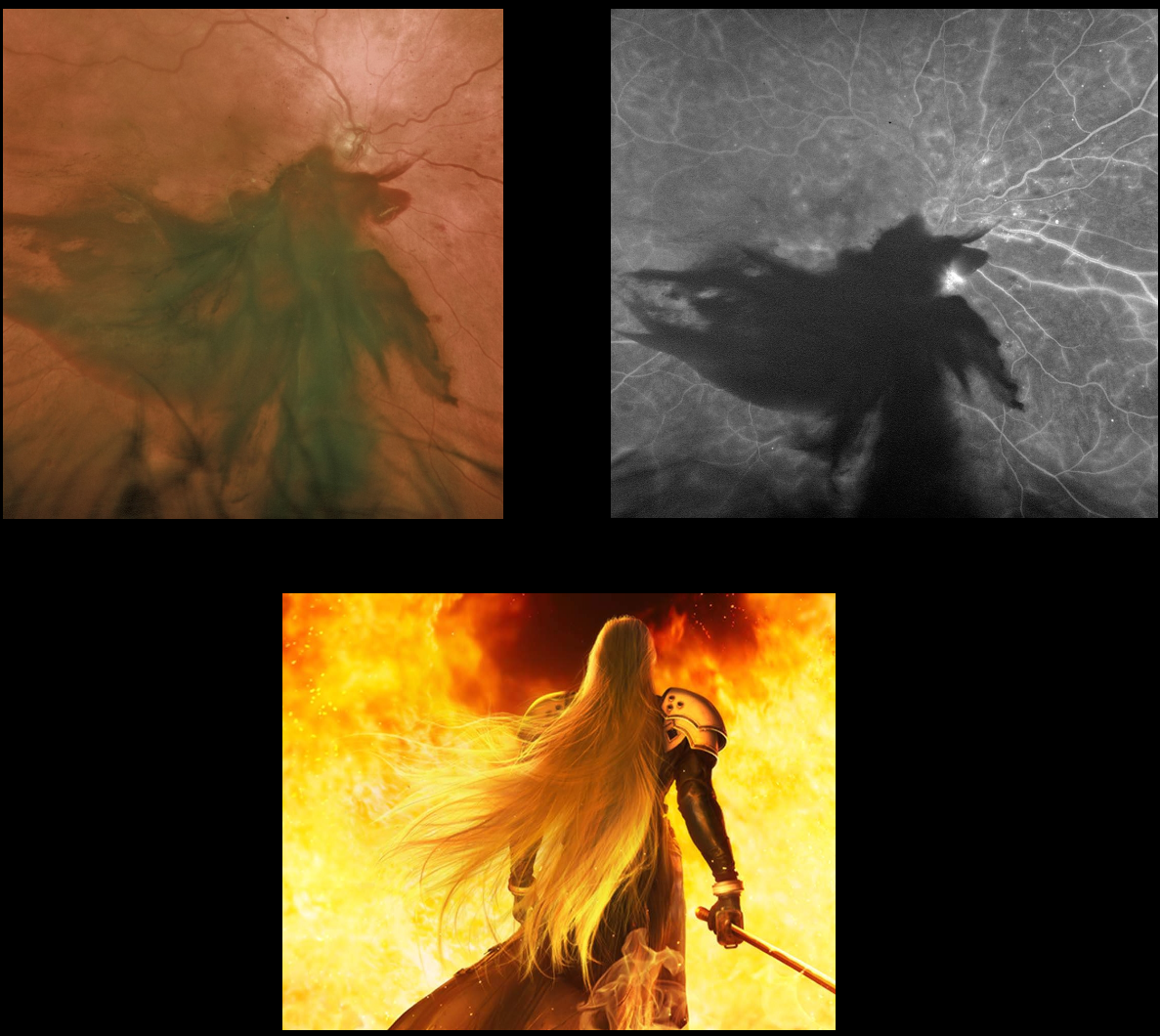
SEPHIROTH RETINOPATHY
Originally posted on @retina.rocks March 25, 2022
We all know it’s a large subhyaloid hemorrhage from proliferative diabetic retinopathy, but check your imagination and tell us what you see.
We see Sephiroth from Final Fantasy!
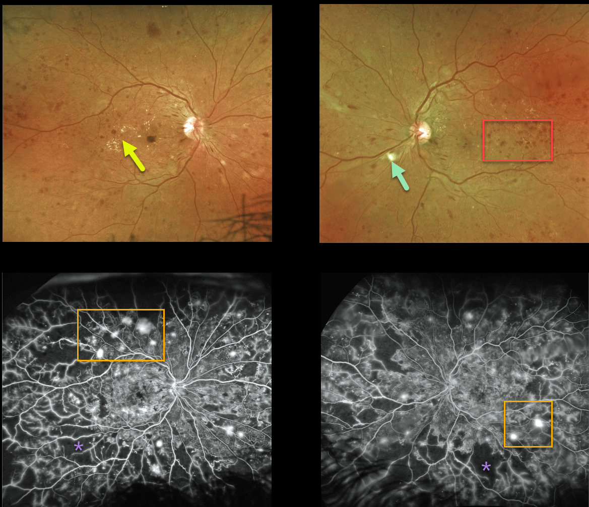
PROLIFERATIVE DIABETIC RETINOPATHY (PDR)
Originally posted on @retina.rocks March 23, 2022
This 41YO female presented with bilateral proliferative diabetic retinopathy (PDR).
Optos ultra-widefield color imaging shows extensive bilateral retinal hemorrhages, along with hard exudates and cotton wool spots.
Fluorescein angiogram shows extensive scattered retinal neovascularization with more peripheral retinal ischemia.
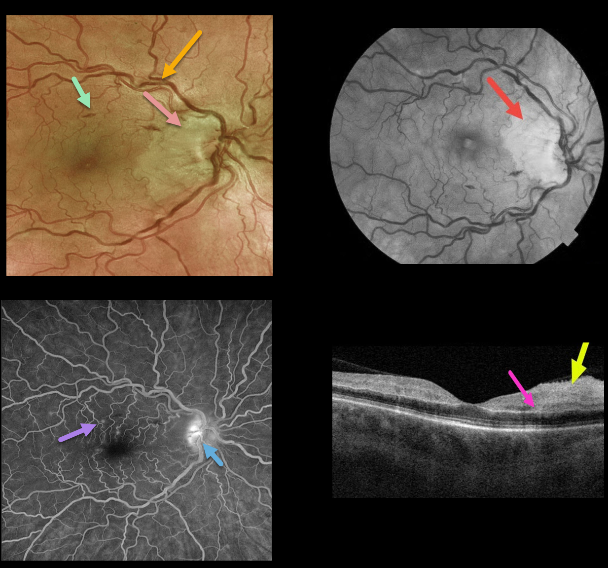
CRVO + RAO + PAMM
Originally posted on @retina.rocks March 4, 2022
This 50YO female presented with counting finger vision in her right eye due to an acute central retinal vein occlusion (CRVO) with a nasal macular retinal artery occlusion (RAO).
Optos fundus photography shows retinal vascular tortuosity, mild retinal hemorrhages, a mildly swollen nerve, and white ischemic nasal macular retina. The ischemic retinal changes are most apparent on the red-free photo.
Fluorescein angiography (FA) shows some mild focal areas of macular leakage with late staining of the temporal disc.
OCT shows areas of inner retinal edema and hyperreflectivity. There is also a small area of mid-retinal hyperreflectivity consistent with paracentral acute middle maculopathy (PAMM).
Learning Points:
Cilioretinal artery occlusion is usually non-embolic and seen in association with giant cell arteritis, secondary to a CRVO, or as an isolated event. The occlusion may be due in part to arterial compression from a swollen nerve.
Pichi et al felt that CRVO PAMM-type lesions, as seen in our patient, are due to hypoperfusion from increased downstream intraluminal pressure (Br J Ophthalmol 2019;103:1137-1145).
We couldn’t find a definite cilioretinal artery in our patient, although the ischemia was within the distribution of one if one were present. Interestingly, the nasal macular capillaries appeared perfused on FA. However, FA captures just the inner retinal capillary plexus.
Although the OCT shows evidence of acute inner capillary plexus ischemia, the angiographically perfused inner retina supports a more PAMM-type mechanism in our case.
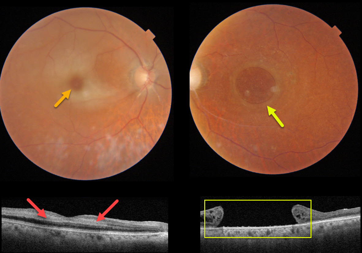
CRAO + MACULAR HOLE
Originally posted on @retina.rocks February 21, 2022
This 82YO female presented with counting fingers vision in her previously healthy right eye. Vision in her left eye had been counting fingers for many years due to a large chronic macular hole measuring 2,870 microns.
An acute central retinal artery occlusion (CRAO) was noted in her right eye. The foveal retina is still perfused by the underlying choroid, resulting in the classic cherry red spot.
Swept-source OCT shows hyperreflectivity from the opaque, ischemic inner retinal layers.
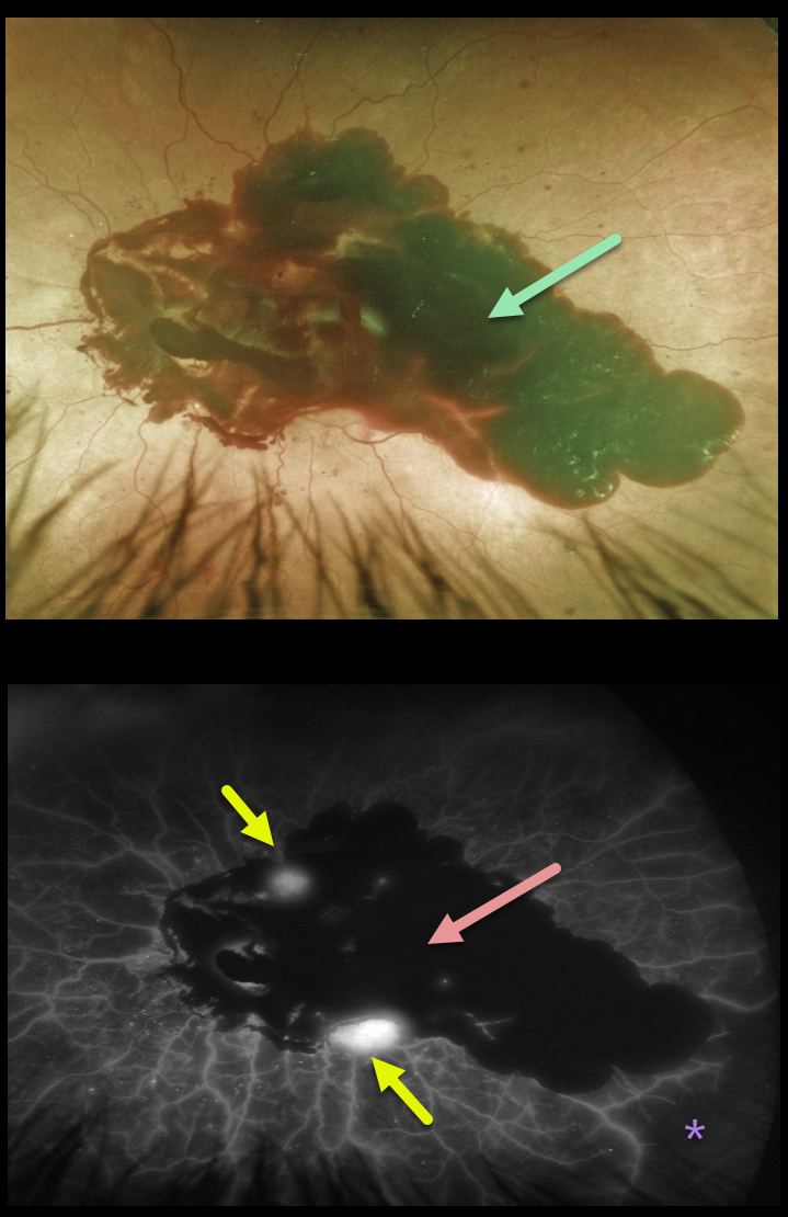
PROLIFERATIVE DIABETIC RETINOPATHY (PDR)
Originally posted on @retina.rocks February 1, 2022
This 69YO male with type 2 diabetes presented with severe acute vision loss in his left eye from a large posterior pole subhyaloid hemorrhage.
The hemorrhage blocks on fluorescein angiography, with a few areas of leaking neovascularization and severe peripheral ischemia.
He was initially treated with intravitreal Avastin, and over the coming months, panretinal photocoagulation (PRP) was performed.
However, he later developed a large vitreous hemorrhage and will likely need vitrectomy surgery.
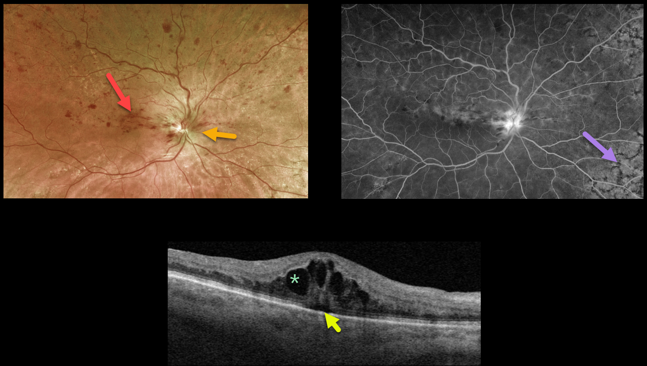
HRVO
Originally posted on @retina.rocks January 13, 2022
This 81YO female presented with 20/60 vision from a symptomatic superior hemiretinal vein occlusion (HRVO). Optos imaging shows foveal and superior inner retinal hemorrhages with a swollen optic nerve, and fluorescein angiography shows a well-perfused occlusion.
Swept-source OCT confirms moderate cystic macular edema with a trace amount of underlying subretinal fluid.
Age-related peripheral reticular degeneration of the RPE is best seen angiographically as an incidental finding.
The patient is receiving ongoing anti-VEGF injections to control her macular edema. Intravitreal injections (anti-VEGF or steroids) are the treatment of choice, although grid laser may help decrease the treatment burden.
Learning Points:
Branch retinal vein occlusions are virtually always caused by a retinal artery compressing a retinal vein within its common adventitial sheath.
Some people are born with an anomalous two-trunked central retinal vein that independently drains the superior and inferior retinal hemispheres.
HRVO, similar to central retinal vein occlusion, is caused by compression of the retinal vein posterior to the lamina cribrosa (see Hayreh, Arch Ophthalmol 1980;98:1600-1609).
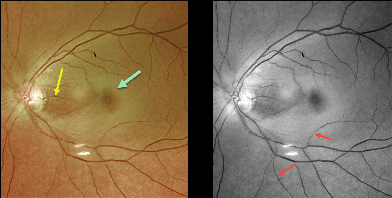
CRAO
Originally posted on @retina.rocks January 7, 2022
This 73YO female presented with counting-fingers vision secondary to an acute central retinal artery occlusion (CRAO).
The foveal retina is still perfused by the underlying choroid, resulting in the classic cherry red spot appearance. The nasal macula is also preserved due to the presence of a cilioretinal artery, which unfortunately does not supply the macular center. The Optos green channel best images the stagnant blood column.
Our patient’s MRI revealed several acute diffuse embolic strokes. Carotid duplex ultrasound showed severe bilateral carotid stenosis. In addition, her blood glucose was 357, and her erythrocyte sedimentation rate was 94. She was also started on systemic prednisone by the ER until giant cell arteritis could more definitively be ruled out.
Learning Points:
Patients with acute retinal ischemia (defined as transient monocular vision loss, acute BRAO, or acute CRAO) need to be emergently referred to a stroke center.
This is especially urgent with an acute CRAO since about 75% of patients have already developed a recent stroke.
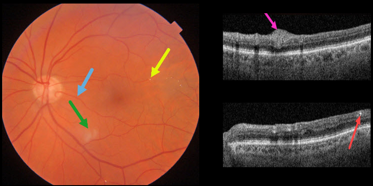
BRAO
Originally posted on @retina.rocks January 6, 2022
This 85YO male has 20/25 vision despite multiple retinal emboli and a resolving branch retinal artery occlusion (BRAO).
Triton swept-source OCT shows residual inner retinal edema and opacification. The embolus on Triton photography is also imaged in the OCT B-scan.
A cilioretinal artery may be responsible for the good vision.
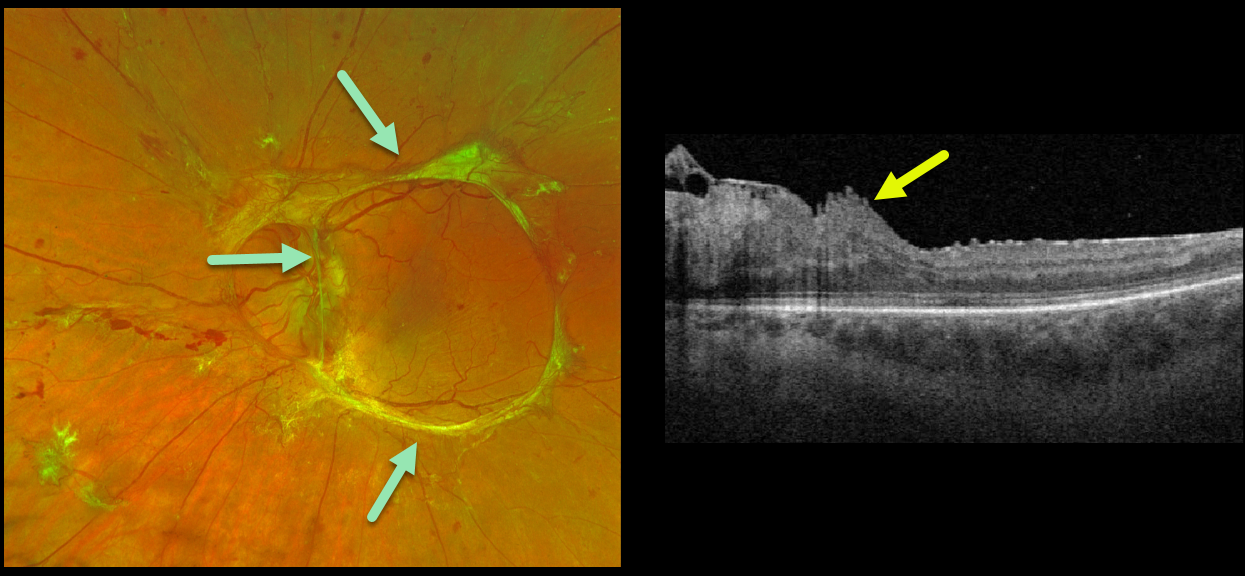
TRACTIONAL RETINAL DETACHMENT
Originally posted on @retina.rocks January 3, 2022
This 32YO female with type 1 diabetes presented with an 18-month history of vision loss in her left eye. Vision was counting fingers.
Optos imaging beautifully captures the traction retinal detachment from mostly fibrosed retinal neovascularization extending circularly from the nerve along the arcades.
Triton swept-source OCT surprisingly shows relatively mild macular distortion despite the severe vision loss, which is most likely due to vitreopapillary traction.
Intravitreal Avastin followed by vitrectomy will be performed.
Learning Points:
Tractional retinal detachment is one of the main complications of untreated or incompletely treated proliferative diabetic retinopathy.
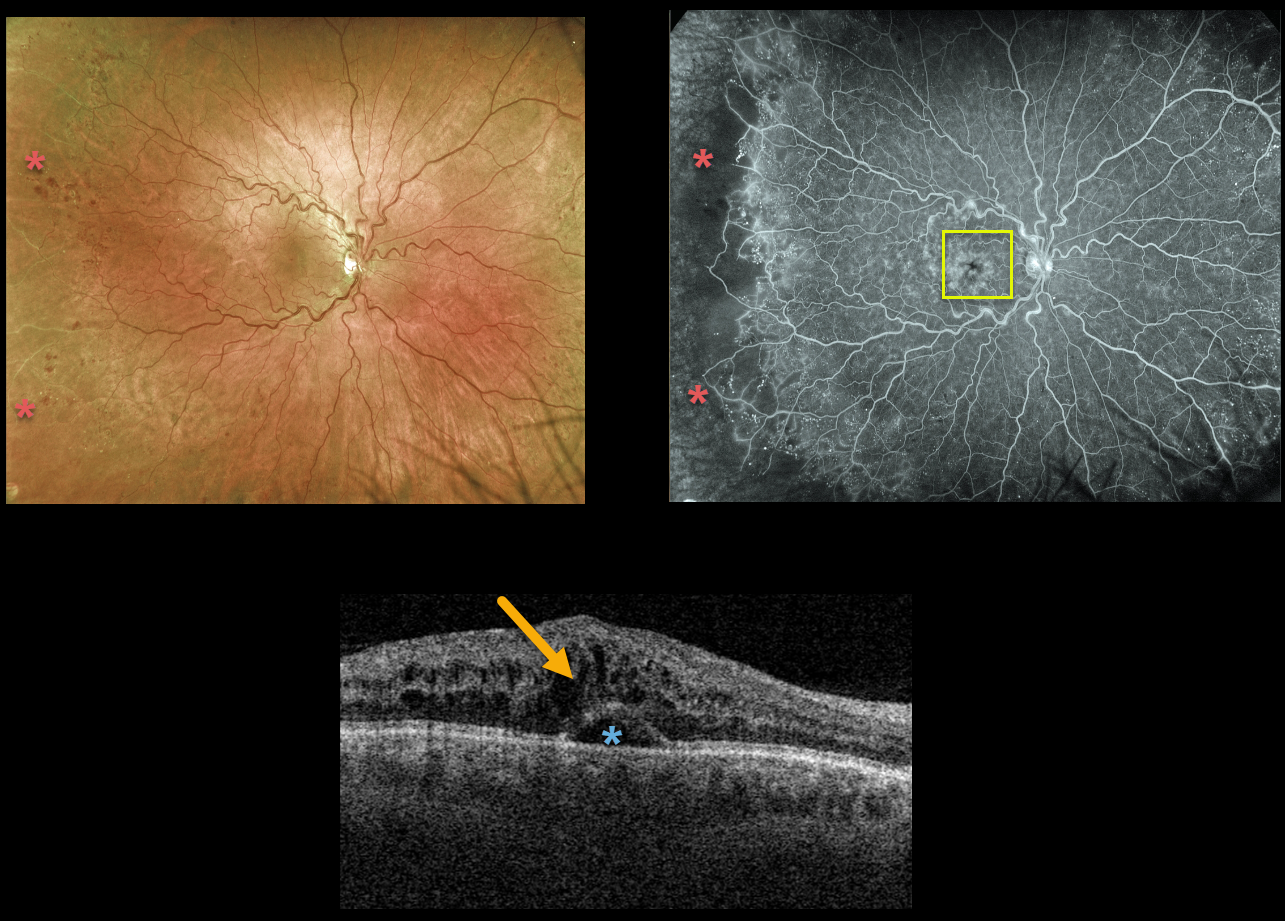
CRVO
Originally posted on @retina.rocks December 1, 2021
This 51YO male presented with 20/200 vision from an acute central retinal vein occlusion (CRVO).
There are virtually no posterior pole hemorrhages. Ultrawidefield Optos fluorescein angiography shows good posterior pole perfusion, although peripheral temporal ischemia is noted. Angiographic cystoid leakage is seen.
Triton swept-source OCT confirmed cystoid edema with some subfoveal fluid. Intravitreal anti-VEGF injections were started.
Learning Points:
The Central Vein Occlusion Study (CVOS) defined ischemic CRVO as having at least 10 disc areas of angiographic retinal nonperfusion as determined by standard photographic views with the Canon (60 degrees) or Topcon (45 degrees) wide-angle fundus camera (Ophthalmology 1995;102:1434-1444).
Although clearly ischemic, our patient would have been classified by the CVOS as a perfused occlusion.
As with diabetic retinopathy (see Brucker, Retina 2021;41:461-463), ultrawidefield imaging and anti-VEGF injections have thrown a literal wrench into how we think about, classify, and treat retinal disorders.
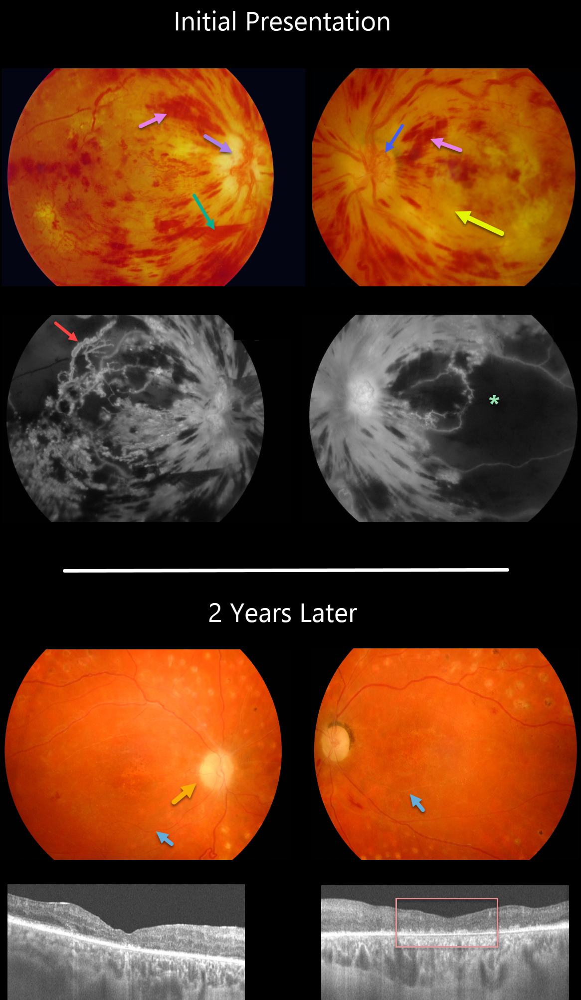
PDR + HYPERTENSIVE RETINOPATHY
Originally posted on @retina.rocks November 30, 2021
This 30YO female presented in 2008 with acute severe bilateral vision loss. She had a history of well-controlled type 1 diabetes.
There were severe bilateral retinal hemorrhages with white, ischemic maculas. A small preretinal hemorrhage was present just inferior to the right nerve. The right optic nerve was swollen. Disc neovascularization was noted in her left eye.
Fluorescein angiography showed severe macular ischemia, especially in her left eye, with near total obliteration of the normal macular vascular anatomy. Prominent staining venous beading was present in the right superotemporal macula.
Her blood pressure was severely elevated. Following emergent hypertensive control, bilateral anti-VEGF therapy was given, followed by panretinal photocoagulation.
Two years later, vision returned to 20/40 OD and 20/400 OS. The optic nerves were pale, the retinal vessels were narrowed and sheathed, and the foveas had dry pigmentary changes.
OCT scanning showed thinned, atrophic maculae with disorganized inner retinal layers (DRIL).
Learning Points:
Malignant hypertension, defined as blood pressure above 180 systolic and/or 120 diastolic, is a life-threatening medical emergency. Eye doctors are in a unique position to often diagnose this condition. Patients will often present with bilateral optic nerve swelling, flame-shaped retinal hemorrhages, and, with more chronic disease, lipid precipitates in the nasal macular outer plexiform layer.
Hypertensive retinopathy can have a similar appearance to diabetic retinopathy, and patients (like this one) can even present with signs of both.
Over time, patients with hypertensive retinopathy will develop near complete regression of all findings following systemic blood pressure treatment and control.
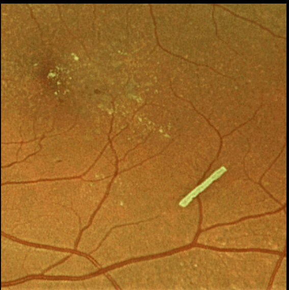
OZURDEX GHOST
Originally posted on @retina.rocks November 26, 2021
This 71YO female is getting Ozurdex intravitreal injections every 2 months to control her macular edema.
Learning Points:
The Ozurdex implant slowly releases the steroid and eventually totally absorbs, with the clinical effect usually lasting about 2 to 4 months.
The ghost-like remnants can persist for many months (see Kim et al, Retina 2020;40;2226-2231).
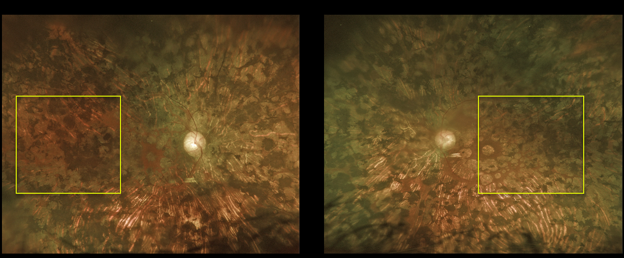
PANRETINAL PHOTOCOAGULATION
Originally posted on @retina.rocks November 11, 2021
This 63YO female underwent incredibly intense, confluent, and posterior panretinal photocoagulation (PRP) and macular laser elsewhere before transferring her care to our office. Vision was hand motion OD and 20/400 OS.
The laser performed on this patient is wrong on so many levels, including spots extending to the macula, touching the nerve, and confluent elsewhere. The laser surgeon essentially gave this patient severe iatrogenic retinitis pigmentosa.
Learning Points:
PRP should begin at least 1 disc diameter from the optic nerve and outside the major temporal arcades. Treatment is performed with a wide-angle lens or an indirect ophthalmoscope and should be applied from the posterior retina towards the periphery to prevent accidental macular burns.
The laser spots should be 200-500 microns in size at the retina, spaced about one burn width apart, and extend as far anteriorly as possible.
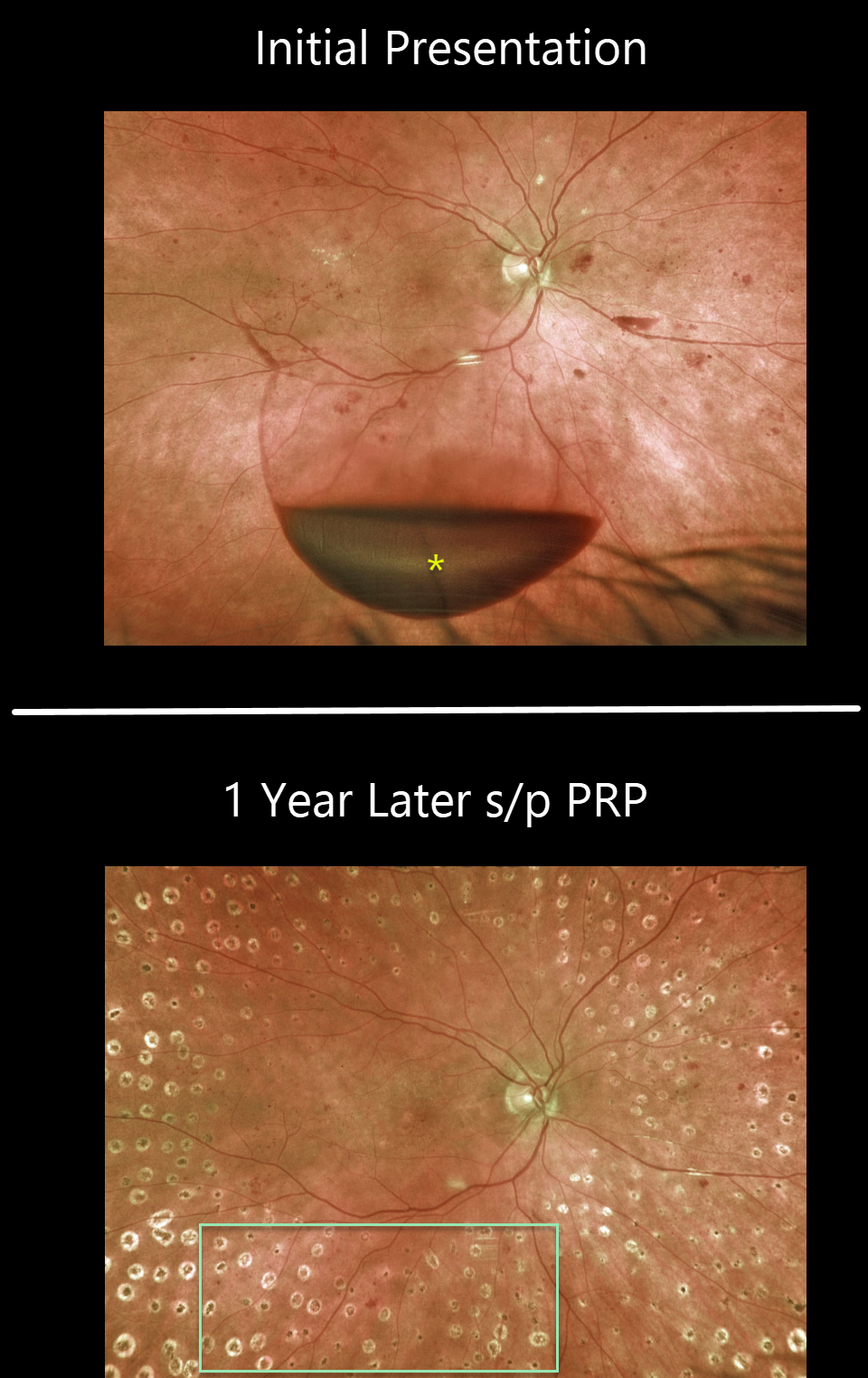
PROLIFERATIVE DIABETIC RETINOPATHY (PDR)
Originally posted on @retina.rocks November 4, 2021
This 65YO female presented with 20/400 vision in her right eye. An acute inferior subhyaloid hemorrhage was noted along with non-central diabetic macular edema. Over the coming months, macular laser followed by panretinal photocoagulation (PRP) was performed.
One year later, vision was 20/50, with complete resolution of the preretinal blood and proliferative disease.
Learning Points:
Before PRP, patients with proliferative diabetic retinopathy (PDR) routinely went blind, and one of the treatments actually included pituitary ablation (for an amazing historical perspective on diabetic laser photocoagulation, see Goldberg and Jampol, Ophthalmology 1987;94;741-746).
PRP originally extended fairly posteriorly, and patients often needed fill-in treatment before vitrectomy and anti-VEGF treatments became available. Today, most laser surgeons will begin treatment well away from the nerve and major arcades to minimize the risks of central and peripheral vision loss.
Once considered a “one and done” treatment, more recent studies show that patients may still need additional laser, anti-VEGF injections, or vitrectomy (see Gonzalez et al, Ophthalmology 2021;128:1448-1457).
Some also advocate anti-VEGF injections as the primary treatment for PDR. However, most still prefer to add PRP at some point due to the risk of patients being lost to follow-up (see Gross et al, DRCR Protocol S, JAMA Ophthalmology 2018;136:1138-1148).
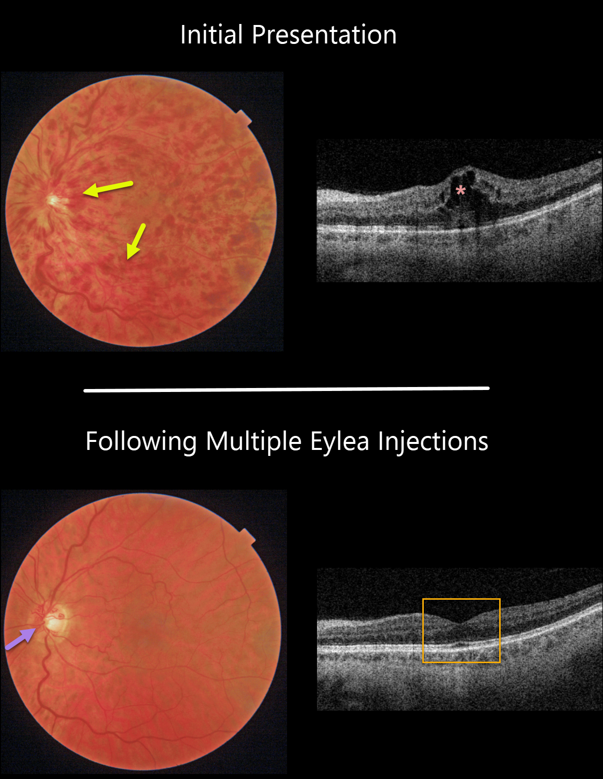
CRVO
Originally posted on @retina.rocks October 25, 2021
This 61YO male patient presented with a non-ischemic central retinal vein occlusion (CRVO), 20/30 vision, and macular edema.
With ongoing Eylea treat-extend protocol, the hemorrhages and macular edema completely resolved. Prominent optic nerve collaterals were noted.
Learning Points:
Before the introduction of intravitreal injections in the early 2000s, there were essentially no treatments for CRVO-related macular edema.
Anti-VEGF injections are now generally the initial treatment of choice, with about 50-60% of eyes gaining at least 3 lines of vision. Intravitreal steroids, usually Ozurdex, are more often used as secondary treatment.
Macular grid laser photocoagulation is not performed because it has been shown to confer no visual benefit in the Central Vein Occlusion Study Group (see Ophthalmology 1995;102;1425-1433).
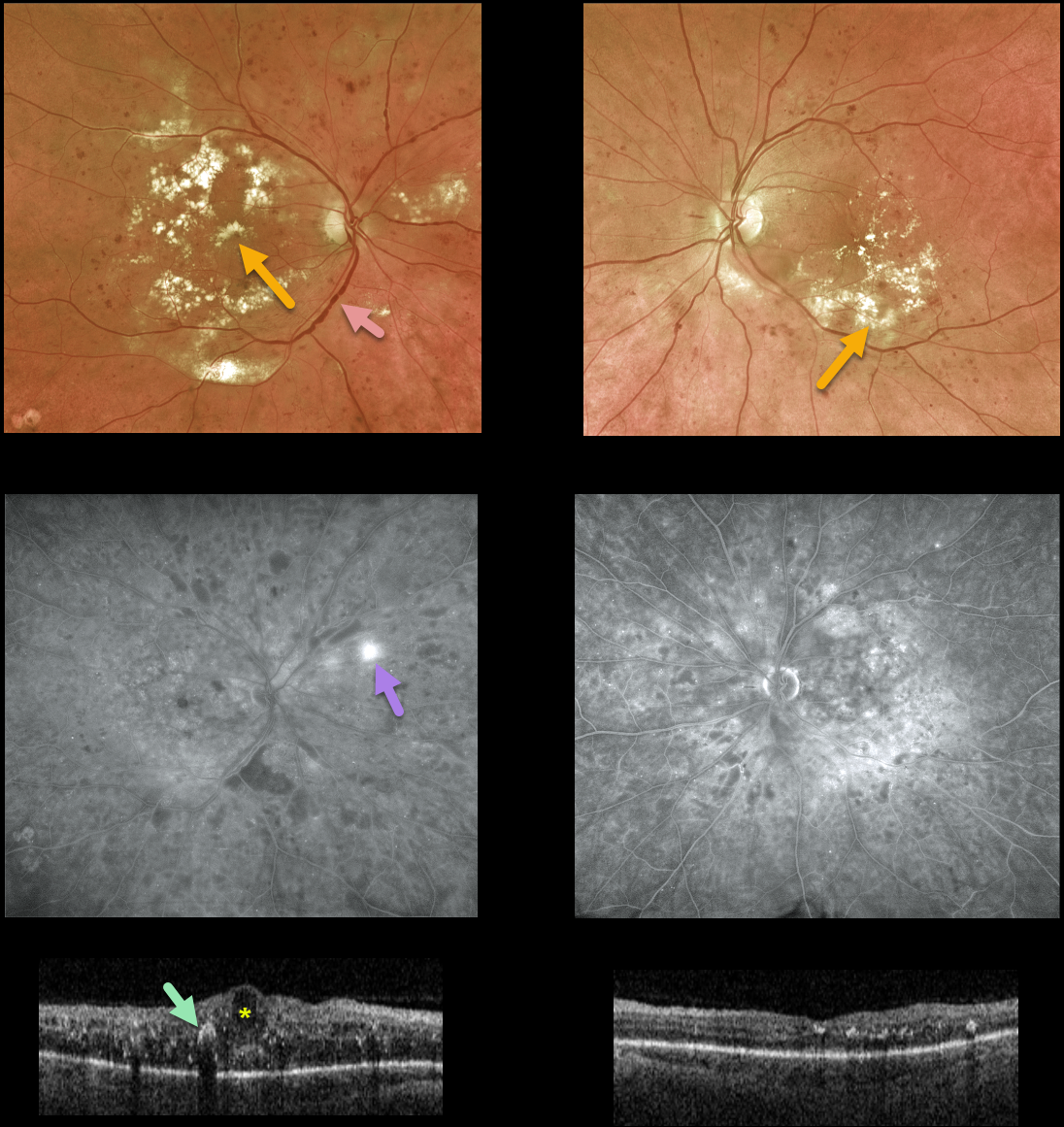
DIABETIC MACULAR EDEMA (DME)
Originally posted on @retina.rocks October 14, 2021
This 59YO male with a history of type 2 diabetes presented with bilateral diabetic macular edema. Vision was counting fingers OD and 20/30 OS.
There was extensive bilateral macular lipid, particularly in his right eye. Venous beading and sausaging were noted OD, indicating severe retinal ischemia.
Fluorescein angiography confirmed severe ischemia with peripheral temporal retinal neovascularization.
Triton swept-source OCT scanning shows severe central-involved diabetic macular edema (CI-DME) with scattered hyperreflective lipid flecks OD, non-central-involved diabetic macular edema (NCI-DME), and milder lipid OS.
Macular laser for the NCI-DME was recommended for his left eye. Monthly anti-VEGF injections were recommended for the CI-DME in his right eye. Remarkably, vision improved to 20/80 so far after a single injection.
Learning Points:
Anti-VEGF injections are the mainstay of treatment for CI-DME. It is expected that these injections will also stabilize, or possibly reverse, this patient’s proliferative disease.
We also plan to begin panretinal photocoagulation once the central edema resolves. There are multiple ways we could have approached his treatment plan. What would you do and why?
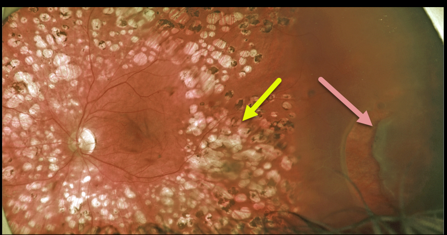
RHEGMATOGENOUS RETINAL DETACHMENT
Originally posted on @retina.rocks September 22, 2021
This 50YO male presented for a routine diabetic follow-up. A new asymptomatic peripheral retinal tear causing a rhegmatogenous retinal detachment was noted anterior to full panretinal photocoagulation (PRP) scarring.
We will often demarcate asymptomatic retinal detachments with laser photocoagulation (Vrabec and Baumel, Ophthalmology 2000;107:1063-1067). This patient essentially had laser demarcation before his retinal detachment, but the end result was the same, with his detachment being contained by the laser scarring.
Learning Points:
We opted to treat this eye with vitrectomy, but one could argue that this detachment could also be closely followed since it was contained by the prior laser.
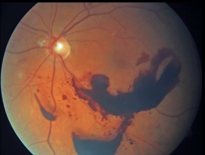
MERMAID MACULOPATHY
Originally posted on @retina.rocks September 8, 2021
This 55YO female with a history of proliferative diabetic retinopathy presented with a symptomatic preretinal hemorrhage. Vision was 20/50.
You don’t need to have an overly active imagination to see this rare finding of mermaid maculopathy.
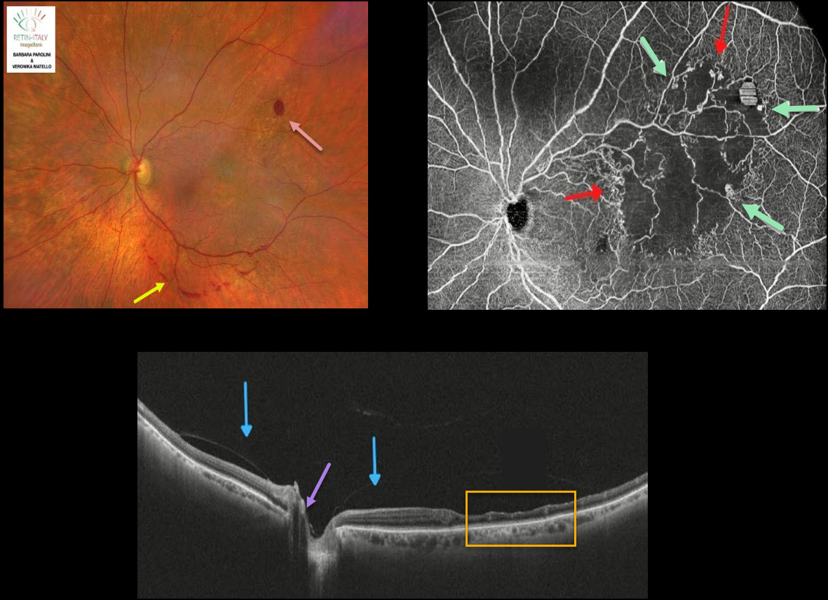
BRVO
Originally posted on @retina.rocks September 7, 2021
This 76YO female was referred for recent floaters. Wide-field color photography shows some retinal hemorrhages in the superotemporal retina, along with a band of preretinal blood below the inferotemporal arcade.
Wide-field SD-OCT shows inner retinal thinning within the area of occlusion temporally. The posterior hyaloid is detached over the posterior pole with a persistent adherence to the optic nerve.
Wide-field OCT angiography (OCTA) shows a broad area of capillary ischemia within the distribution of the branch retinal vein occlusion (BRVO), along with areas of retinal neovascularization.
Learning Points:
The endothelial cells in retinal neovascularization lack the tight junctions found in the normal retinal vasculature and therefore leak profusely on fluorescein angiography.
OCTA highlights retinal vessels not with a traditional fluorescing dye, but by detecting the movement of red blood cells within the vessel. This is why OCTA images offer beautiful detail but no leakage.
The role of OCTA versus traditional fluorescein angiography is still being determined, but growing evidence suggests it may provide similar clinical information without the need for dye injection (see Spaide et al, Progress Retinal Eye Research 2018;54;1-55).
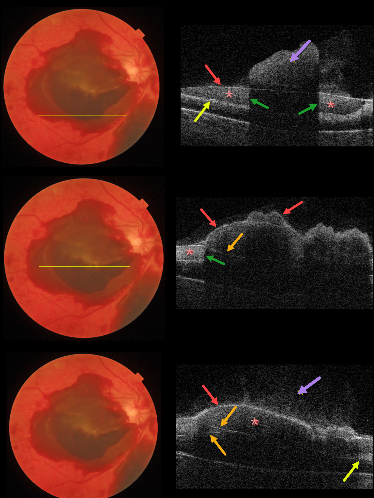
PROLIFERATIVE DIABETIC RETINOPATHY (PDR)
Originally posted on @retina.rocks August 23, 2021
This 53YO female with type 2 diabetes presented with acute vision loss from previously undiagnosed proliferative diabetic retinopathy (PDR).
Color imaging shows multiple layers of preretinal blood. The blood is located above the neurosensory retina but is separated into several pockets due to posterior hyaloid splitting (vitreoschisis), rather than cleanly separating into a single plane.
Using the Triton swept-source OCT, we’ve taken four B-scans through different portions of the macula to highlight the complex anatomy of the preretinal blood.
Vitreous blood variably floats above the posterior hyaloid. The red arrows show the anterior edge of the posterior hyaloid. In some sections, the posterior hyaloid is split, separating the preretinal blood into several compartments.
Blood reflects and absorbs the OCT laser, making it difficult to see underlying retinal details (shadowing). The retinal nerve fiber layer is variably visible posterior to the blood.
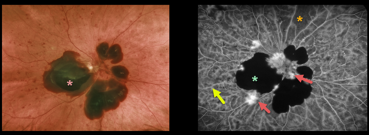
PROLIFERATIVE DIABETIC RETINOPATHY (PDR)
Originally posted on @retina.rocks August 10, 2021
This 53YO female with type 2 diabetes presented with acute vision loss from previously undiagnosed proliferative diabetic retinopathy.
Optos color imaging shows patches of mostly subhyaloid blood in the macula and peripapillary retina.
Fluorescein angiography beautifully exhibits various angiographic characteristics for hypo- and hyperfluorescence. We can see both blocking and filling defects, in addition to diffuse vascular staining and leakage of the disc and adjacent to the subhyaloid blood.
Learning Points:
Angiographically, hypofluorescence is classified as either a blockage or a filling defect. Blood, which masks the underlying retinal and choroidal fluorescence, causes blockage. There is diffuse capillary loss and ischemia, appearing as glassy-dark areas of hypofluorescence throughout, representing filling defects. Filling defects are virtually never as dark as areas of blockage.
Angiographic hyperfluorescence is classified into window defects, pooling, staining, leakage, and neovascularization, and our case exhibits several of these features.
The endothelial cells of the larger vessels in areas of capillary dropout become ischemic, leading to breakdown of the inner retinal barrier. The larger vessels, therefore, often show late staining in these regions as fluorescein dye leaks into and stains the vessel wall. Vascular staining is evident throughout the entire imaged fundus.
The new blood vessels growing on the disc and adjacent to the subhyaloid blood lack endothelial tight junctions, which is why neovascularization shows profound angiographic leakage.
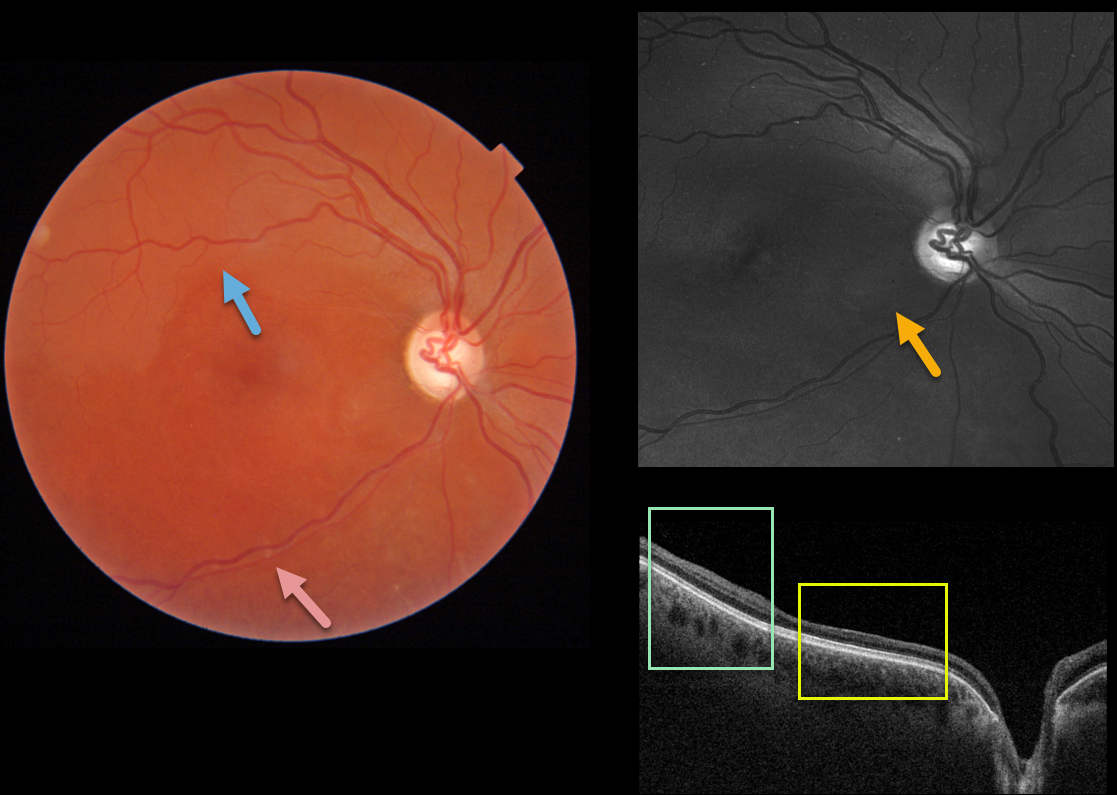
SUSAC SYNDROME
Originally posted on @retina.rocks July 20, 2021
This healthy 30YO female was referred for poor vision in her right eye. She mentioned losing vision in this eye during high school, but couldn’t provide any further details. She also gave a history of hearing loss, which had never been evaluated. Vision was HM OD and 20/30 in her normal left eye.
Triton imaging shows faint retinal whitening in the superior macula. The retinal arterioles are narrowed and somewhat sheathed inferiorly.
Swept-source OCT shows normal inner retinal architecture temporally, with inner retinal atrophy and disorganization of the normal architecture more nasally. Right inferotemporal nerve fiber loss is quite dramatic on Optos green imaging.
We diagnosed her with a resolved inferior branch retinal artery occlusion (BRAO), although this may represent Susac’s syndrome. The involved retina appeared redder than the uninvolved superior retina, accounting for the whitish superior retina noted above.
Subsequent work-up, including hypercoagulable labs and MRI, was negative. She denied prior intravenous drug use. To complete the workup, we will be obtaining carotid Doppler and cardiac ultrasonography.
Learning Points:
Susac’s syndrome, also known as retinocochleocerebral vasculopathy, is a rare microangiopathic disease characterized by a triad of encephalopathy, sensorineural hearing loss, and branch retinal artery occlusions with a predilection for younger females.
In the presence of Susac’s syndrome, MRI findings should show corpus callosum involvement. However, the full triad commonly takes months to develop, complicating diagnosis.
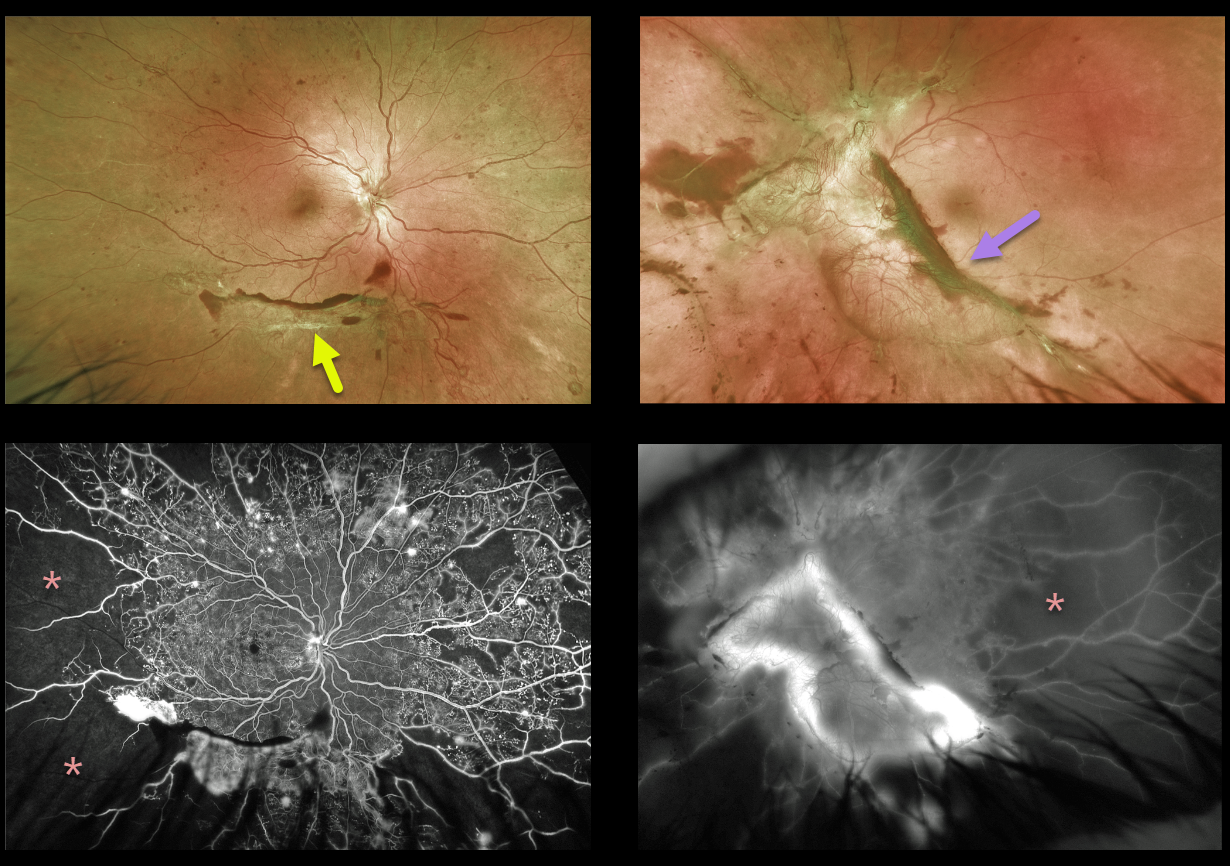
TRACTIONAL RETINAL DETACHMENT
Originally posted on @retina.rocks July 19, 2021
This 34YO female presented with 20/20 vision OD and 20/80 vision OS. Optos imaging of her right eye shows inferior fibrotic neovascularization with marked peripheral ischemia, with more extensive fibrovascular proliferation and ischemia in her left eye.
The relatively good vision in her left eye is deceiving, with potentially blinding traction lurking just outside the fovea. Triton swept-source OCT imaging revealed a relatively normal, flat central macula (not shown here).
Panretinal photocoagulation was started in her right eye. She is scheduled for primary vitrectomy in her left eye.
Learning Points:
Tractional retinal detachments are one of the complications that can develop from proliferative diabetic retinopathy.
Although described as a specific manifestation in sickle cell disease (see Han, Retina 2021; 41;1361-1363), we think the left eye resembles a sea fan.
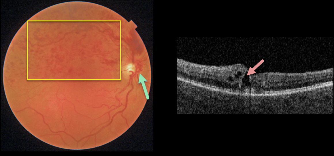
HRVO
Originally posted on @retina.rocks July 5, 2021
This 62YO female presented with 20/30 vision from a symptomatic superior hemiretinal vein occlusion (HRVO). Fundus imaging shows moderate retinal hemorrhages in the superior retinal hemisphere and a prominent collateral vessel along the nasal aspect of the optic nerve. There is superior macular edema on OCT.
Learning Points:
Branch retinal vein occlusions (BRVO) are virtually always caused by a retinal artery compressing a retinal vein within its common adventitial sheath. Some people are born with an anomalous two-trunked central retinal vein that independently drains the superior and inferior retinal hemispheres.
HRVO, similar to central retinal vein occlusion, is caused by compression of the retinal vein posterior to the lamina cribrosa (see Hayreh, Arch Ophthalmol 1980;98:1600-1609).
Collateral vessels are dilated, pre-existing vessels that bypass an occlusion. These are most commonly seen in the posterior pole following retinal venous occlusions, but can also occur on the optic disc with optic nerve meningiomas.
The endothelial tight junctions are intact, so there’s no leakage on angiography. This helps differentiate them from neovascularization, which does leak.
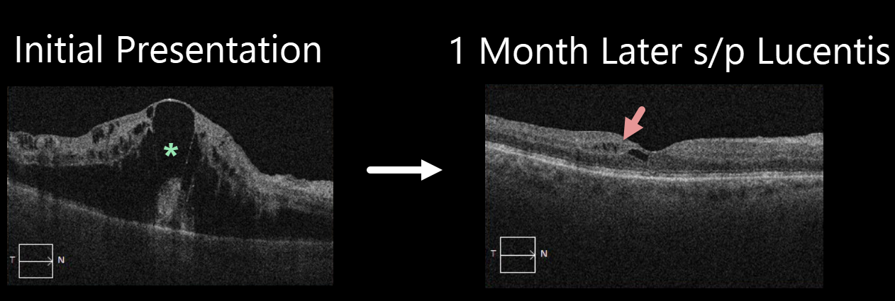
CRVO
Originally posted on @retina.rocks June 30, 2021
This 72YO male presented with marked macular edema and 20/150 vision from a nonischemic central retinal vein occlusion (CRVO).
Four weeks later, following a single Lucentis injection, vision improved to 20/70, and the edema almost completely resolved. Although most patients respond favorably to anti-VEGF therapy, our patient’s dramatic, rapid improvement following a single injection is quite unusual.
CRVO macular edema usually requires ongoing injections, and we are currently following a treat-and-extend protocol to maintain this improvement while minimizing the treatment burden.
Learning Points:
Prior to the introduction of intravitreal injections in the early 2000s, there were essentially no treatments for CRVO-related macular edema. Anti-VEGF injections in general are now the initial treatment of choice, followed by intravitreal steroids (usually Ozurdex).
Macular grid laser photocoagulation is not performed because it has been shown to confer no visual benefit in the Central Vein Occlusion Study Group (see Ophthalmology 1995;102;1425-1433).
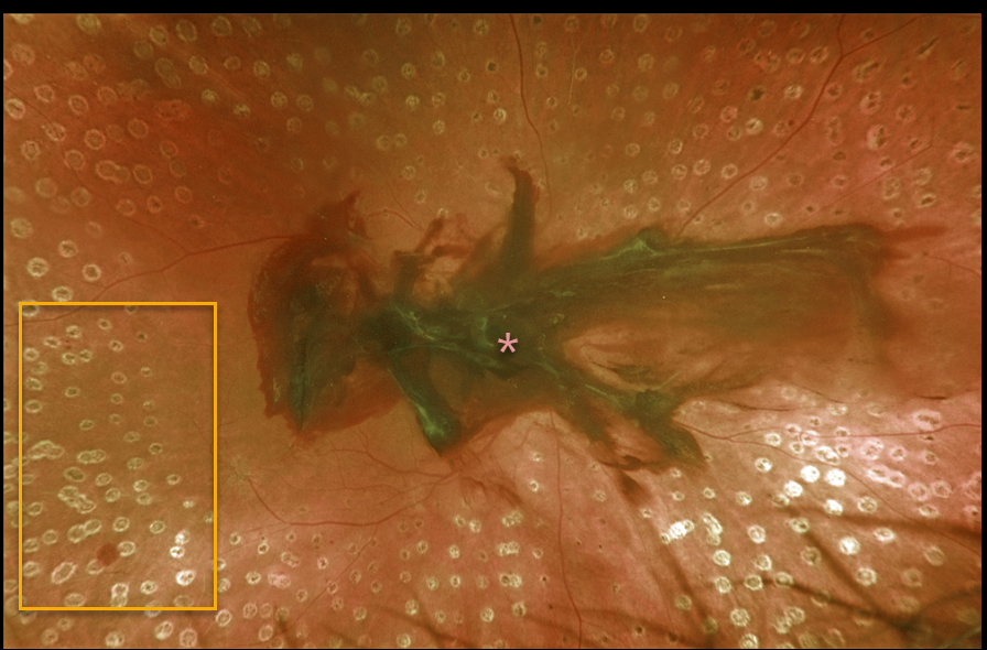
PROLIFERATIVE DIABETIC RETINOPATHY (PDR)
Originally posted on @retina.rocks June 28, 2021
Despite full panretinal photocoagulation, this 57YO patient with proliferative diabetic retinopathy developed an acute premacular hemorrhage. Her vision was 20/300 in the right eye.
Learning Points:
Panretinal photocoagulation has been around since the 1960s and still remains the mainstay for treating proliferative disease. Often considered a “one and done,” patients can still develop breakthrough hemorrhages.
Intravitreal anti-VEGF injections are often helpful, but some will require vitrectomy.
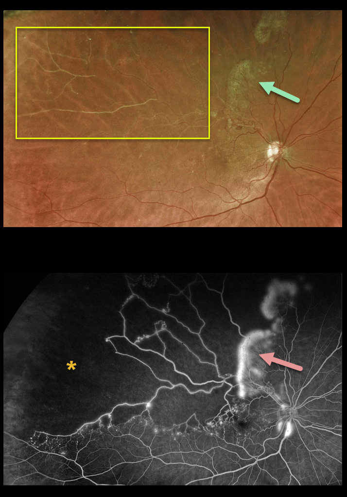
BRVO
Originally posted on @retina.rocks June 14, 2021
This 53YO male presented with a major superotemporal branch retinal vein occlusion (BRVO) and 20/40 vision.
Despite the deceptively good vision, Optos ultra-widefield imaging shows marked ischemia throughout the distribution of the occlusion, which fortunately spares the central macula. There is a broad zone of mostly fibrosed retinal neovascularization at the border of the occlusion superiorly.
Angiography demonstrates ischemia and leakage corresponding to the fibrosed retinal neovascularization.
Scatter laser throughout the area of ischemia was recommended.
Learning Points:
In 1986, the Branch Vein Occlusion Study Group reported that scatter laser decreased the risk of vitreous hemorrhage from retinal neovascularization in BRVO, and suggested that laser should be applied once neovascularization develops (Arch Ophthalmol 1986;104:34-41).
Thirty-five years later, scatter laser remains the gold standard, although some may prefer to stabilize neovascularization with anti-VEGF therapy before laser.
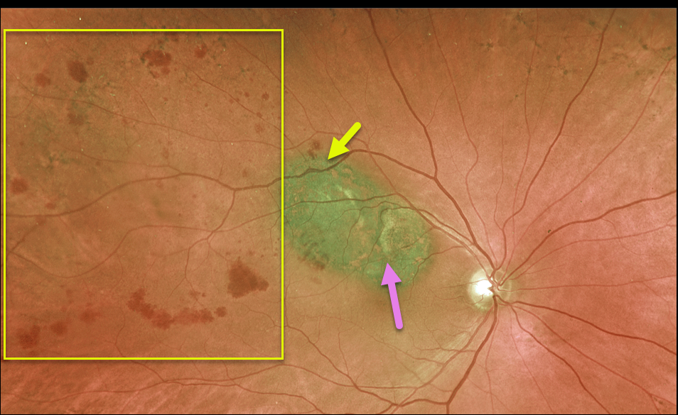
UVEAL MELANOMA + BRVO
Originally posted on @retina.rocks May 18, 2021
This 78YO male has a history of uveal malignant melanoma (MM), which was successfully treated with initial photodynamic therapy (PDT) (due to the presence of subretinal fluid) followed by transpupillary thermotherapy (TTT) once dry.
He developed a secondary branch retinal vein occlusion (BRVO), likely due to thermal damage from the TTT.
Vision is 20/30, and we continue to follow him closely. A scatter laser will be needed if he develops increasing ischemia with neovascularization from the BRVO.
Learning Points:
BRVOs are virtually always caused by an artery compressing a vein at a shared arteriovenous crossing.
However, rarely, they can occur due to other causes, including inflammation (most commonly sarcoidosis) or secondary to interventions such as TTT.
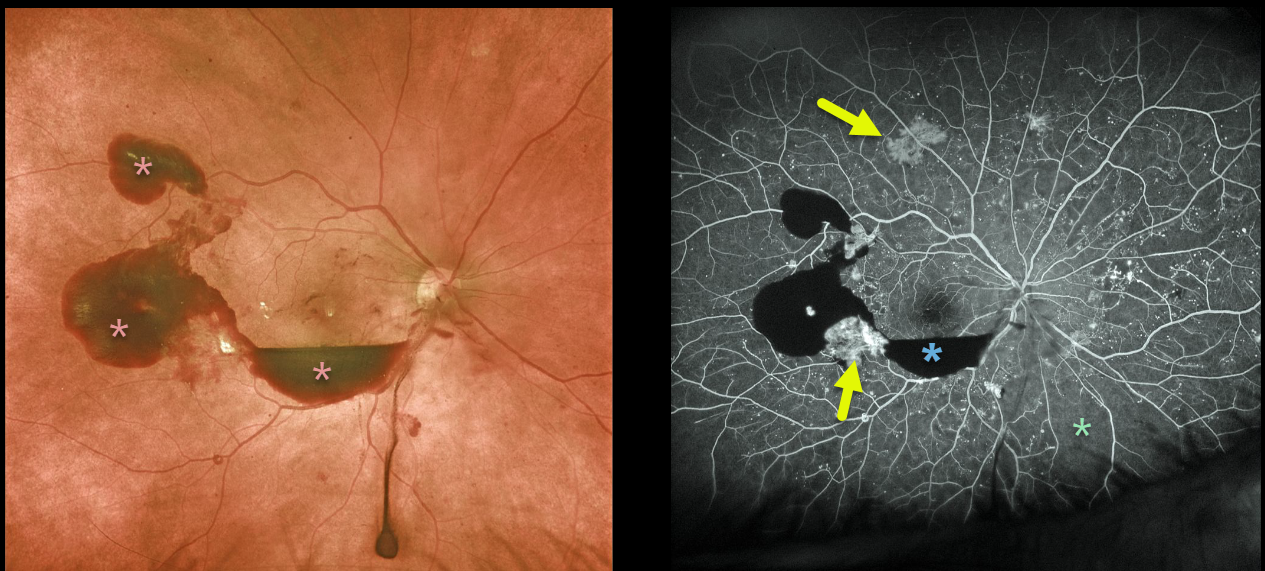
PROLIFERATIVE DIABETIC RETINOPATHY (PDR)
Originally posted on @retina.rocks May 17, 2021
The 58 yo male with proliferative diabetic retinopathy presented with preretinal blood. Optos fluorescein angiography shows blockage from this blood with scattered areas of midperipheral neovascularization and peripheral ischemia, especially inferonasally.
Learning Points:
Angiographically, hypofluorescence is classified as either a blockage or a filling defect. Blood, which masks the underlying retinal and choroidal fluorescence, causes blockage. There is diffuse capillary loss and ischemia, appearing as glassy-dark areas of hypofluorescence throughout, representing filling defects. Filling defects are virtually never as dark as areas of blockage.
Angiographic hyperfluorescence is classified into window defects, pooling, staining, leakage, and neovascularization. The new blood vessels adjacent to the subhyaloid blood and in the mid-periphery lack endothelial tight junctions, which is why neovascularization shows profound angiographic leakage.
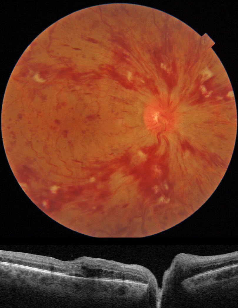
CRVO
Originally posted on @retina.rocks May 5, 2021
This patient presented with 20/70 vision from an acute central retinal vein occlusion (CRVO) with mild macular edema.
The mild vision reduction and the well-perfused fluorescein angiogram support the diagnosis of a non-ischemic CRVO.
He is currently undergoing intravitreal injections for the macular edema.
Learning Points:
Patients with CRVO are at risk of vision loss due to macular edema or anterior segment neovascularization. Macular edema is common in both non-ischemic and ischemic occlusions, although rubeosis is found almost exclusively with ischemic occlusions.
Iris neovascularization with neovascular glaucoma develops in about two-thirds of patients with untreated ischemic occlusions. Rubeosis is initially treated with an anti-VEGF injection, followed by aggressive, complete panretinal photocoagulation.
Some patients will need a glaucoma tube or a cyclodestructive procedure if the IOP remains too high.
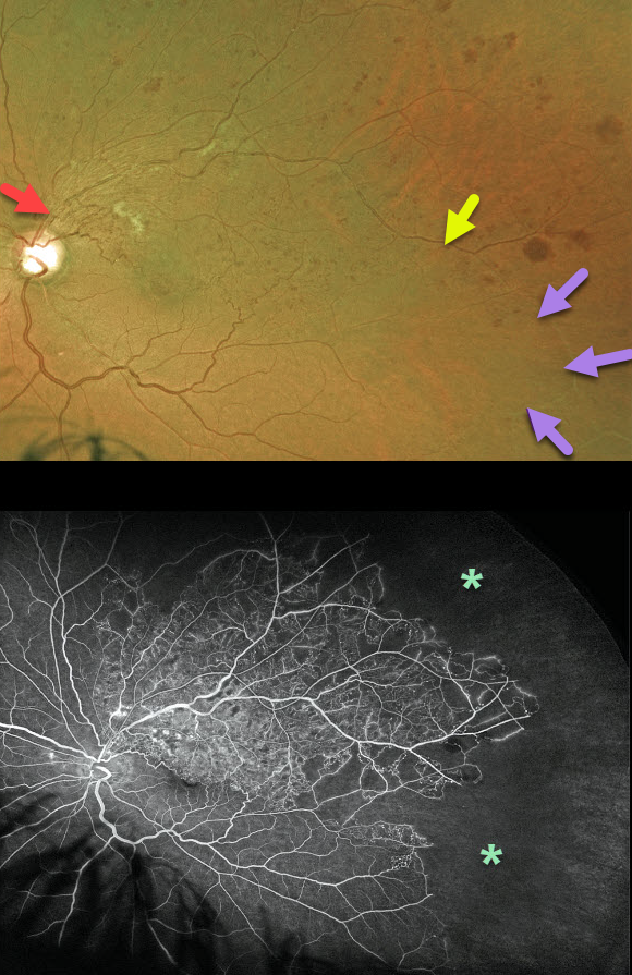
BRVO
Originally posted on @retina.rocks April 30, 2021
This is a 54YO male presented with a major branch retinal vein occlusion (BRVO). Vision was 20/80, and Eylea injections were started for associated macular edema.
The Optos color image shows a major superotemporal BRVO with moderate retinal hemorrhages, some nerve fiber layer infarcts, and collateral vessels. The involved retinal vein is red in the midperiphery. However, it becomes white as it continues into the inferotemporal periphery.
Optos fluorescein angiography shows extensive peripheral capillary dropout.
Usually, one would expect a BRVO to involve one quadrant (retinal vessels virtually always respect the horizontal meridian).
The occluded vein in our patient most likely represents an atypical retinal venous malformation (RVM), which is a congenital anomalous vessel that, by definition, crosses the horizontal.
Learning Points:
Retinal venous malformations were originally described by Gary Brown et al as congenital retinal macrovessels (Arch Ophthalmology 1982;100:1430-1436). These vessels are usually located in the macular region and are virtually always venous.
These lesions are now called retinal venous malformations (RVMs) since about one-quarter of patients have associated venous anomalies in the brain (Pichi et al., JAMA Ophthalmol 2018;136;372-379).
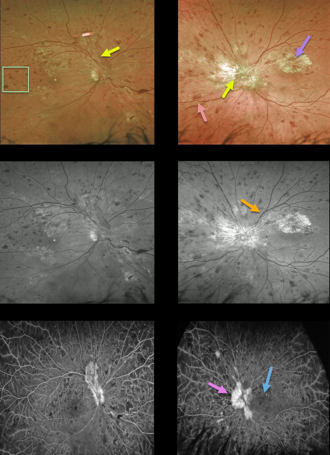
PROLIFERATIVE DIABETIC RETINOPATHY (PDR)
Originally posted on @retina.rocks April 29, 2021
This 40YO man presented with bilateral proliferative diabetic retinopathy (PDR). Vision was 20/60 OD and 20/200 OS.
Optos color and green channel imaging show prominent bilateral fibrovascular disc neovascularization, obscuring the left disc.
A few smaller areas of more peripheral neovascularization are present nasally in the OS. Severe retinal hemorrhages are noted in all quadrants; the retinal veins are beaded OU superiorly, along with some perivascular lipid superotemporally OD and nasally OS. The retinal vascular abnormalities are best highlighted with the green channel image.
There also appears to be an area of fibrosed neovascularization in the superior left macula. However, these findings are visible beneath the retinal vessels, indicating they are subretinal and composed of lipid. Lipid is usually angiographically invisible but, when dense, can obscure the underlying choroidal fluorescence, as in this case.
Fluorescein angiography shows leakage from the neovascularization, diffuse vascular staining, and mostly peripheral retinal nonperfusion.
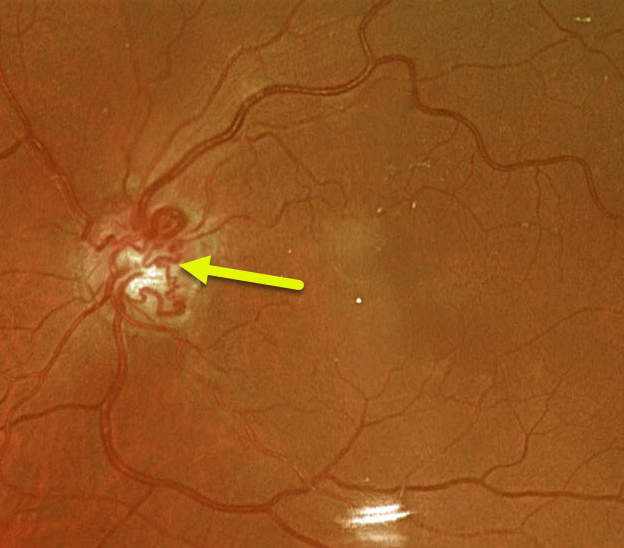
CRVO
Originally posted on @retina.rocks April 22, 2021
This 74YO female has large optic nerve collaterals from an old non-ischemic central retinal vein occlusion. Vision was 20/30.
Learning Points:
Collateral vessels are dilated, pre-existing vessels that bypass an occlusion. These are most commonly seen in the posterior pole, following retinal venous occlusions, often on the optic disc.
In collateral vessels, the endothelial tight junctions are intact, so they don’t leak angiographically. This helps differentiate them from neovascularization, which does leak.
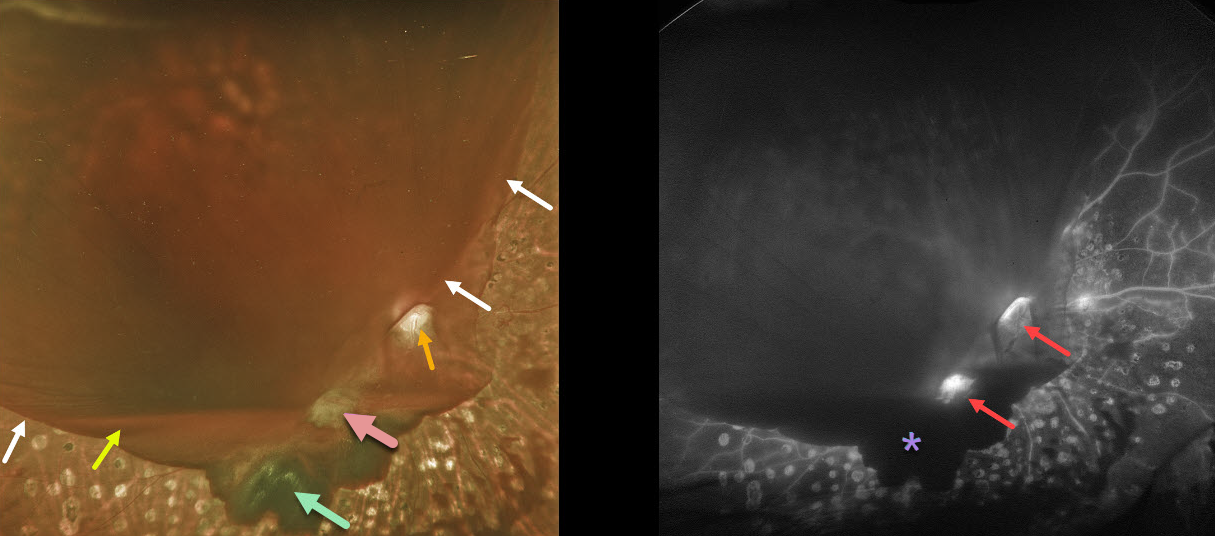
PROLIFERATIVE DIABETIC RETINOPATHY (PDR)
Originally posted on @retina.rocks April 2, 2021
This diabetic patient developed a symptomatic hemorrhage in his right eye despite partial panretinal photocoagulation (PRP) treatment. The blood in our patient highlights the various layers of vitreous separation.
The reason why the far inferior and nasal retina and PRP scarring are visible is that the hyaloid is completely attached here. The hyaloid is shallowly elevated inferiorly, which gives a scalloped appearance to the dark red subhyaloid blood.
Elsewhere, the hyaloid is completely detached and highly elevated, allowing blood to pool in this space (white arrows mark the junction between the attached and detached hyaloid). A layer of blood pools inferiorly at the base of the hyaloid separation.
The optic disc and fibrosed neovascularization are well visualized because the vitreous remains tethered, preventing blood from accumulating.
Fluorescein angiography highlights areas of neovascularization and the blockage of the underlying retina by various layers of blood.
Learning Points:
At birth, the vitreous is diffusely adherent to the entire retina. But over the years, the vitreous begins to separate, first in the midperiphery, then in the peripheral macula, then the central macula, and finally the optic nerve.
For a great review of posterior vitreous detachment (PVD) evolution and complications, see Johnson AJO 2010;149:371-382.
Retinal neovascularization arises from the inner retina and extends onto the back surface of the vitreous; without this scaffold, it will not develop. A complete PVD is therefore protective against posterior segment retinal neovascularization.
Since the vitreous in the posterior pole is most adherent around the optic nerve and major arcades, this is where diabetic neovascularization most commonly develops. It is also why the vitreous often remains tethered to these structures as the proliferation progresses.
The posterior hyaloid continues to detach and contract, as does the neovascularization, which can cause vitreous hemorrhage and traction retinal detachment.
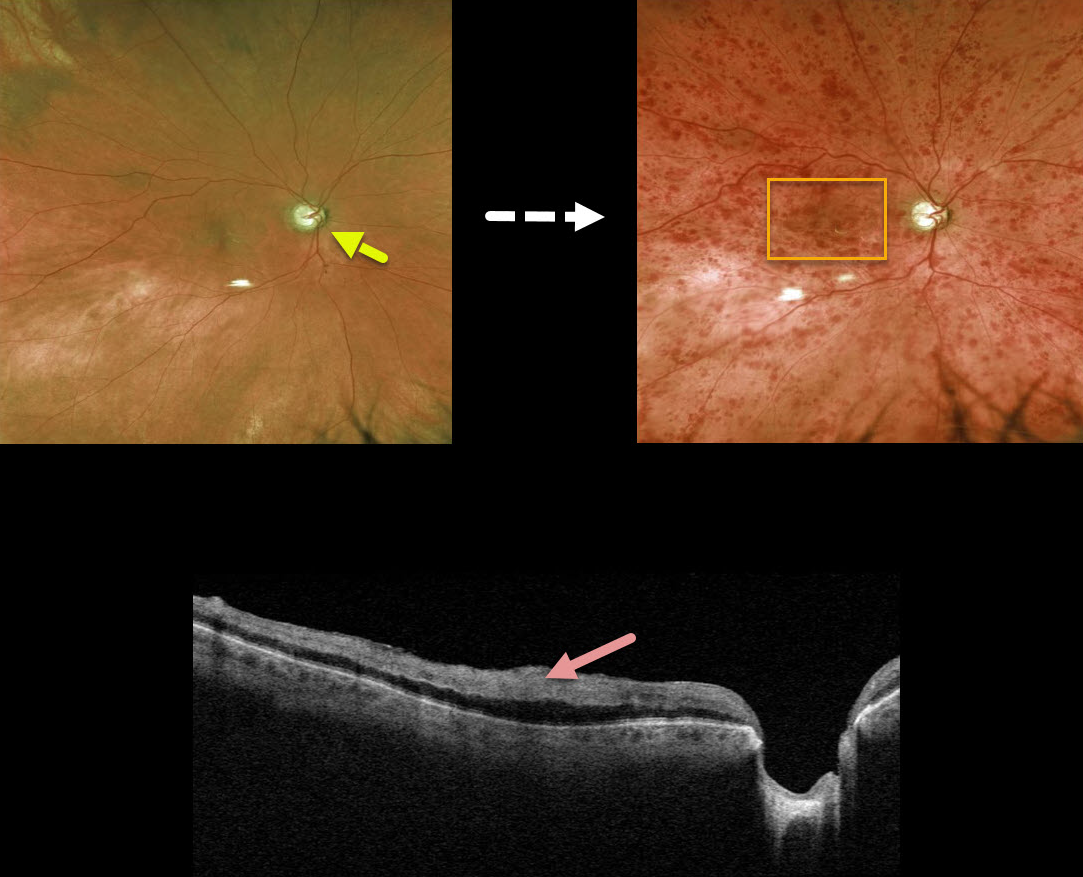
CRVO
Originally posted on @retina.rocks March 23, 2021
This 79YO male with a history of chronic open-angle glaucoma (COAG) presented with counting fingers vision from an acute nonischemic central retinal vein occlusion.
Even though his COAG was well-controlled with topical therapy, we felt that his glaucoma was a significant risk factor for the occlusion. A fundus photo from before the CRVO shows that significant glaucomatous cupping developed after the CRVO.
Triton swept-source OCT scanning shows marked inner retinal hyperreflectivity from the confluent macular blood.
Learning Points:
Patients with CRVO are at risk of vision loss due to macular edema or anterior segment neovascularization.
Macular edema is common in both non-ischemic and ischemic occlusions, although rubeosis is found almost exclusively with ischemic occlusions. Inner macular blood, as in this patient, can also cause vision loss.
Iris neovascularization with neovascular glaucoma develops in about two-thirds of patients with untreated ischemic occlusions. Rubeosis is treated initially with an anti-VEGF injection, followed by aggressive, complete panretinal photocoagulation.
Some patients will need a glaucoma tube or a cyclodestructive procedure if the IOP remains too high.
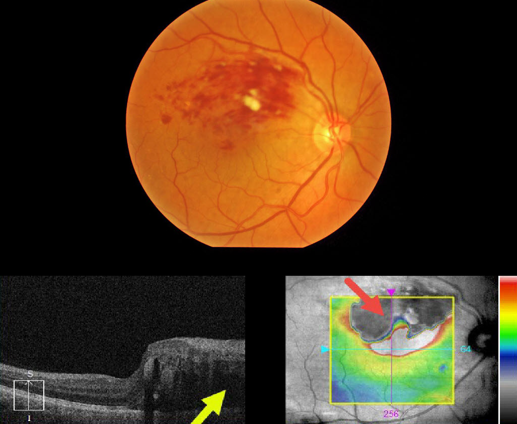
BRVO
Originally posted on @retina.rocks March 8, 2021
This 57YO male presented with 20/400 vision in his right eye from a superior macular branch retinal vein occlusion.
The OCT at first glance seems to just show superior macular edema extending through the macular center, but why is the superior retinal thickness map black, and what does that mean?
The retinal thickness map is supposed to represent the retinal thickness from the internal limiting membrane (ILM) to the tip of the outer segments just above the apical retinal pigment epithelium (RPE).
Our very expensive OCT machines usually accurately identify the ILM, but often don’t do such a great job of finding the RPE, especially when the OCT beam is attenuated as it passes through edematous retina or increased inner retinal reflectivity. In this case, the confluent inner retinal blood is highly reflective, causing posterior shadowing of the OCT signal.
Learning Points:
Intuitively, most of us would realize the black portion of the thickness map is an artifact, but now we know exactly what is going on. These artifacts are quite common if one looks for them.
The OCT cannot accurately locate the RPE signal and therefore incorrectly localizes it to the outer margin of the inner retinal blood.
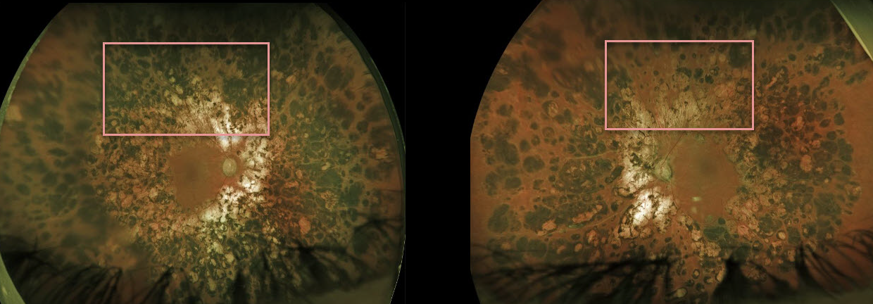
PANRETINAL PHOTOCOAGULATION
Originally posted on @retina.rocks March 5, 2021
This 51YO male has heavy bilateral panretinal photocoagulation (PRP) scarring for proliferative diabetic retinopathy (PDR). All laser treatments were done years earlier at an outside practice.
The burns are confluent and extend into the arcades. Thankfully, the treating surgeon did not quite burn the optic nerves. However, the visual fields are severely constricted with essentially iatrogenic retinitis pigmentosa.
Learning Points:
Although possibly appropriate before the advent of modern vitrectomy surgery and anti-VEGF therapy, this degree of laser should never be seen in today’s patients.
Laser should begin well outside the arcades and away from the optic nerve, extending as anteriorly as possible, using 200- to 500-micron retinal burns spaced about one burn width apart.
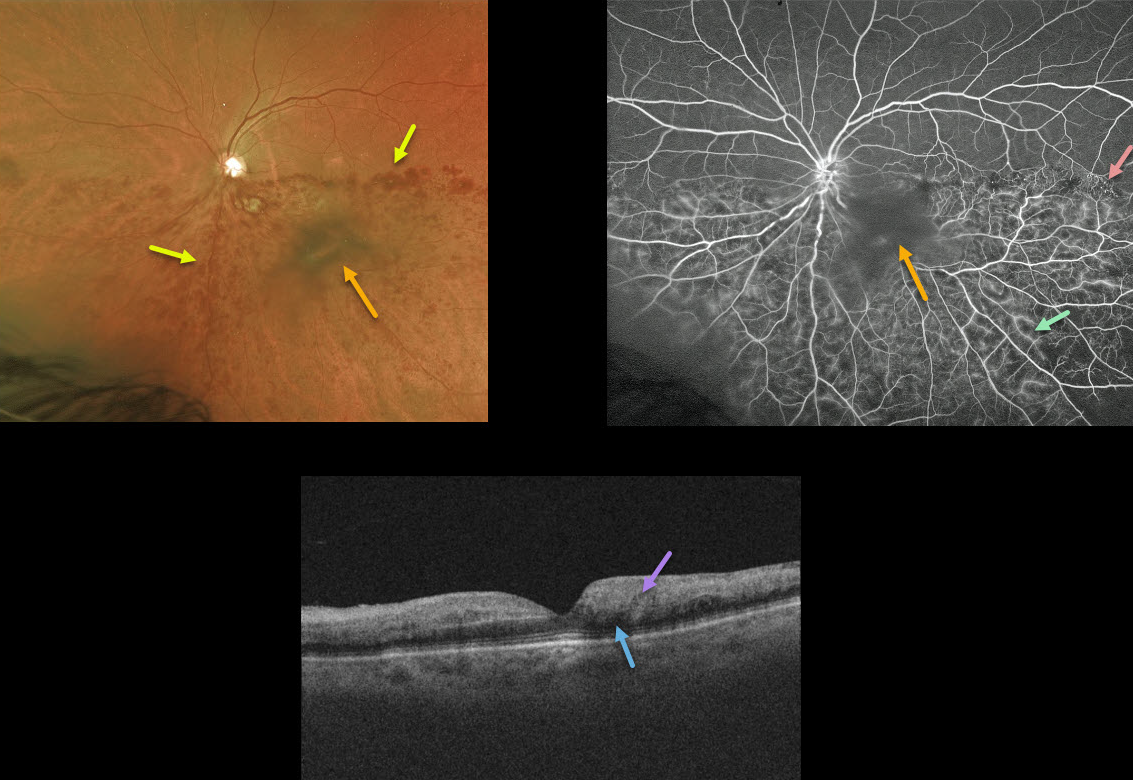
HRVO
Originally posted on @retina.rocks March 2, 2021
This 78YO female presented with diffuse intraretinal hemorrhages in the inferior half of her posterior pole from an inferior hemiretinal vein occlusion. She had a long history of uncontrolled hypertension, although her blood pressure had not been acutely elevated.
Fluorescein angiography shows good perfusion, some areas of vascular staining/leakage, and some telangiectasia at the border of the occlusion temporally. The large central blob in both the Optos image and the angiogram is clinically noted as a dense vitreous condensation, not blood.
OCT shows no macular edema with mild outer retinal shadowing due to the hyperreflective inner retina. Since there is no edema, we are currently observing the patient and are hoping the vision will improve as the retinal blood absorbs.
Learning Points:
Over time, patients can develop retinal atrophy from ischemic BRVO. In addition, collateral vessels are a common finding seen in venous occlusions.
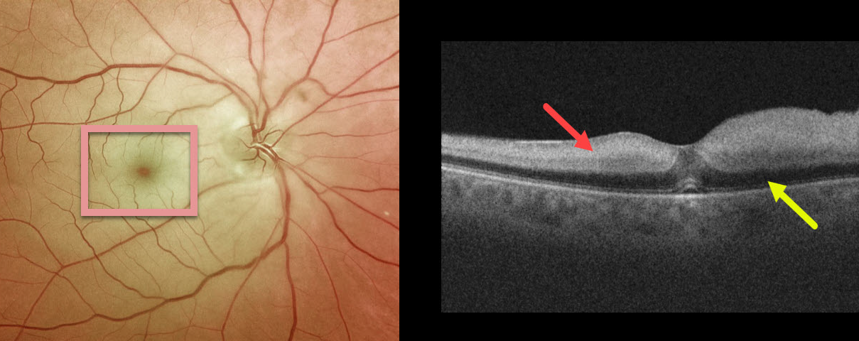
CRAO
Originally posted on @retina.rocks February 26, 2021
This patient presented with counting-finger vision and classic findings of a white retina and a cherry-red spot from an acute central retinal artery occlusion (CRAO). The cherry red spot is caused by the unaffected foveal retina (receiving intact choroidal blood flow) being surrounded by more distal white and ischemic retina no longer receiving circulation from the central retinal artery.
On Triton swept-source OCT, the inner two-thirds of the acutely ischemic retina were thickened with marked hyper-reflectivity. Less light reaches the deep retina, making it darker than normal and producing pseudo-edema of the outer nuclear layer.
She was immediately sent to the nearest stroke center, where she was found to have had an acute but asymptomatic lacunar stroke.
Learning Points:
Patients with acute retinal ischemia (acute CRAO, acute branch retinal artery occlusion, or transient monocular vision loss) should be sent immediately to the nearest stroke center due to the risk of a pre-existing or recent asymptomatic stroke or impending stroke (see Biousse et al, Ophthalmology 2018;125;1597-1607).
About 75% of patients with an acute CRAO will be found to have a concurrent, often asymptomatic recent stroke, as was the case with this patient.
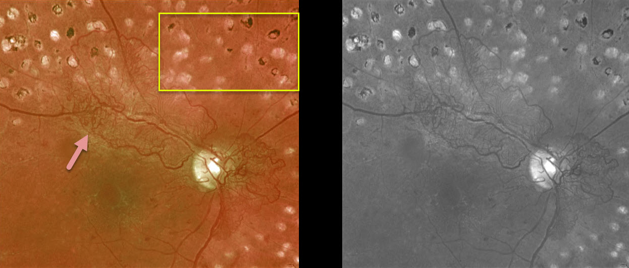
PROLIFERATIVE DIABETIC RETINOPATHY (PDR)
Originally posted on @retina.rocks February 25, 2021
This is a 55YO diabetic patient who had been treated with complete panretinal photocoagulation (PRP). Vision was 20/40. Despite PRP, the patient still had significant residual posterior neovascularization.
Since it is not uncommon for neovascularization to persist following full laser, we are observing for now.
Learning Points:
Panretinal photocoagulation has been around since the 1960s and still remains the mainstay for treating proliferative disease. Often considered a “one and done,” patients can still develop breakthrough hemorrhages. Intravitreal anti-VEGF injections are often helpful, but some will require vitrectomy.
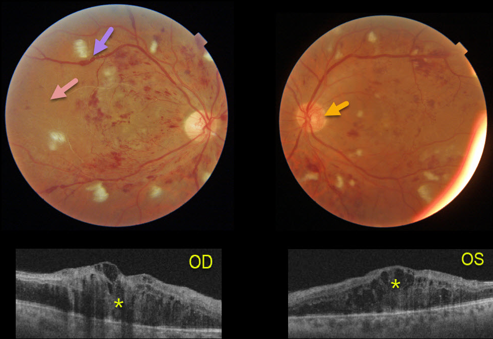
HYPERTENSION + PROLIFERATIVE DIABETIC RETINOPATHY (PDR)
Originally posted on @retina.rocks February 2, 2021
This 41YO male presented after a recent hospitalization for severe hypertension with vision of counting fingers OD and 20/60 OS.
The bilateral nerve fiber layer infarcts (cotton-wool spots) are more characteristic of hypertension than of diabetes.
Severe retinal hemorrhages and disc neovascularization are noted in each eye. The right eye, in particular, is severely ischemic, with Triton imaging showing a featureless, ischemic retina and white vessels, most pronounced in the temporal retina.
The venous sausaging is a clinical sign of severe ischemia, and when present, virtually always shortly precedes or is indicative of concurrent proliferative disease. Triton swept-source OCT shows severe bilateral center-involving diabetic macular edema.
He subsequently received multiple bilateral anti-VEGF injections followed by panretinal photocoagulation, with marked improvement of the central edema.
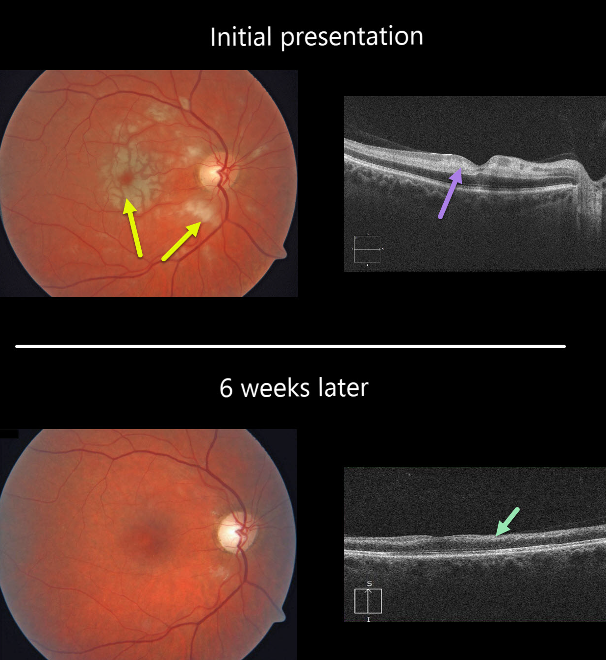
CRAO
Originally posted on @retina.rocks December 24, 2020
This healthy 49YO female presented with severe, sudden vision loss in her right eye.
There were multiple white macular patches similar to Purtscher’s retinopathy, but without retinal hemorrhages.
Optical coherence tomography (OCT) showed multiple mid-retinal hyperreflective paracentral acute macular maculopathy (PAMM) lesions. Since we felt this could represent an incomplete central retinal artery occlusion (CRAO), we sent her immediately to the nearest stroke center for further assessment.
At her six-week follow-up, the retinal appearance virtually normalized. However, OCT showed severe diffuse thinning of the inner non-photoreceptor retinal layers consistent with a resolved CRAO. Her stroke workup was unremarkable.
Learning Points:
Purtscher’s retinopathy was first described by Otmar Purtscher in 1910. Although originally described in a man who fell from a tree with cranial trauma, these white patches of retinal ischemia and hemorrhages are found in numerous other etiologies, including pancreatitis, chest trauma, and collagen vascular diseases.
The findings are called Purtscher’s retinopathy when due to trauma, and Purtscher ‘s-like retinopathy when seen from other causes.
The underlying cause of Purtscher’s is unknown, but is thought to involve leukoembolization due to complement activation. The areas of ischemia include more superficial nerve fiber layer infarcts (cotton wool spots) and deeper ischemia involving the middle retinal layers (PAMM).
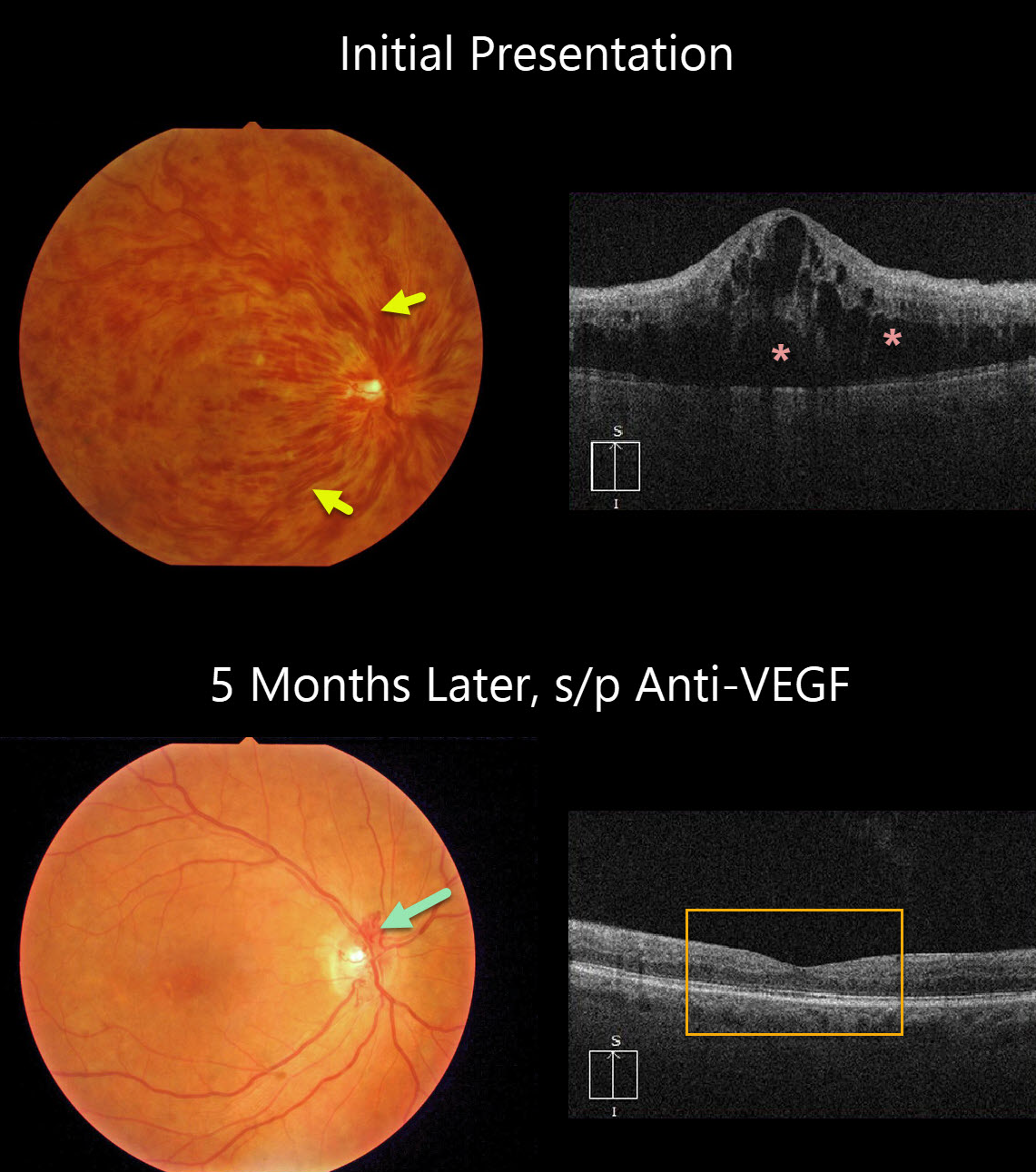
CRVO
Originally posted on @retina.rocks December 21, 2020
This patient presented with counting-finger vision from an acute hemorrhagic central retinal vein occlusion (CRVO) with marked macular edema. The poor vision and extensive retinal hemorrhages suggest that this is an ischemic CRVO.
After five monthly anti-VEGF injections, vision improved to 20/50, the retinal hemorrhages resolved dramatically, optic disc collaterals formed, and OCT showed complete resolution of all macular fluid. This degree of improvement is quite unusual.
Learning Points:
Patients with CRVO are at risk of vision loss due to macular edema or anterior segment neovascularization. Macular edema is common in both non-ischemic and ischemic occlusions, and this responds best to anti-VEGF therapy.
Rubeosis is found almost exclusively with ischemic occlusions, developing in about two-thirds of patients with untreated ischemic occlusions. This was common in the pre-anti-VEGF days.
However, since most patients with CRVO have macular edema and are receiving ongoing anti-VEGF therapy, neovascularization is now rarely seen unless patients present with untreated CRVO or therapy is discontinued.
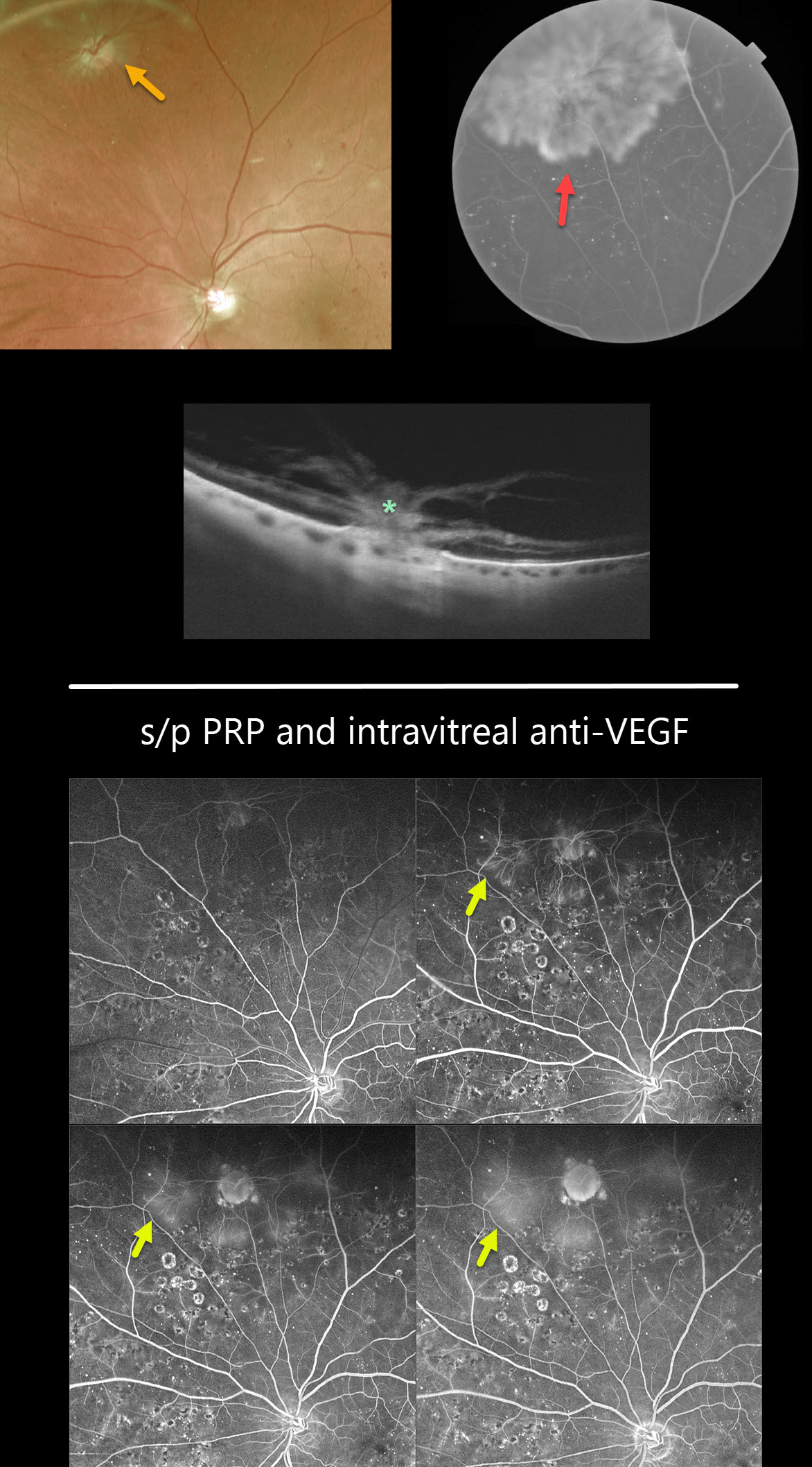
OPTIC NERVE PSEUDO-DUPLICATION WITH NVD ELSEWHERE
Originally posted on @retina.rocks December 18, 2020
Is that another optic nerve?
This is a pseudo-duplicated optic disc from peripheral neovascularization emanating from a disc-sized mid-peripheral chorioretinal scar.
The OCT reveals the area of choriovitreal neovascularization.
Learning Points:
The neovascularization regressed following panretinal photocoagulation (PRP) supplemented with intravitreal Eylea injections.
Fluorescein angiography following PRP and anti-VEGF injections shows virtually no leakage from the neovascularization.
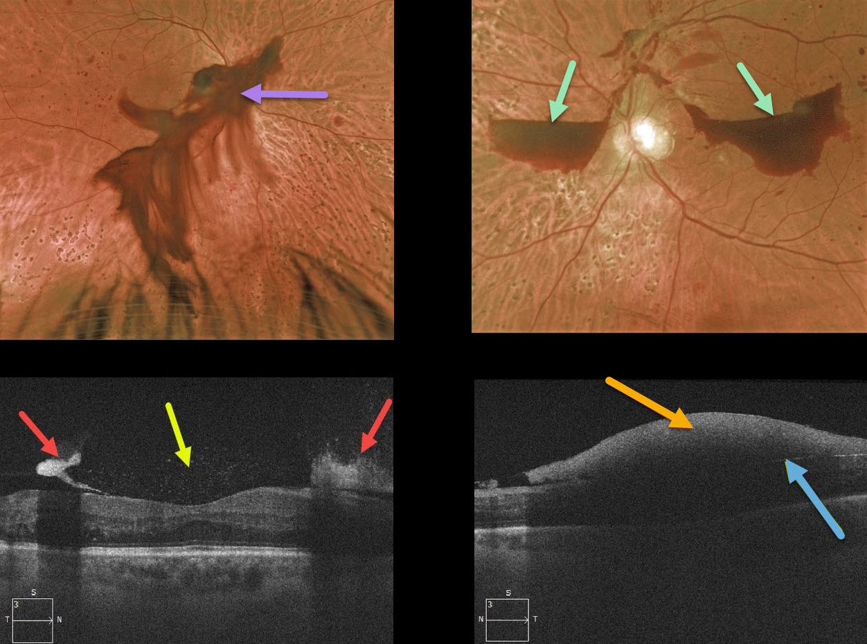
PROLIFERATIVE DIABETIC RETINOPATHY (PDR)
Originally posted on @retina.rocks December 11, 2020
This patient presented with a posterior pole vitreous hemorrhage OD and localized subhyaloid hemorrhages OS.
The OCT of the right eye highlights a thickened hyaloid on either side of the fovea with specks of hyperreflective pre-retinal blood. The left eye shows the subhyaloid blood blocking most of the underlying retinal detail.
This patient has already received partial panretinal photocoagulation bilaterally and will receive additional laser therapy.
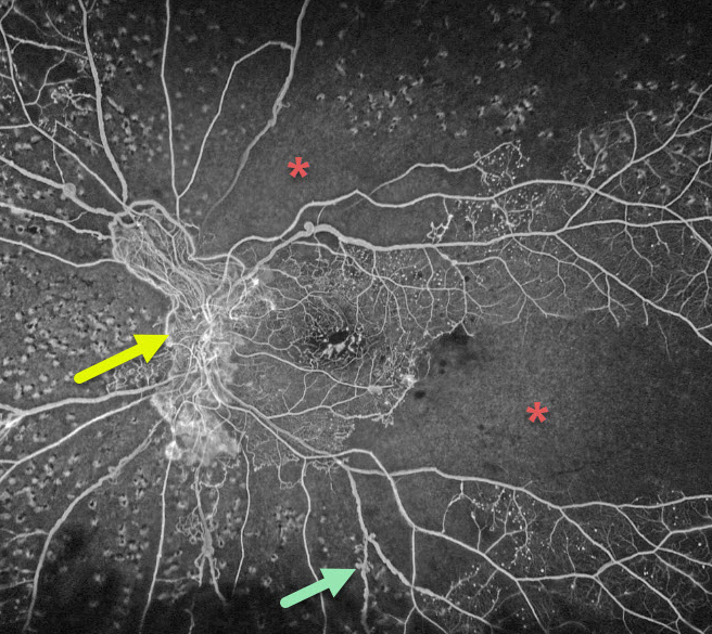
PROLIFERATIVE DIABETIC RETINOPATHY (PDR)
Originally posted on @retina.rocks November 10, 2020
This is an Optos ultra-widefield fluorescein angiogram of a patient with proliferative diabetic retinopathy. Despite recent panretinal photocoagulation (PRP), there is persistent large disc neovascularization. Extensive capillary nonperfusion is noted superotemporal to the disc and temporarily. Prominent venous beading is also noted inferotemporally. Additional PRP was added.
Learning Points:
Panretinal photocoagulation has been around since the 1960s and still remains the mainstay for treating proliferative disease. Often considered a “one and done,” patients can still develop breakthrough hemorrhages. Intravitreal anti-VEGF injections are often helpful, but some will require vitrectomy.
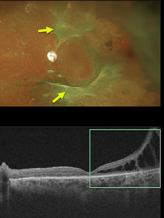
TRACTIONAL RETINAL DETACHMENT
Originally posted on @retina.rocks October 22, 2020
For many years, we have been following this 55YO male with 20/50 vision and involuted proliferative diabetic retinopathy with a stable extrafoveal traction retinal detachment in his left eye.
His right eye was enucleated following several failed vitrectomy surgeries years ago, before he came to our practice.
Although completely stable funduscopically, OCT shows some subclinical, progressive tractional schisis near the temporal macular center.
After a lengthy discussion, the patient chose to avoid vitrectomy for now, given his total lack of symptoms and prior experience with his right eye. We continue to follow him extremely closely since surgery may be necessary in the near future.
Learning Points:
Tractional retinal detachment is one of the main complications of untreated or incompletely treated proliferative diabetic retinopathy.
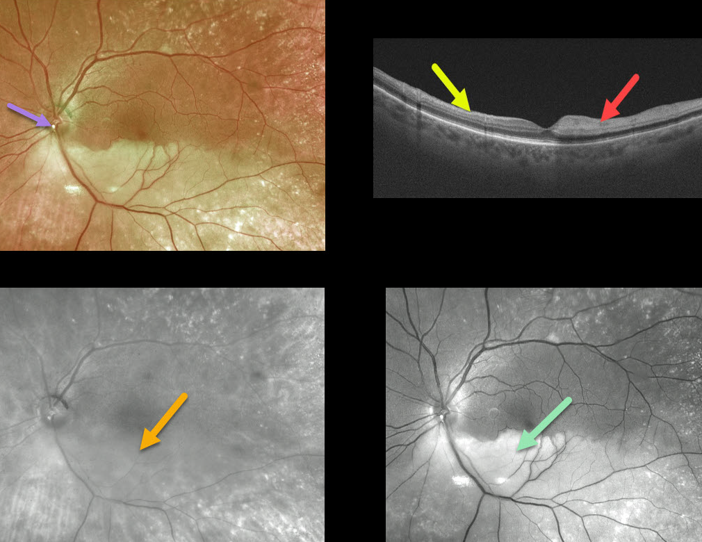
BRAO
Originally posted on @retina.rocks October 19, 2020
This is an 80YO female who presented with a branch retinal arterial occlusion (BRAO) and 20/40 vision.
The causative calcific embolus is seen overlying the inferior optic nerve. The opaque retina is especially well visualized with the Optos green channel but virtually invisible in the Optos red channel.
The vertical B-scan contrasts the normal superior retina with the ischemic inferior retina (marked hyperreflectivity of the inner retina, causing shadowing of the outer retina and choroid).
Learning Points:
This patient had acute retinal ischemia (defined as acute BRAO, central retinal artery occlusion, or transient monocular vision loss) and was immediately referred to the nearest stroke center, which is the standard of care.
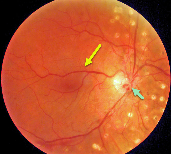
RETINAL VENOUS MALFORMATION
Originally posted on @retina.rocks October 12, 2020
This patient has a retinal venous malformation (formerly known as retinal macrovessel), in addition to full panretinal photocoagulation (PRP) for proliferative diabetic retinopathy. Residual flat disc neovascularization remains despite full PRP.
Learning Points:
A retinal venous malformation is an anomalous retinal vessel that crosses the horizontal raphe (remember that normal retinal vessels respect the horizontal meridian).
These vessels are technically arterio-venous malformations (AVM). Since patients with AVMs can have additional venous anomalies in the brain (24% compared to up to 6% of the population), a brain MRI is recommended to rule out any intracranial abnormalities.
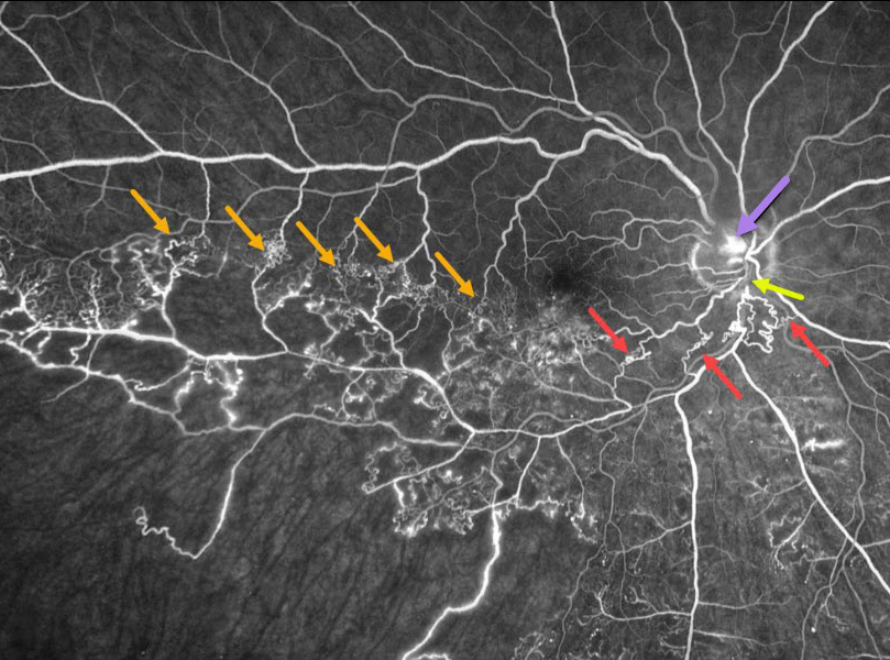
BRVO
Originally posted on @retina.rocks August 31, 2020
This patient has an ischemic branch retinal vein occlusion (BRVO) with extensive collateral vessels.
Occlusion occurs when an artery compresses the underlying vein where the vessels share a common adventitial sheath. Extensive collateral vessels cross the temporal horizontal raphe as well as around the occlusion more posteriorly. A small area of disc neovascularization leaks.
Learning Points:
Collateral vessels are dilated, pre-existing vessels that bypass an occlusion. These are most commonly seen in the posterior pole following retinal venous occlusions, but can also occur on the optic disc with optic nerve meningiomas.
The endothelial tight junctions are intact, so there’s no leakage on angiography. This helps differentiate them from neovascularization, which does leak.
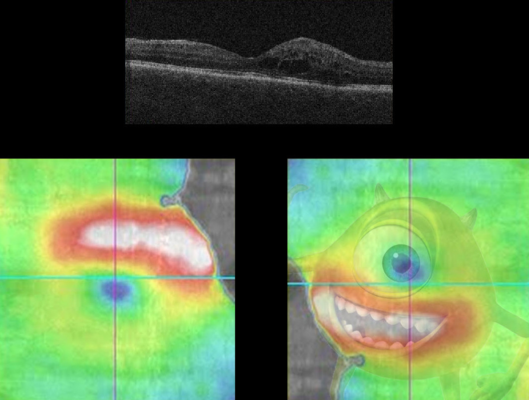
MIKE WAZOWSKI MACULOPATHY
Originally posted on @retina.rocks August 28, 2020
Here we see a patient with the ever-elusive Mike Wazowski Maculopathy (for you Baby Boomers, he stars in Monsters, Inc.).
The OCT image, rotated 180 degrees, details a classic Mike Wazowski pattern of edema.
In reality, the patient has some non-central diabetic macular edema.
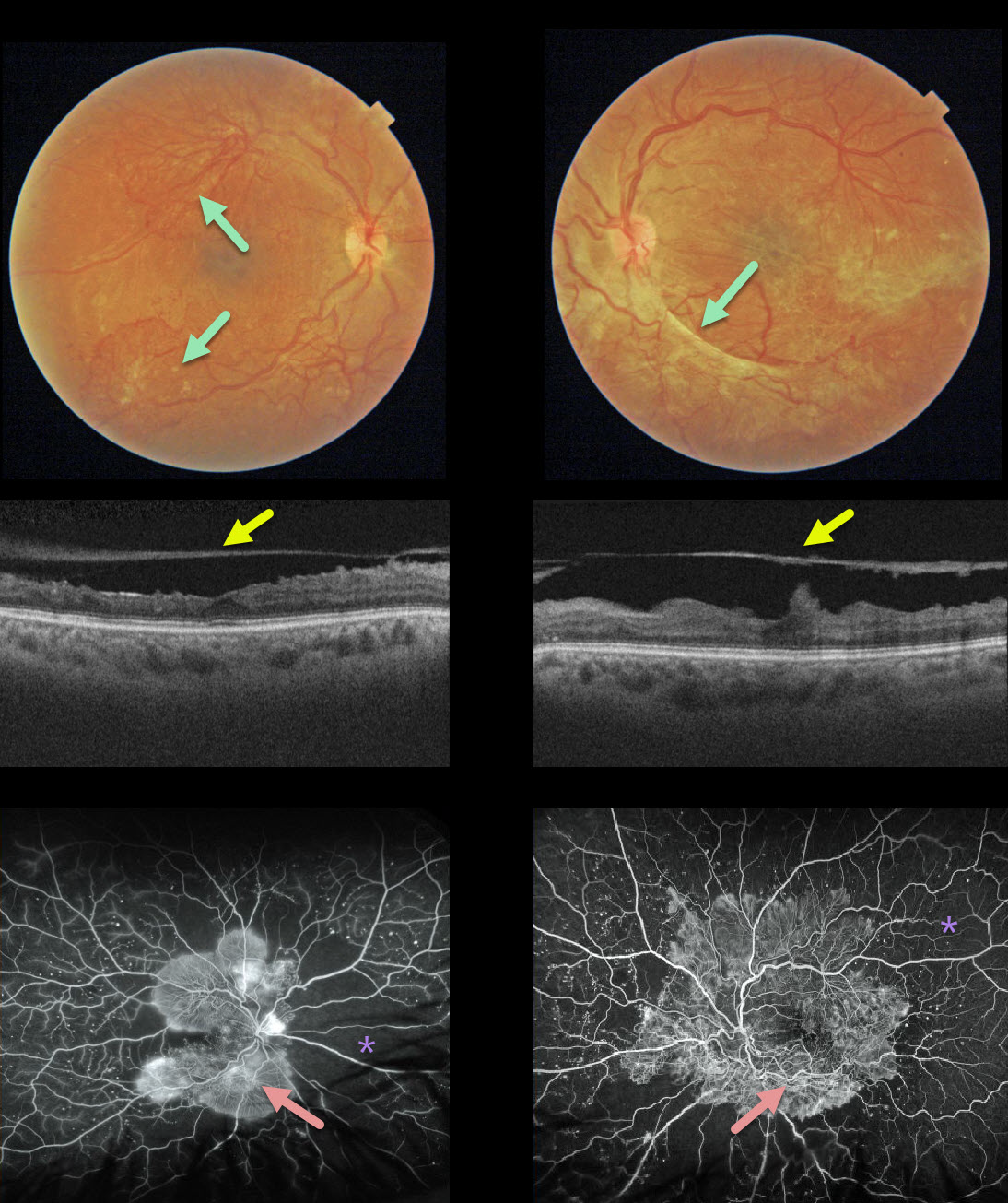
PROLIFERATIVE DIABETIC RETINOPATHY (PDR)
Originally posted on @retina.rocks August 12, 2020
This 32YO female presented with 20/400 vision bilaterally, a hemoglobin A1C over 12%, and a blood glucose level over 300. Fibrosed neovascularization variably covers each posterior pole.
OCT shows that the posterior hyaloid has spontaneously detached from each central macula, which has a rippled contour. This appearance is also often seen following vitrectomy for tractional detachment with peeling of the hyaloid.
Fluorescein angiography shows extensive posterior pole neovascularization with peripheral ischemia.
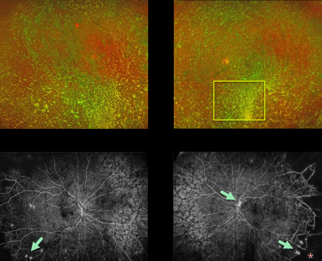
ASTEROID HYALOSIS
Originally posted on @retina.rocks July 29, 2020
Although this diabetic patient had normal vision with absolutely no symptoms, what we saw on clinical examination and Optos color imaging was nothing but asteroid hyalosis.
Fluorescein angiography (FA) showed bilateral proliferative diabetic retinopathy with areas of leakage and peripheral capillary dropout. In-office panretinal photocoagulation (PRP) was not possible due to the dense asteroid.
We discussed ongoing anti-VEGF injections versus vitrectomy + PRP to clear the visual axis. She decided to proceed with vitrectomy in both eyes.
Learning Points:
Asteroid hyalosis is caused by the accumulation of benign calcium-lipid particles suspended in the vitreous.
Fluoroscein angiography and OCT can image through asteroid hyalosis.
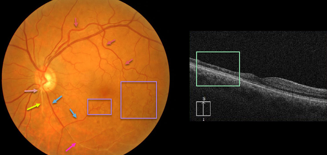
RETINAL ARTERIOVENOUS MALFORMATION
Originally posted on @retina.rocks July 28, 2020
At first or quick glance, this looks like an old inferotemporal branch retinal vein occlusion (BRVO).
On closer inspection, there is a white-sheathed vessel arising from the nerve inferiorly. Numerous collateral vessels are noted inferior and temporal to the fovea, and there is a sheathed vessel in the inferior arcade.
OCT shows marked inner retinal atrophy with preservation of the photoreceptor layers, consistent with a prior branch retinal artery occlusion. So the collateral vessels (virtually always a sign of a prior venous occlusion) indicate an old BRVO, and the atrophic and sheathed vessel indicates an old BRAO, both in the same quadrant. Right?
Tracing the large branch of the superotemporal arcade inferotemporally, we see that the collaterals appear to be arterial. Inferior to the nerve, we see an arteriole that crosses the sheathed vessel, indicating that the sheathed vessel is not an artery but a vein. Moreover, the large vessel off the nerve, inferotemporal in location, appears to be a vein that eventually connects to the collateral vessels we previously identified as arterial.
Our best guess is that this may be a low-grade arteriovenous malformation, possibly with a prior secondary BRAO/BRVO. Unfortunately, we don’t have a fluorescein angiogram, which may help better define the vascularization.
Learning Points:
Remember that arteries don’t cross arteries and veins don’t cross veins. Tracing vessels can be a helpful key to identifying retinal vascular disorders.
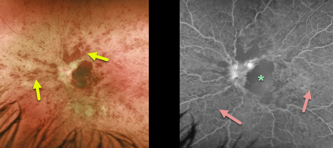
CRVO
Originally posted on @retina.rocks July 22, 2020
This patient is a 28YO who presented with hand motion vision from an acute central retinal vein occlusion (CRVO).
The poor vision and extent of retinal hemorrhages would suggest an ischemic occlusion. However, fluorescein angiography shows good peripheral perfusion, indicating a non-ischemic CRVO.
Angiography also shows marked foveal hypofluorescence, which may be due to a filling defect (ischemia) or a blockage. The contour of the dark, hypofluorescent macula precisely corresponds to the macular hemorrhage noted on the color photograph, indicating that this is due to blockage rather than ischemia.
Learning Points:
Although CRVOs can occur in younger patients, it is sometimes prudent to look for other systemic causes, including hypercoagulable conditions and hyperviscosity syndromes.
See Liu et al. Retina 2018;38;1175-1179 and Bremond-Gignac et al Retina 2020;40;1339-1343 for further discussion of the debate over the need for a medical workup in younger patients with CRVO.
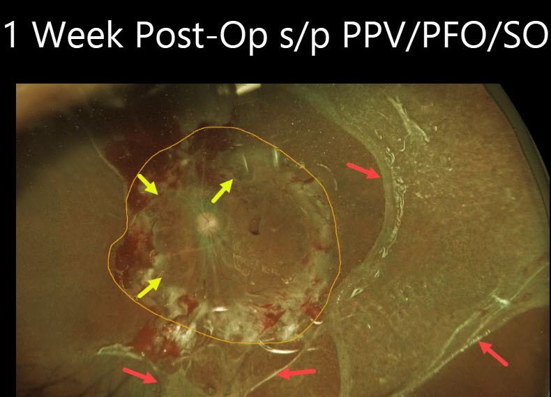
TRACTIONAL RETINAL DETACHMENT
Originally posted on @retina.rocks July 13, 2020
This patient had hand motion vision in her left eye from a closed funnel, combined with diabetic tractional and rhegmatogenous retinal detachment.
Along with vitrectomy, a 360-degree retinectomy was made anterior to the macula. The posterior retina was then flattened with Perfluoron, followed by silicone oil. A large macular hole was also noted intraoperatively.
This image was taken 1 week postoperatively and shows the extent of the retinectomy. Although intraoperatively we felt that we were excising full-thickness retina, once the retina flattened, a remaining outer layer of retina was variably present temporally and inferiorly.
Although Perfluoron is completely removed during surgery, complex cases such as this often have residual subretinal bubbles.
Functionally blind before surgery, she was able to fully care for herself in her apartment and use her microwave and cell phone at 1 week.
Learning Points:
Sometimes during vitrectomy, the only way to partially flatten the retina is to do a retinectomy to excise the extremely thick, stiff, and scarred retina anterior to the macula.
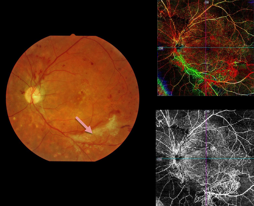
PROLIFERATIVE DIABETIC RETINOPATHY (PDR)
Originally posted on @retina.rocks July 2, 2020
This patient has proliferative diabetic retinopathy. The fundus photo shows scattered red and fibrosed neovascularization.
The color OCTA image is a composite showing all layers of the retinal vasculature: red is the inner retina, green is the mid-retinal layer, and blue is the outer retina (photoreceptors, which are usually devoid of intrinsic vasculature). The bottom right image isolates the inner retina, showing regions of retinal ischemia with neovascularization.
Learning Points:
OCTA is a relatively new and non-invasive modality able to quickly identify diabetic retinal vascular abnormalities. Its role in screening for retinal ischemia and proliferative disease is still being defined.
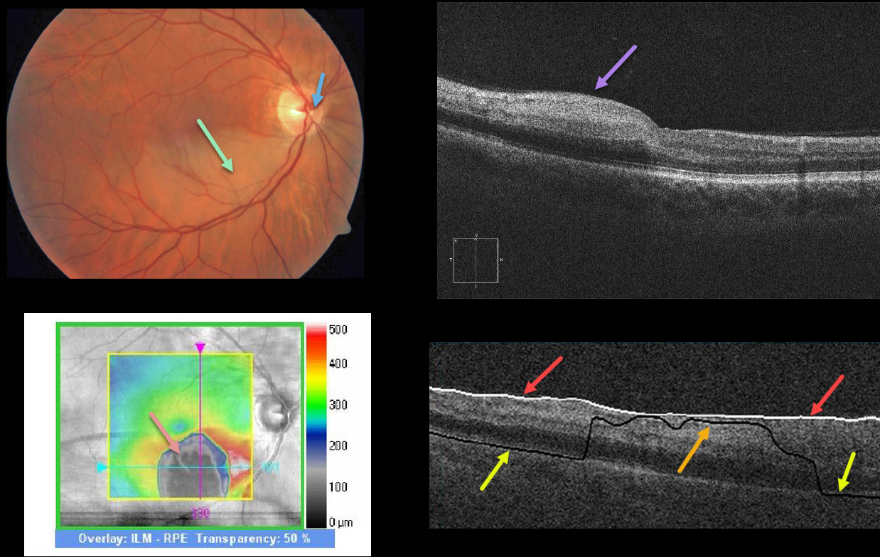
BRAO
Originally posted on @retina.rocks June 30, 2020
This patient has a subtle white, opaque retina in the inferior macula due to an acute branch retinal artery occlusion (BRAO). The causative embolus is the tiny white lesion just nasal to the nerve’s center.
OCT shows hyperreflectivity from the acutely ischemic inner retinal layers. The black blob on the retinal thickness map reflects an incorrectly identified RPE detachment (PED) caused by the algorithm misidentifying the RPE due to the marked hyperreflectivity of the inner retinal layers.
This phenomenon is demonstrated by the B-scan, which shows that the ILM is correctly identified by the white line. The black line is supposed to identify the RPE, which is correctly identified nasally and temporally but misidentified centrally due to said hyperreflectivity.
Learning Points:
An acute BRAO can be subtle clinically but more dramatic on OCT. Acute inner retinal OCT hyperreflectivity can lead to misidentification of retinal layers, resulting in artifacts.
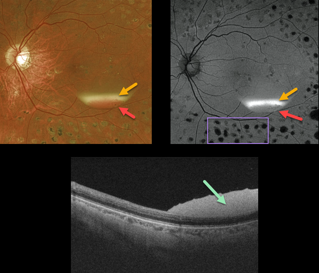
PROLIFERATIVE DIABETIC RETINOPATHY (PDR)
Originally posted on @retina.rocks June 16, 2020
This patient, previously treated for proliferative diabetic retinopathy with panretinal photocoagulation (PRP), presented with an asymptomatic resolving two-toned subhyaloid hemorrhage. The devitalized yellow blood indicates chronicity, although some red blood remains.
The OCT confirms the subhyaloid location of the hyperreflective blood.
Red blood is dark on fundus autofluorescence (FAF), although yellow blood is extremely hyperautofluorescent. Lipofuscin within the RPE is the main fluorophore in FAF. The laser PRP scars are hypoautofluorescent due to loss of the RPE.
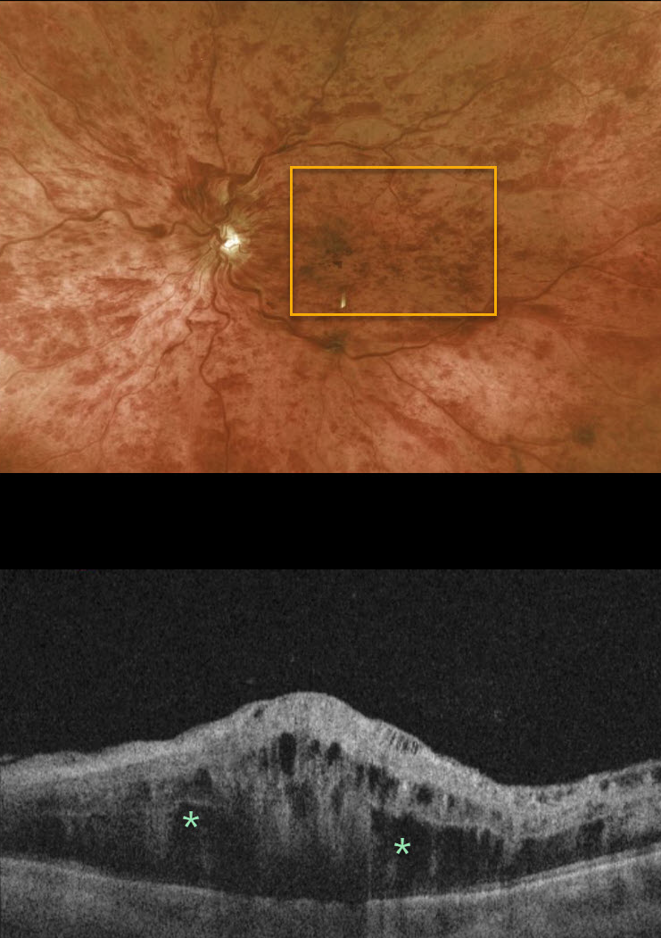
CRVO
Originally posted on @retina.rocks June 15, 2020
This 76YO male presented with 20/200 vision, the presence of an afferent pupillary defect, and extensive retinal hemorrhages. He had an acute ischemic central retinal vein occlusion (CRVO) with macular edema.
Patients with CRVO are at risk of vision loss due to macular edema or anterior segment neovascularization. Macular edema is common in both non-ischemic and ischemic occlusions, although rubeosis is found almost exclusively with ischemic occlusions.
Learning Points:
Intravitreal therapy in the early 2000s brought about the first successful treatment for CRVO-related macular edema. Anti-VEGF drugs are the first-line therapy, with the majority of patients gaining several lines of vision. Treatment is often ongoing, as it is for wet macular degeneration.
Although rubeosis was a common finding in the past (and is still seen if patients present with an untreated ischemic occlusion), this is now a rare occurrence since the vast majority of these patients have macular edema that is treated with ongoing anti-VEGF injections. Interestingly, if the injections are stopped several years later, rubeosis can still develop.
Iris neovascularization with neovascular glaucoma develops in about two-thirds of patients with untreated ischemic occlusions. Rubeosis is treated initially with an anti-VEGF injection, followed by aggressive, complete panretinal photocoagulation. Some patients will need a glaucoma tube or a cyclodestructive procedure if the IOP remains too high.
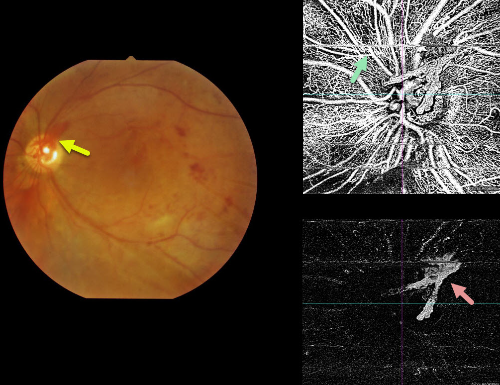
PROLIFERATIVE DIABETIC RETINOPATHY (PDR)
Originally posted on @retina.rocks June 3, 2020
This patient has PDR and prominent neovascularization of the disc (NVD), which is well-captured by the OCT angiogram.
Movement artifacts are noted throughout the scan. The bottom-right image isolates the vitreoretinal interface, showing only the NVD.
Learning Points:
The OCT angiogram captures blood vessels from the full-thickness retina, including the superficial radial peripapillary capillary network, which is not imaged with traditional fluorescein angiography.
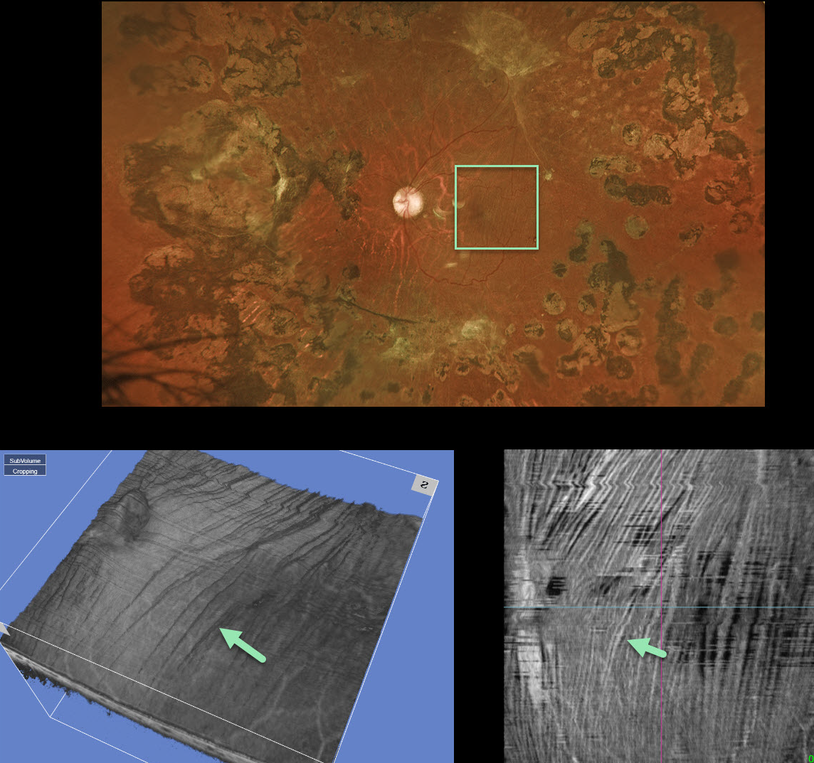
RETINAL STRIAE FOLLOWNG VITRECTOMY FOR DIABETIC TRD
Originally posted on @retina.rocks May 21, 2020
This 35 YO male had a severe diabetic tractional retinal detachment with subsequent pars plana vitrectomy a few years prior.
Now, macular striae are apparent in the inner retina and are especially dramatic on en face imaging (image 4). Remarkably, this patient maintains 20/40 vision. It is quite common for these postoperative traction detachments to have residual retinal striae, which usually don’t adversely affect vision.
Learning Points:
En face OCT is extremely helpful in evaluating and managing retinal disease, offering a unique perspective for viewing pathology.
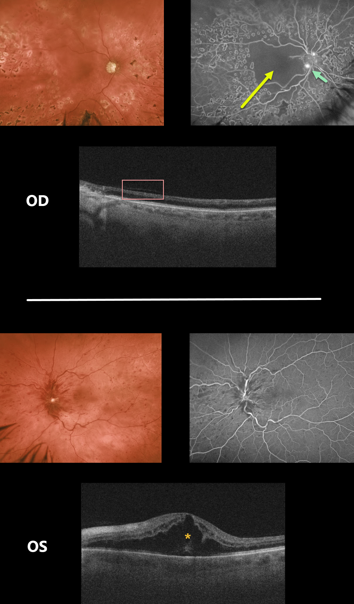
CRVO
Originally posted on @retina.rocks May 12, 2020
This patient had counting fingers vision OD due to a longstanding ischemic CRVO complicated by macular ischemia, disc neovascularization and neovascular glaucoma.
Fluorescein angiography shows profound capillary loss throughout the macula along with leaking disc neovascularization.
Due to the profound retinal ischemia there is marked OCT thinning of the inner two-thirds of the retina, giving a similar appearance to that seen with central retinal arterial occlusion.
She recently developed a new non-ischemic CRVO in her left eye with macular edema and 20/80 vision.
Learning Points:
This patient highlights several common and uncommon findings with CRVO. About 10% of patients will develop bilateral occlusions.
Eighty percent of occlusions are non-ischemic, although about one-third of these will convert to ischemic occlusions within several years. About 60% of ischemic occlusions will develop anterior segment neovascularization with neovascular glaucoma.
For some reason, posterior segment neovascularization rarely occurs (see the classic Hayreh 1983 paper Ophthalmology 1983;90;488-506).
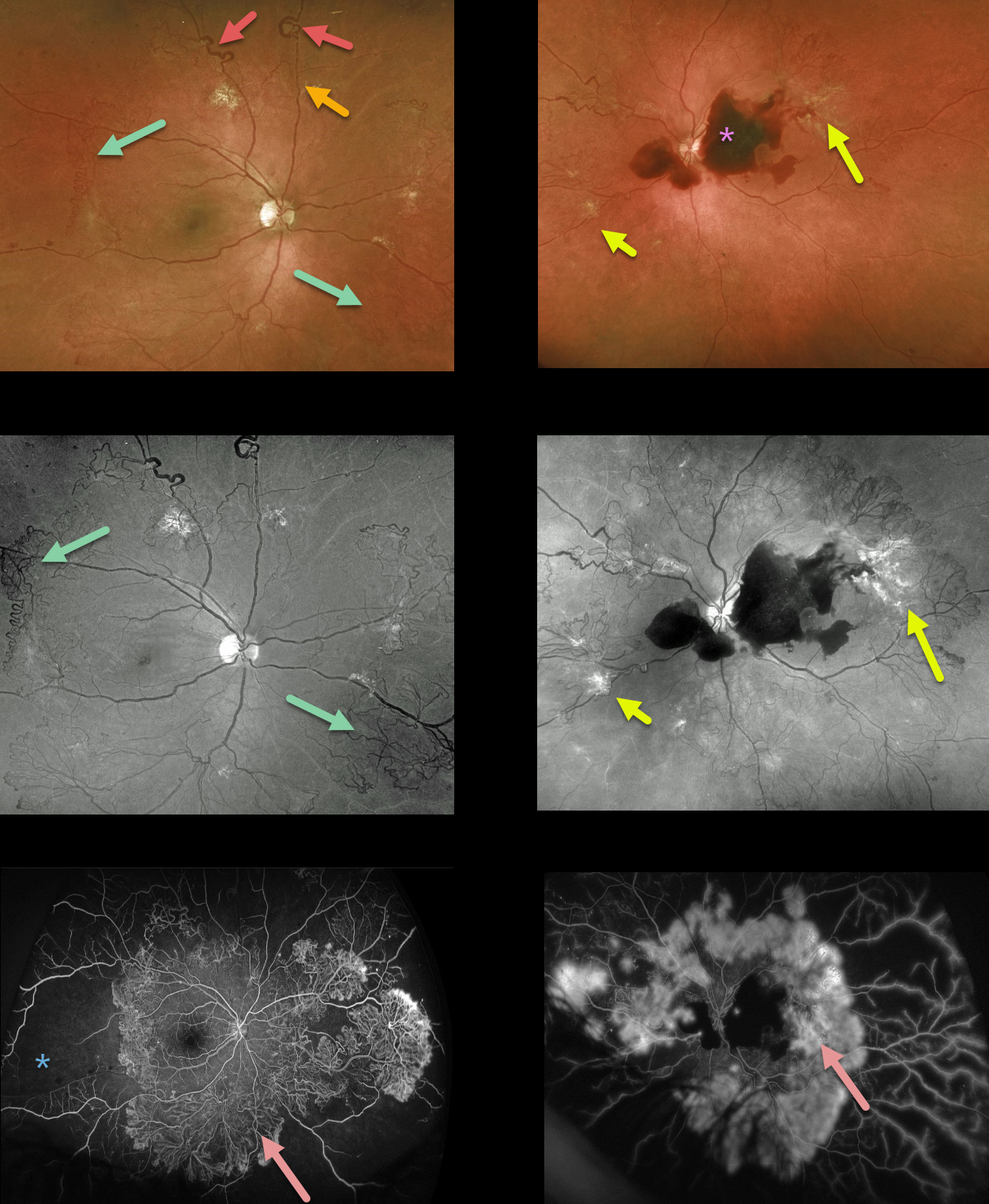
PROLIFERATIVE DIABETIC RETINOPATHY (PDR)
Originally posted on @retina.rocks May 4, 2020
This 39YO presented with severe PDR in both eyes, highlighting nearly all of the key features of proliferative disease. Surprisingly, vision was 20/30 OD and 20/50 OS.
The right eye shows great examples of venous loops and venous beading and extensive scattered NVE, especially highlighted by the green channel Optos.
The left eye has a central subhyaloid hemorrhage along with venous beading and scattered NVE.
Fluorescein angiography shows dramatic and extensive neovascularization encircling both posterior poles with marked anterior ischemia.
Learning Points:
This case further emphasizes the importance of regular diabetic dilated examinations with peripheral retinal examination to unveil the true extent of the retinopathy.
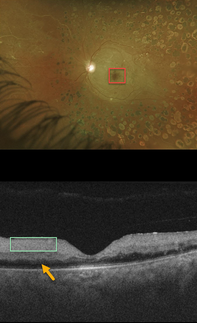
CRAO
Originally posted on @retina.rocks April 30, 2020
This patient presented with acute vision loss from a fresh central retinal artery occlusion (CRAO). Of note, this patient also has involuted PDR following full PRP.
The cherry red spot is caused by the unaffected foveal retina (receiving intact choroidal blood flow) being surrounded by more distal white and ischemic retina no longer receiving circulation from the central retinal artery.
Although the retina is somewhat thickened on OCT, the acutely ischemic inner two-thirds of the retina, which is supplied by the central retinal artery, is extremely hyperreflective. Less light is available to illuminate the deep retina, which is therefore darker than normal with “pseudo-edema.”
Learning Points:
Patients with acute retinal ischemia (acute CRAO, acute branch retinal artery occlusion or transient monocular vision loss) need to be sent immediately to the nearest stroke center due to the risk of pre-existing recent asymptomatic stroke or impending stroke (see Biousse et al, Ophthalmology 2018;125;1597-1607 or our post https://odsonfb.com/amaurosis-brao-and-crao-the-medical-emergency-many-eye-doctors-still-dont-know-about/).
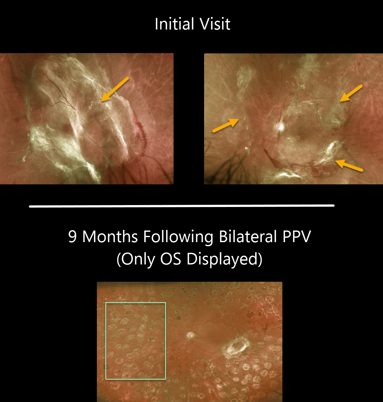
TRACTIONAL RETINAL DETACHMENT
Originally posted on @retina.rocks April 28, 2020
This patient presented with bilateral diabetic traction detachments with counting fingers OD and 20/400 OS. The fibrotic neovascularization is particularly severe in his right eye, obscuring the entire posterior pole.
He subsequently underwent bilateral pars plana vitrectomy. An image of the left eye is shown about 9 months later. All neovascular tissue was removed intraoperatively and endolaser was done. Vision remarkably improved to 20/60.
We were unable to get a photo of the right eye at that visit due to a recurrent vitreous hemorrhage. The blood subsequently resolved with an intravitreal Avastin injection, and vision was 20/200 OD at the most recent visit.
Learning Points:
Tractional retinal detachment is one of the main complications of untreated or incompletely treated proliferative diabetic retinopathy.
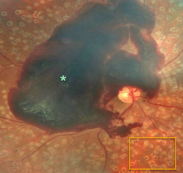
PROLIFERATIVE DIABETIC RETINOPATHY (PDR)
Originally posted on @retina.rocks April 24, 2020
This patient still developed a large foveal subhyaloid hemorrhage despite very full prior panretinal photocoagulation (PRP).
Learning Points:
PRP has been around since the 1960s and still remains the mainstay for treating proliferative disease. Often considered a “one and done,” patients can still develop break-through hemorrhages. Intravitreal anti-VEGF injections are often helpful, but some will require vitrectomy.
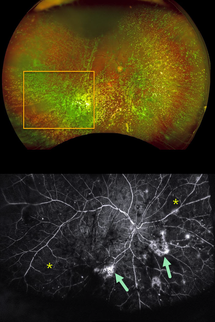
ASTEROID HYALOSIS OBSCURING OCCULT PROLIFERATIVE DIABETIC RETINOPATHY (PDR)
Originally posted on @retina.rocks April 17, 2020
Examining the fundus with dense asteroid hyalosis, as in this diabetic patient, can be extremely challenging.
Fluorescein angiography (FA) and OCT are great modalities since they’re both able to visualize the retina when the clinical exam can’t.
FA was critical in diagnosing proliferative retinopathy with peripheral ischemia in this patient.
Learning Point:
Asteroid hyalosis is caused by calcium-lipid complexes suspended throughout the vitreous. These white opacities resemble stars on a night sky – hence the name.
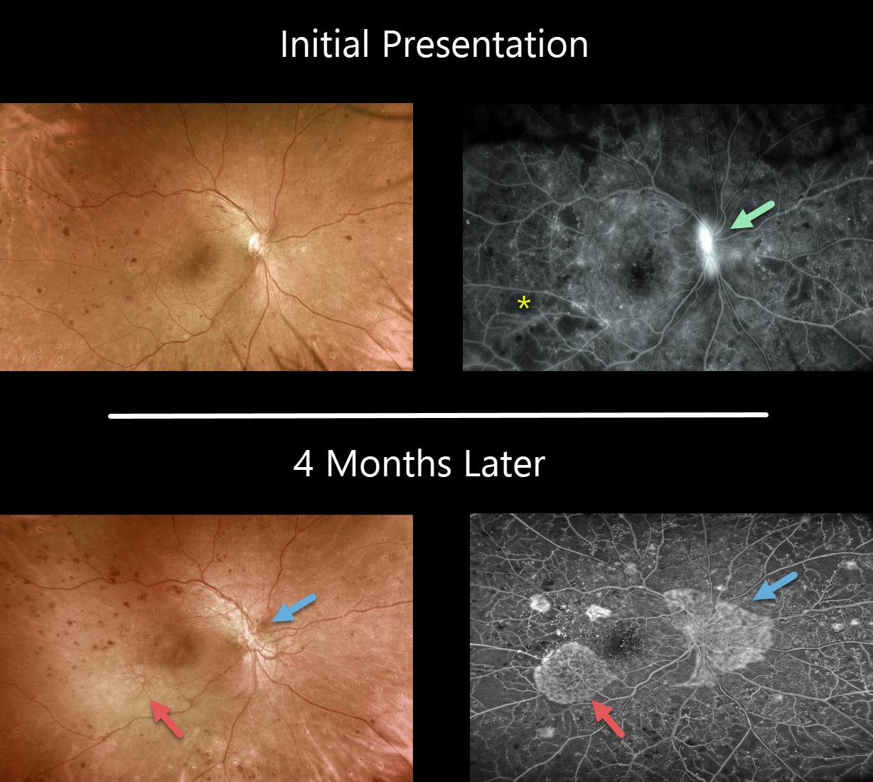
PROLIFERATIVE DIABETIC RETINOPATHY (PDR)
Originally posted on @retina.rocks April 13, 2020
This patient, who was in the process of undergoing panretinal photocoagulation for proliferative diabetic retinopathy, was originally seen with NVD and severe nonperfusion.
Unfortunately, she was lost to follow-up for 4 months and presented again with increased NVD and new NVE. The Optos fluorscein angiogram shows the complex of fine neovascular vessels and capillary dropout in exquisite detail.
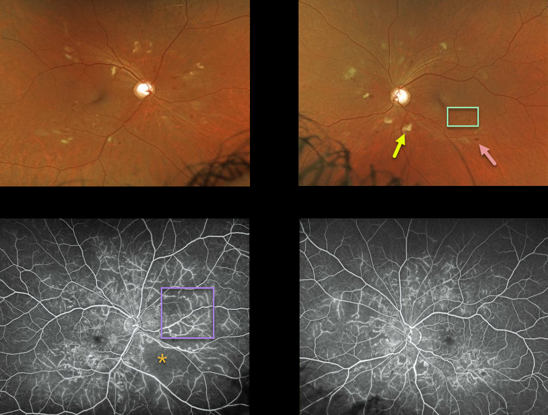
NON-PROLIFERATIVE DIABETIC RETINOPATHY (NPDR)
Originally posted on @retina.rocks March 17, 2020
The fundus photos demonstrate severe NDPR with cotton wool spots, microaneurysms, and intraretinal hemorrhages.
Our patient’s ultra wide-field fundus fluorescein angiography (UW-FFA) shows significant areas of non-perfusion with vascular staining but no neovascularization.
Learning Points:
In some cases of moderate or severe retinopathy, a baseline fluorescein angiography is performed. UW-FFA can be useful to detect clinically silent proliferative disease and monitor these patients for conversion from NPDR to PDR.
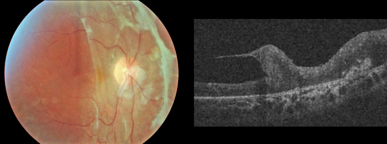
HUMMINGBIRD MACULOPATHY
Originally posted on @retina.rocks February 28, 2020
This rare condition was discovered after a diabetic TRD repair. Thankfully, no birds or retinas were harmed in this process.
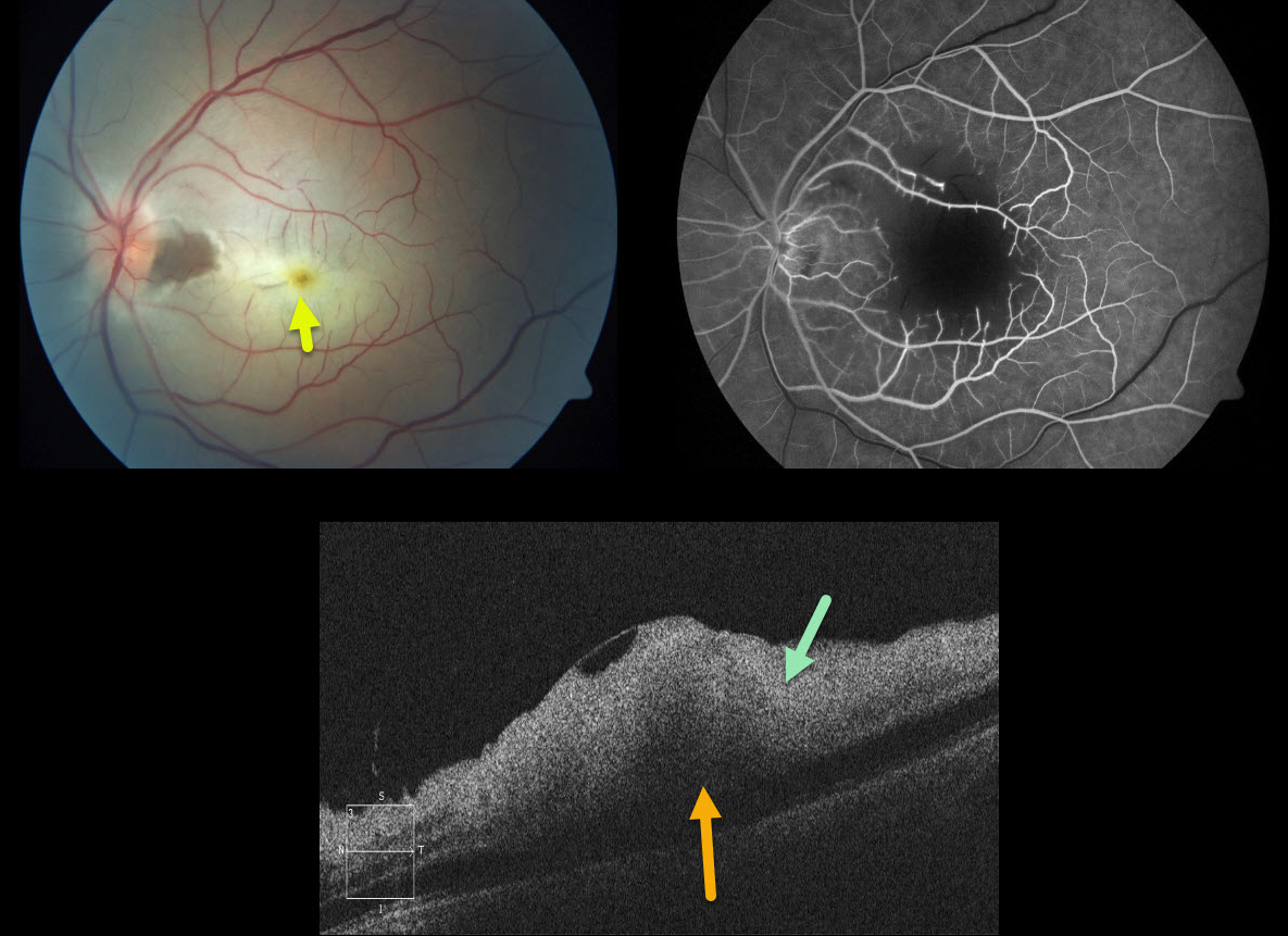
CRAO
Originally posted on @retina.rocks February 24, 2020
This is a 49 yo M who presented with acute 20/400 monocular vision loss and retinal whitening.
The OCT shows marked inner retinal hyperreflectivity from the acute ischemia, with pseudo-edema of the outer retina due to decreased transmission of light through the inner opaque retina.
The outer retina is still receiving oxygen from the choroid, as evidenced by sparing of the fovea (the yellow xanthophyll is prominently seen).
Learning Points:
This patient was sent to the emergency room for a stroke protocol workup (including electrocardiography, MRI, carotid non-invasive, cardiac echography, hypercoagulable labs, and inflammatory labs). The evaluation was totally negative.
This case looks very similar to gentamicin retinal toxicity. Intravitreal gentamicin can cause severe macular ischemic retinal necrosis. The macular capillaries have a characteristic cut-off appearance, as is seen in our patient who had no gentamicin exposure.
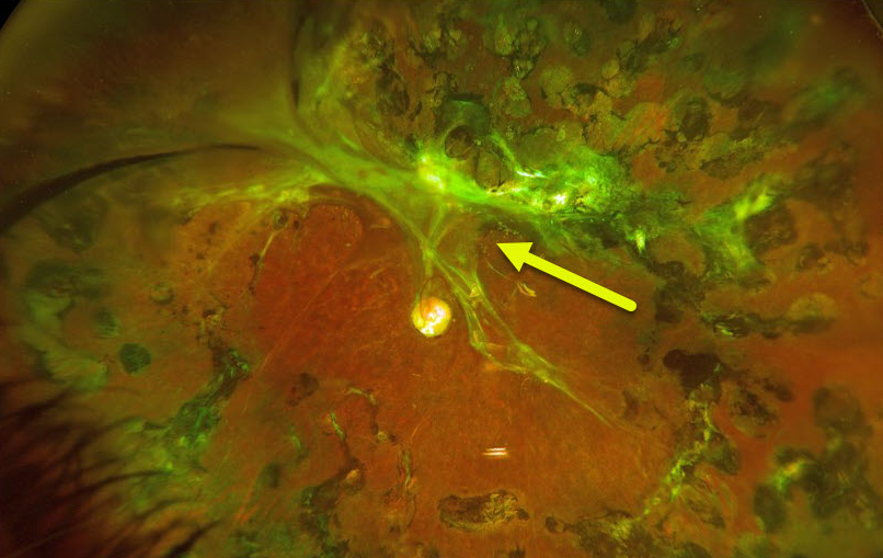
TRACTIONAL RETINAL DETACHMENT
Originally posted on @retina.rocks February 20, 2020
This patient underwent vitrectomy in 2013 for a severe table-top diabetic traction retinal detachment (TRD). We were able to free up all significant traction except superiorly and superotemporally, shown in the Optos image.
Despite this, the patient has been quite stable anatomically, maintaining 20/30 vision.
Learning Points:
Tractional retinal detachment is one of the main complications of untreated or incompletely treated proliferative diabetic retinopathy.
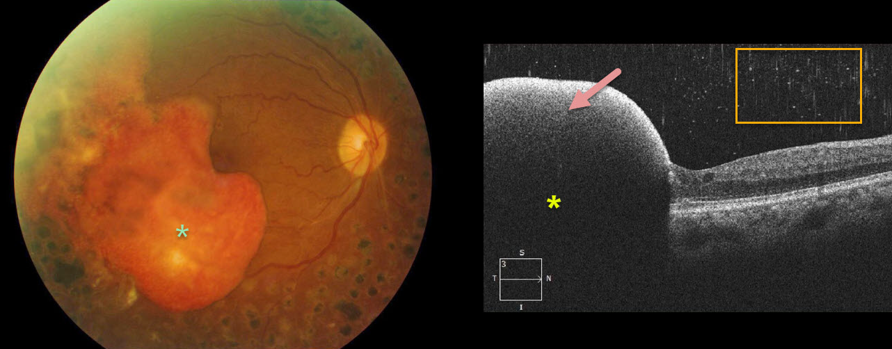
PROLIFERATIVE DIABETIC RETINOPATHY (PDR)
Originally posted on @retina.rocks February 6, 2020
This patient with proliferative diabetic retinopathy has a dense subhyaloid hemorrhage. The yellowing subhyaloid blood indicates that this has been present for at least several months.
The OCT highlights the subhyaloid hemorrhage that blocks all underlying retinal detail and the hyper-reflective vitreous red blood cells gives a “snowy” appearance.
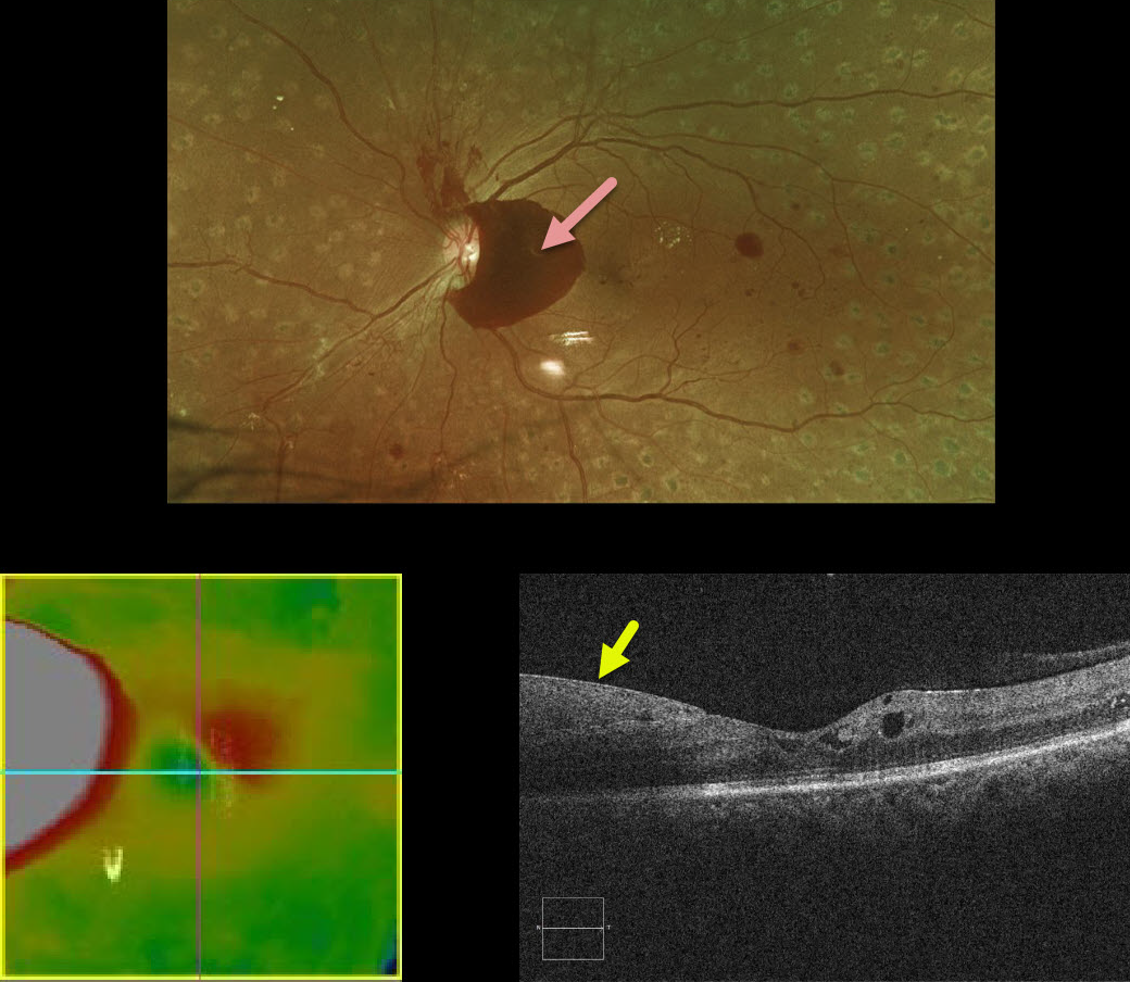
PROLIFERATIVE DIABETIC RETINOPATHY (PDR)
Originally posted on @retina.rocks January 31, 2020
This patient with a history of involuted proliferative diabetic retinopathy following complete panretinal photocoagulation presented with what appeared on OCT to be new diabetic macular edema.
A closer inspection of the B-scan shows the retina to be normal with the pseudo-edema representing subhyaloid blood.
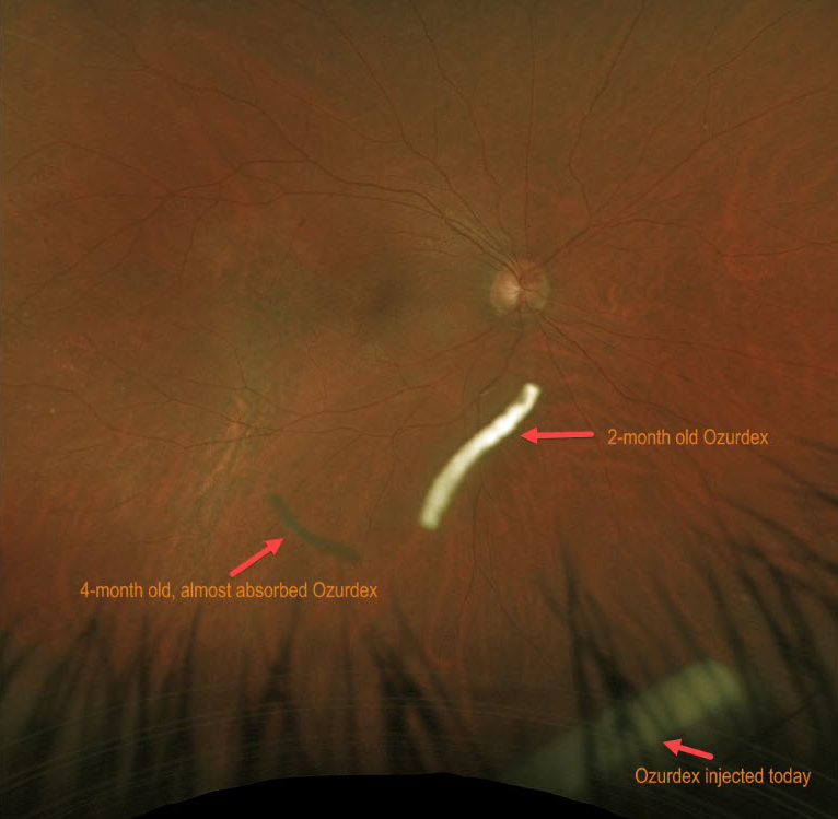
BRVO
Originally posted on @retina.rocks January 27, 2020
This patient is receiving Ozurdex (dexamethasone 0.7%) intravitreal injections every 2 months to control macular edema from a branch retinal vein occlusion (BRVO).
The Ozurdex implant slowly releases the steroid and eventually totally absorbs, with the clinical effect lasting about 2 to 3 months. This Optos image shows 3 Ozurdex implants in various stages of their life cycles.
Learning Points:
In addition to Ozurdex, other possible treatment modalities for BRVO macular edema include anti-VEGF or triamcinolone injections, and macular laser photocoagulation.
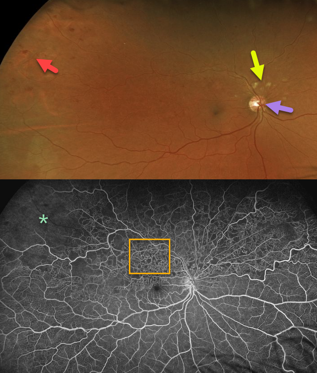
HEMI-RETINAL VEIN LCCUSION
Originally posted on @retina.rocks January 21, 2020
This patient has a superior hemi-reinal vein occlusion with superotemporal intraretinal hemorrhages and cotton wool spots superior to the optic nerve.
The fluorescein angiogram (FA) shows extensive retinal vascular telangiectasia and peripheral ischemia, indicating that this patient is at risk for developing retinal
neovascularization.
Learning Points:
Collateral vessels of the optic nerve can develop with retinal vein occlusions. Prominent optic nerve collateral vessels are seen in the fundus photo. These don’t leak angiographically proving that they’re not neovascular.
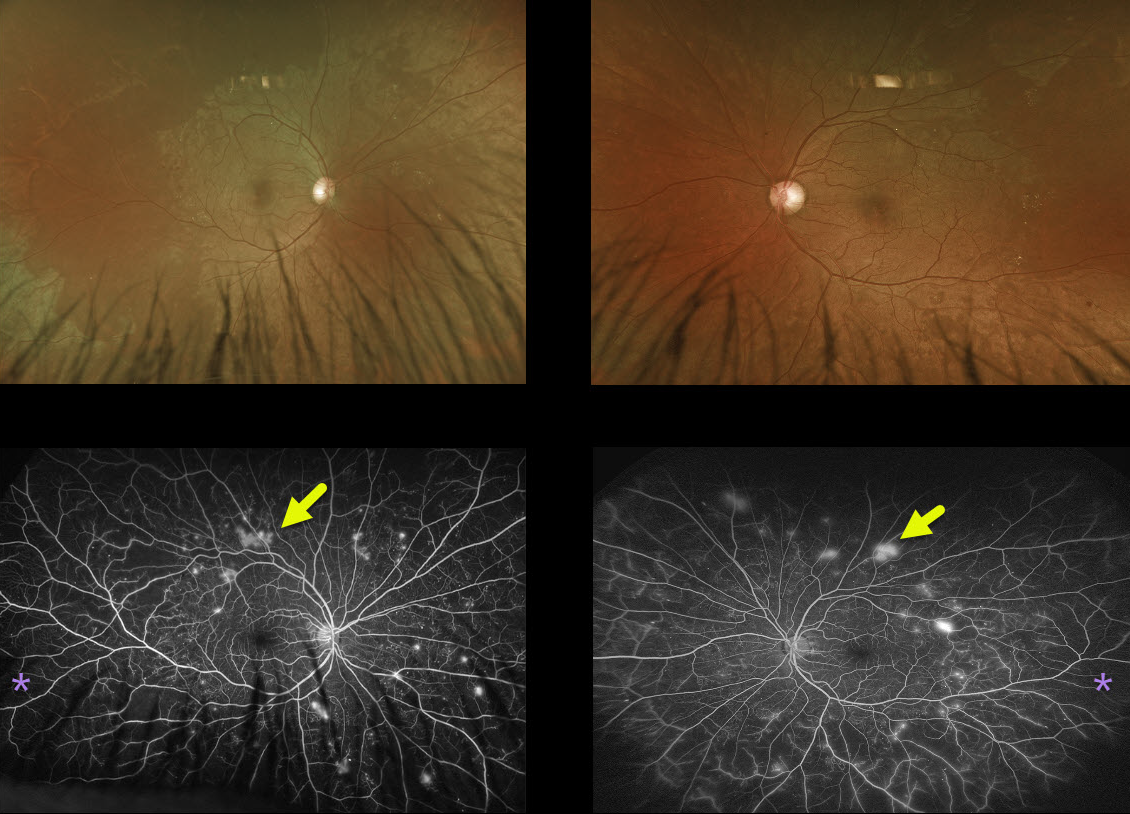
PROLIFERATIVE DIABETIC RETINOPATHY
Originally posted on @retina.rocks January 10, 2020
This patient had relatively normal posterior poles with extensive peripheral ischemia and neovascularization on ultra-widefield fluorescein angiography.
Learning Points:
We see this fairly frequently. Since it is so easy to miss proliferative disease, it is not unreasonable for patients with apparent moderate NDPR to have a baseline and periodic ultra-widefield angiogram to rule out subclinical PDR.
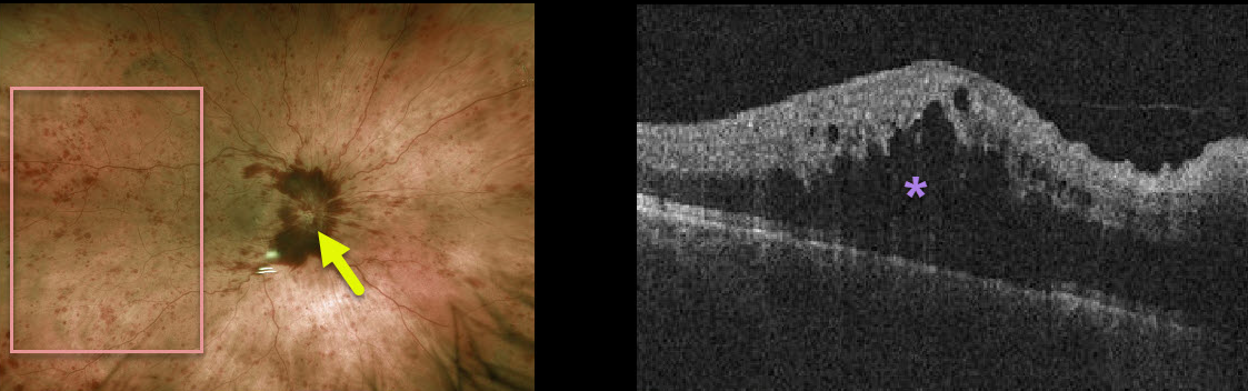
CRVO
Originally posted on @retina.rocks January 7, 2020
This patient has a non-ischemic central retinal vein occlusion (CRVO) with macular edema.
The fundus photo shows diffuse intraretinal hemorrhages, disc hemorrhages, and optic disc edema of the right eye. The OCT demonstrates marked macular edema.
Learning Points:
About 10% of patients with CRVO will develop bilateral occlusions. Eighty percent of occlusions are non-ischemic, although about one-third of these will convert to ischemic occlusions within several years.
About 60% of ischemic occlusions will develop anterior segment neovascularization with neovascular glaucoma. For some reason, posterior segment neovascularization rarely occurs (see the classic Hayreh 1983 paper Ophthalmology 1983;90;488-506).
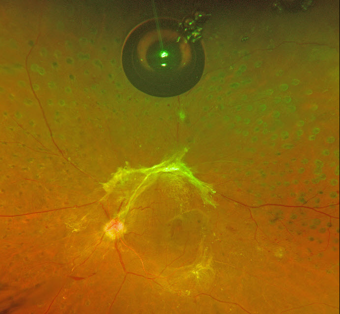
SILICONE OIL DROPLET
Originally posted on @retina.rocks January 2, 2020
This patient with proliferative diabetic retinopathy with tractional detachment was noted to have a large asymptomatic silicone oil bubble following multiple prior Avastin injections.
Learning Points:
The older insulin syringe needles provided by compounding pharmacies for the Avastin (bevacizumab) injections were lubricated with silicone.
Many eyes receiving intravitreal Avastin developed small vitreous silicone droplets, although the droplet is unusually large in this patient. Since then, these insulin syringe needles have been discontinued.
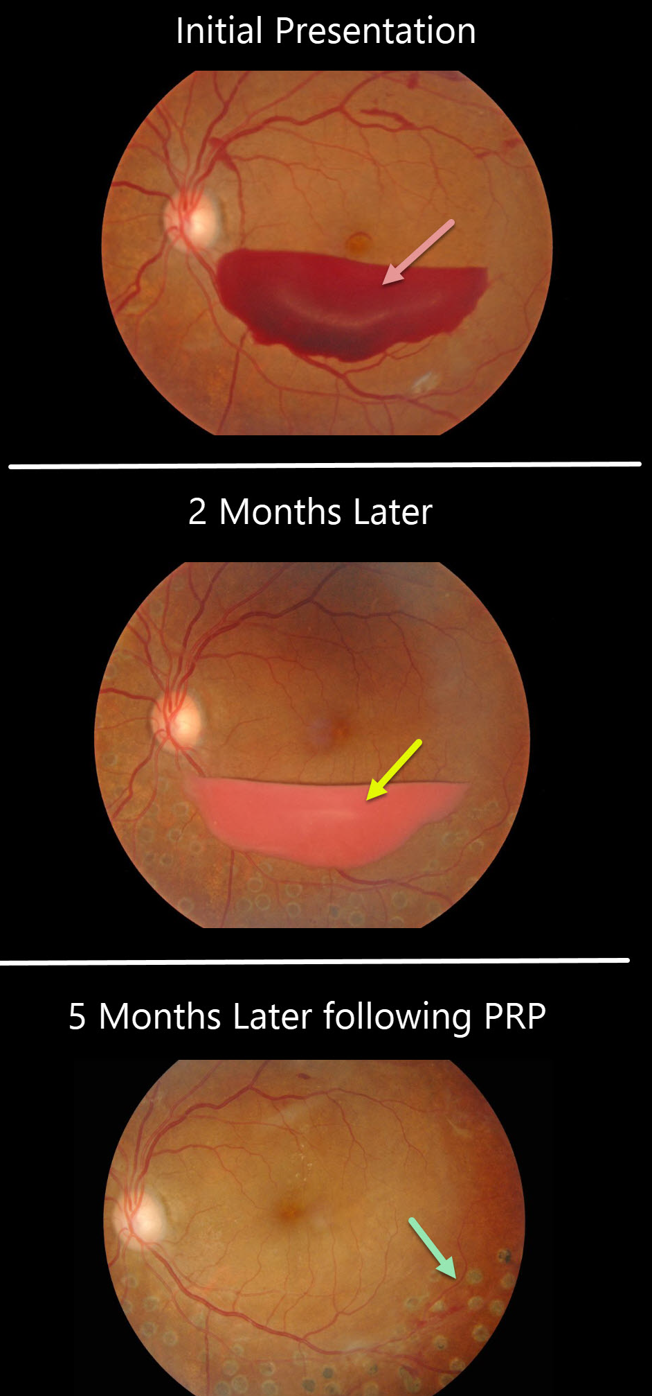
PROLIFERATIVE DIABETIC RETINOPATHY
Originally posted on @retina.rocks December 24, 2019
This patient presented with a sub-hyaloid hemorrhage due to proliferative diabetic retinopathy.
A subsequent photo two months later shows devitalized (yellowing) sub-hyaloid hemorrhage. He received panretinal photocoagulation (PRP) in the left eye and the sub-hyaloid hemorrhage completely resolved three months later. Anti-VEGF therapy was not used.
Learning Points:
PRP has been around since the 1960s and still remains the mainstay for treating proliferative disease. Often considered a “one and done,” patients can still develop break-through hemorrhages. Intravitreal anti-VEGF injections are often helpful, but some will require vitrectomy.


