

Torpedo Maculopathy (RPE Nevus)
<< Back to Cases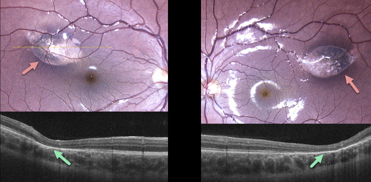
BILATERAL TORPEDO MACULOPATHY
Mattie Adams
Originally posted on @retina.rocks October 2, 2025
This 10YO boy was referred for asymptomatic retinal lesions. He was adopted, so the family history was unknown. The patient and guardian reported a longstanding history of persistent diarrhea that at times required hospitalization. Vision was 20/20 bilaterally.
Color photography shows bilateral torpedo-shaped areas of depigmentation in each superotemporal macula. The lesions were hypo-autofluorescent (not shown). OCT scanning shows outer retinal atrophy through each lesion.
Learning Points:
Torpedo maculopathy was originally described by Gass as a solitary hypopigmented nevus of the RPE (Arch Ophthalmology 1992;110:1358-1359). These benign lesions are teardrop-shaped and are almost always located in the horizontal meridian in the temporal macula. Vision is almost always normal. There is a small risk of macular neovascularization.
The lesions are virtually always unilateral, and we found only a single case report of bilateral lesions in a PubMed search (Richez et al., J Fr Ophthalmol 2010;33:296). Given our patient’s GI symptoms and bilaterality, genetic testing was recommended, but he was unfortunately immediately lost to follow-up.
TORPEDO MACULOPATHY
Originally posted on @retina.rocks March 6, 2025
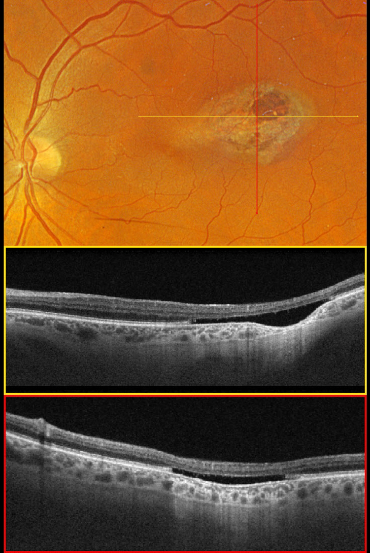
This 66YO male was referred for this unilateral, asymptomatic, variably pigmented lesion in his right eye. Vision was 20/30.
Optos color RGB imaging shows a torpedo-shaped lesion in the temporal macula. Triton swept-source OCT shows an outer retinal cavitation with loss of retinal tissue deep to the external limiting membrane. There is also focal choroidal thinning within the temporal aspect of the lesion.
Learning Points:
Torpedo maculopathy was originally described by Gass as a solitary hypopigmented nevus of the RPE (Arch Ophthalmology 1992;1358-1359). These benign, unilateral lesions are teardrop-shaped and almost always located along the horizontal meridian of the temporal macula. OCT scanning shows attenuation of the RPE and outer retinal layers. Wong et al classified these lesions into type 1 (‘mild outer retinal disturbance’) and type 2 with an outer retinal cavitation as seen in our patient (Clinical Exper Ophthalmology 2015;43:342-348). Vision is almost always normal, although there is a small risk for macular neovascularization. There are no known systemic associations.
TORPEDO MACULOPATHY
Originally posted on @retina.rocks October 14, 2024
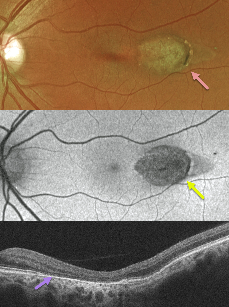
This healthy 44YO female was found to have this asymptomatic retinal finding in her left eye during a routine examination. The right eye was completely normal.
Optos color RG imaging shows a torpedo-shaped area of depigmentation in the temporal macula.
The lesion is hypo-autofluorescent. OCT scanning shows variable outer retinal and RPE atrophy.
Learning Points:
Torpedo maculopathy was originally described by Gass as a solitary hypopigmented RPE nevus (Arch Ophthalmology 1992;110:1358-1359). These benign, unilateral lesions are tear-drop shaped and are almost always located in the horizontal meridian in the temporal macula. Vision is almost always normal. There is a small risk of macular neovascularization.
TORPEDO MACULOPATHY
Zachary Morrison
Originally posted on @retina.rocks May 16, 2023
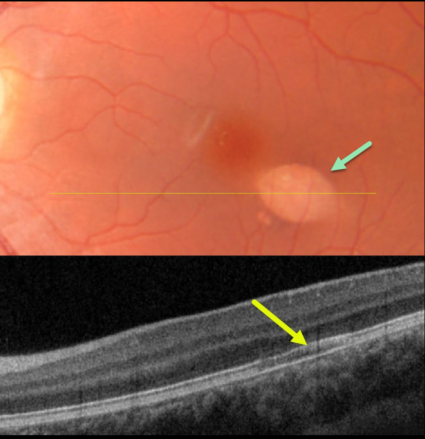
This healthy 12YO girl was found to have this asymptomatic retinal finding in her left eye during a routine examination. The right eye was completely normal.
Color imaging shows a torpedo-shaped area of depigmentation just inferotemporal to the left fovea. OCT scanning through this lesion shows an area of hyporeflectivity between the ellipsoid zone and RPE.
Learning Points:
Torpedo maculopathy was originally described by Gass as a solitary hypopigmented nevus of the RPE (Arch Ophthalmology 1992;110:1358-1359). These benign, unilateral lesions are teardrop-shaped and almost always located in the horizontal meridian of the temporal macula.
Our case is a bit atypical in that OCT scanning classically shows attenuation of both the RPE and outer retinal layers. Vision is almost always normal. There is a small risk of macular neovascularization.
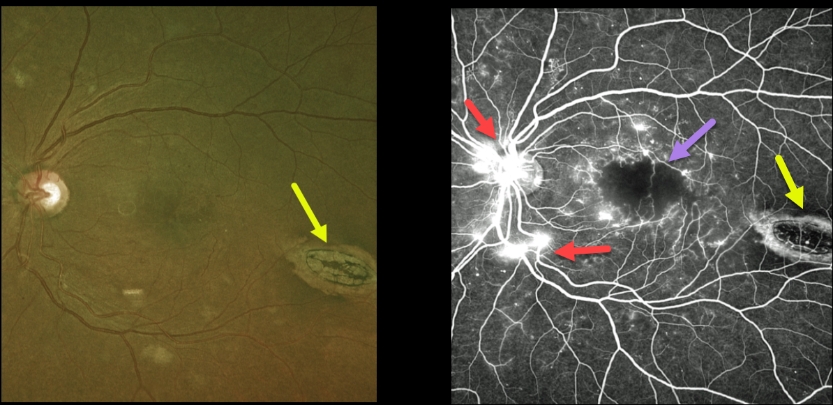
DIABETIC MACULAR ISCHEMIA WITH TORPEDO MACULOPATHY
Originally posted on @retina.rocks June 9, 2022
This 45YO female with type 2 diabetes presented with bilateral proliferative diabetic retinopathy (PDR). Vision in her left eye was 20/200 due to severe foveal ischemia, which is best seen on fluorescein angiography. Leaking disc and peripheral neovascularization are also seen angiographically. The macula was also edematous on OCT scanning (not shown).
An incidental finding completely unrelated to her diabetic retinopathy was an oval-shaped chorioretinal lesion in the distal inferotemporal macula known as torpedo maculopathy.
Although anti-VEGF therapy was started in her left eye for the macula edema, we doubt vision will significantly improve due to the foveal ischemia.
Learning Points:
Gass originally described torpedo maculopathy as a solitary hypopigmented nevus of the RPE (Arch Ophthalmology 1992;1358-1359). These benign, unilateral lesions are teardrop-shaped and almost always located in the horizontal meridian of the temporal macula.
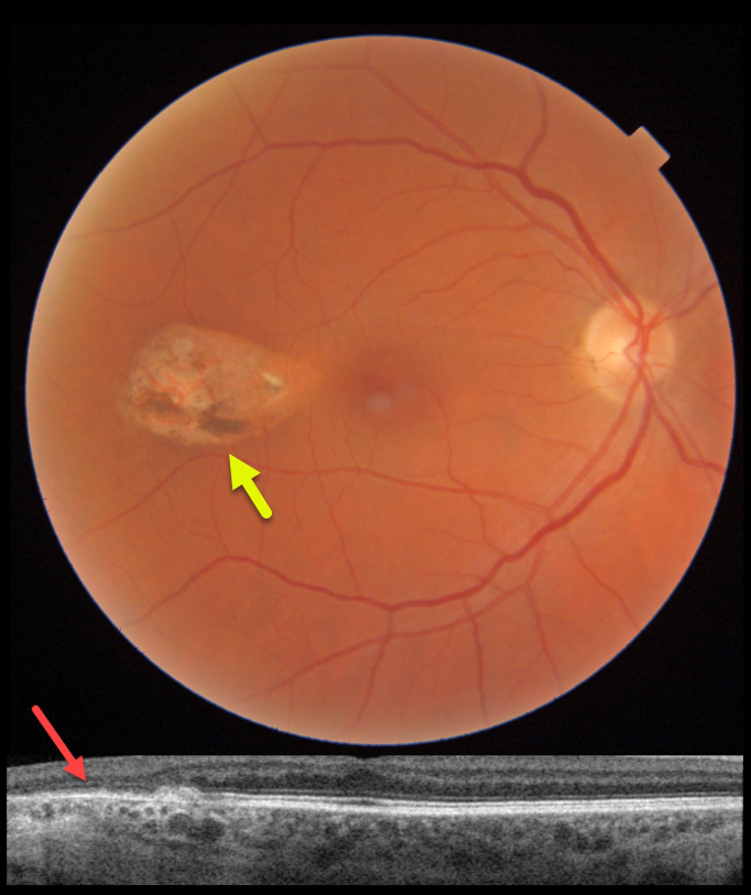
TORPEDO MACULOPATHY
Originally posted on @retina.rocks December 13, 2021
This 58YO female was referred for this unilateral, asymptomatic, variably pigmented lesion in her right eye. Vision was 20/25. Triton swept-source OCT shows outer retinal and RPE atrophy.
Learning Points:
Torpedo maculopathy was originally described by Gass as a solitary hypopigmented nevus of the RPE (Arch Ophthalmology 1992;1358-1359). These benign, unilateral lesions are teardrop-shaped and almost always located in the horizontal meridian in the temporal macula.
OCT scanning shows attenuation of the RPE and outer retinal layers. Vision is almost always normal, although there is a small risk of macular neovascularization. There are no known systemic associations.
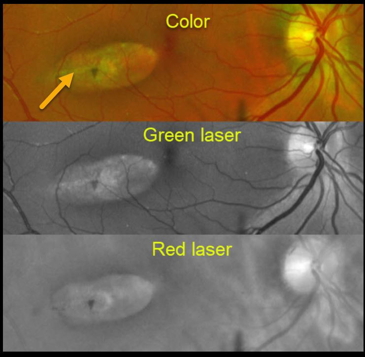
TORPEDO MACULOPATHY
Originally posted on @retina.rocks July 13, 2021
This patient has torpedo maculopathy.
This Optomap montage shows how the same lesion appears in color, green laser, and red laser.
OCT scanning, not available for this patient, would show attenuation of the RPE and outer retinal layers. Vision is almost always normal.
Learning Points:
Torpedo maculopathy was originally described by Gass as a solitary hypopigmented nevus of the RPE (Arch Ophthalmology 1992;1358-1359). These benign, unilateral lesions are teardrop-shaped and almost always located in the horizontal meridian of the temporal macula. There is a small risk of macular neovascularization.
These are congenital lesions with no systemic associations. For a great discussion to help differentiate torpedo maculopathy from other RPE lesions such as CHRPE and bear tracks, see Villegas et al, Ophthalmic Surg Lasers Imaging Retina 2014;45:222-226.
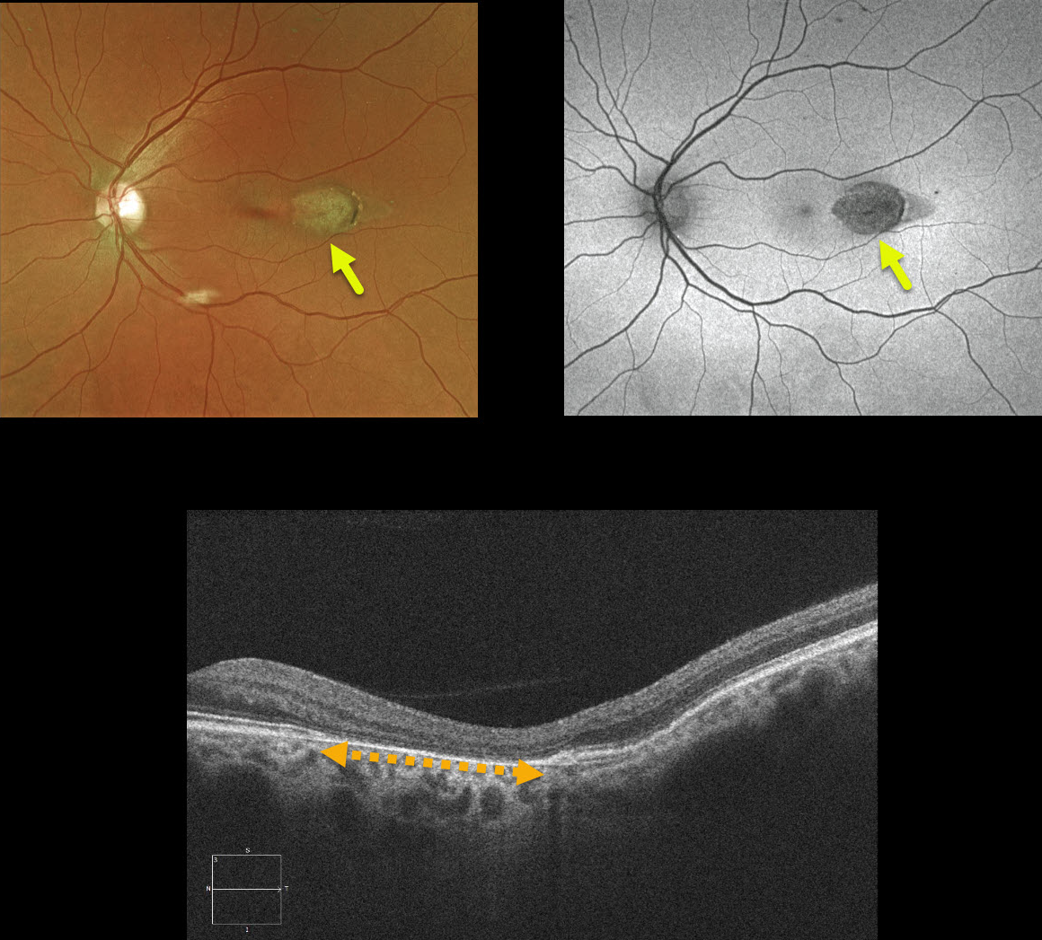
TORPEDO MACULOPATHY
Originally posted on @retina.rocks August 27, 2020
This patient has torpedo maculopathy, originally described by Dr. Gass as a solitary hypopigmented nevus of the RPE.
These benign, unilateral lesions are teardrop-shaped and almost always located in the horizontal meridian of the temporal macula. OCT shows attenuation of the RPE and outer retinal layers.
Vision is almost always normal.
Learning Points
These torpedo lesions are congenital with no systemic associations. Congenital hypertrophy of the RPE and Gardner’s syndrome-associated RPE lesions can mimic torpedo maculopathy.
See Villegas et al, Ophthalmic Surg Lasers Imaging Retina 2014;45:222-226, for a great discussion on distinguishing these RPE lesions.


