

Toxemia
<< Back to Cases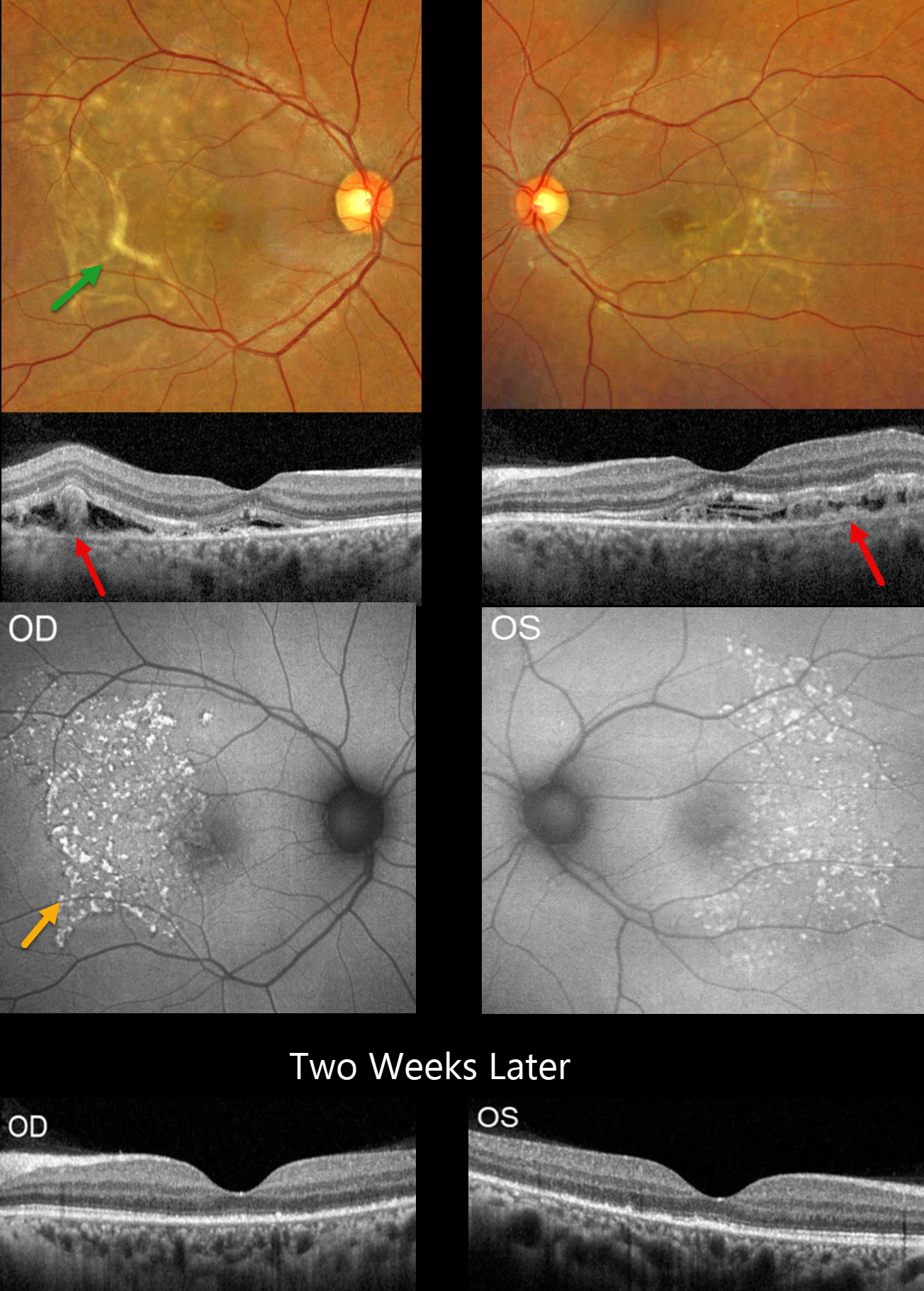
TOXEMIA
The European VitreoRetinal Society (EVRS), Berk Onaral and Ece Özdemir Zeydanlı
Originally posted on @retina.rocks May 1, 2026
This 31YO postpartum female presented with sudden vision loss 3 days following delivery, which was complicated by severe preeclampsia (blood pressure was up to systolic 215 mmHg). Vision was 20/50 bilaterally.
Optos color RG imaging shows bilateral subretinal yellow bands and drusen-like changes. OCT shows disruption of the outer retinal bands with variable amorphous hyperreflective material and fluid. The RPE is variably thinned/absent as indicated by a visible Bruch’s membrane band. Fundus autofluorescence (FAF) shows extensive hyperreflective deposits in the temporal maculae.
Two weeks later, OCT shows resolved fluid with some restoration of the outer photoreceptor architecture.
Learning Points:
Posterior segment findings in toxemia of pregnancy, which develop in up to one-third of patients, are primarily due to choroidal ischemia with secondary RPE dysfunction and exudative retinal detachments. Outer retinal ischemia can cause bizarre and pathognomonic OCT changes as seen in our patient (Song et al, Graefes Arch Clin Exp Ophthalmol 2013;251:2647-2650).
The clinical and OCT findings recover remarkably within weeks following normalization of blood pressure. Upon resolution, the fundi can appear normal or reveal telltale subretinal pigment clumps (Elschnig spots), streaks (Siegrist streaks), or broad areas of RPE pigmentary changes with intraretinal migration that can mimic prior blunt trauma or rhegmatogenous retinal detachment.
TOXEMIA
Nilesh Kumar
Originally posted on @retina.rocks January 21, 2026
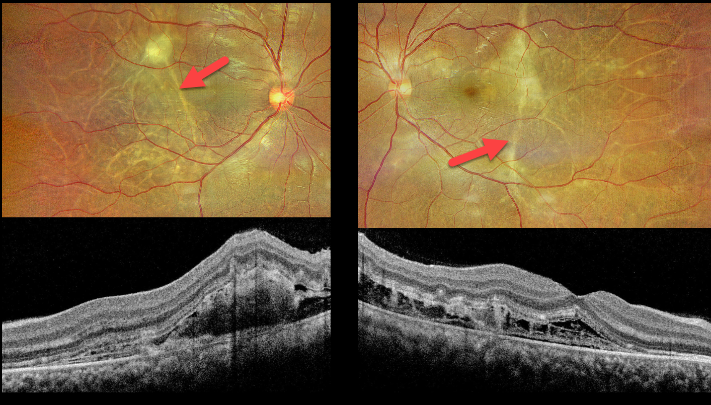
This 25YO female presented 10 days postpartum, complaining of bilateral blurred vision that began shortly following delivery, which was complicated by toxemia (preeclampsia). Blood pressure in our office was 180/90 mmHg. Vision was 20/30 bilaterally.
Optos color RG imaging shows multiple depigmented, wormlike subretinal tracks throughout each posterior pole. A larger contracting triangular-shaped lesion is noted in the superior left macula. Exudative fluid is present in the nasal maculas. OCT scanning shows outer retinal disorganization, an increased number of outer retinal bands, and pockets of variably hyperreflective fibrinous outer retinal fluid.
Learning Points:
Posterior segment findings in toxemia of pregnancy, which develop in up to one-third of patients, are primarily due to choroidal ischemia with secondary RPE dysfunction and exudative retinal detachments. Outer retinal ischemia can cause bizarre and pathognomonic OCT changes as seen in our patient (Song et al, Graefes Arch Clin Exp Ophthalmol 2013;251:2647-2650).
The clinical and OCT findings recover remarkably within weeks following normalization of blood pressure. Upon resolution, the fundi can appear normal or reveal telltale subretinal pigment clumps (Elschnig spots), streaks (Siegrist streaks), or broad areas of RPE pigmentary changes with intraretinal migration that can mimic prior blunt trauma or rhegmatogenous retinal detachment.
TOXEMIA
Originally posted on @retina.rocks November 9, 2020
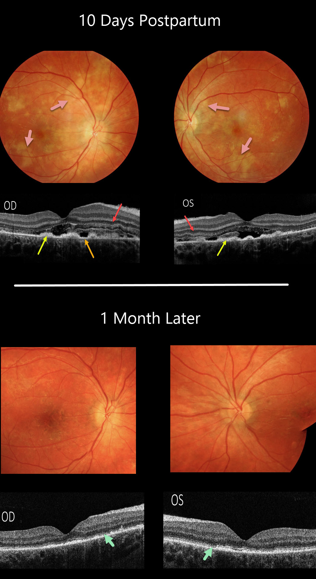
This patient was toxemic before delivery, with rapid bilateral vision loss and severely elevated blood pressure. When we examined her 10 days postpartum, her vision was counting fingers bilaterally.
There are multiple patches of creamy white, ischemic RPE and outer retina scattered throughout both posterior poles.
OCT scanning shows normal inner retinal bands extending from the internal limiting membrane to the inner nuclear layer, but the photoreceptor layers showed multiple additional hypo- and hyper-reflective bands, outer retinal disorganization, retinal edema, thickened RPE, and RPE loss with visible Bruch’s membrane.
One month later, vision remarkably improved to 20/25 bilaterally. The creamy multifocal lesions resolved, with residual pigmentary changes.
The prior OCT outer retinal changes resolved, with residual outer retinal and RPE atrophy.
Learning Points:
Toxemia causes choroidal ischemia with secondary RPE dysfunction, which can cause exudative retinal detachments.
Acute outer retinal ischemia causes these transient, bizarre, and pathognomonic OCT changes.
TOXEMIA
Originally posted on @retina.rocks October 7, 2020
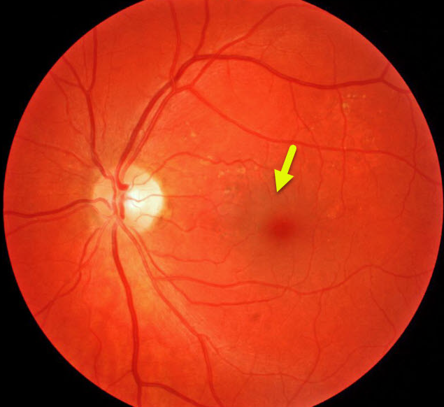
Our patient was referred for asymptomatic macular pigmentary changes. Vision was 20/20 OD and 20/25 OS. On further questioning, she reported severe vision loss with toxemia just before her now 16 YO daughter was born.
Learning Points:
Preeclampsia (toxemia of pregnancy), which usually develops in the third trimester, manifests as severe hypertension, swelling of the legs, feet, and hands, and proteinuria. When seizures develop, the condition is called eclampsia. This may lead to serious and possibly fatal effects for both the mother and child.
Patients may develop hypertensive retinopathy. Choroidal ischemia can cause infarction of the RPE with exudative and hemorrhagic retinal detachment.
These changes usually resolve rapidly with delivery, although they can cause chorioretinal changes that mimic numerous entities, including central serous retinopathy and macular/retinal dystrophies.


