

Toxocara
<< Back to Cases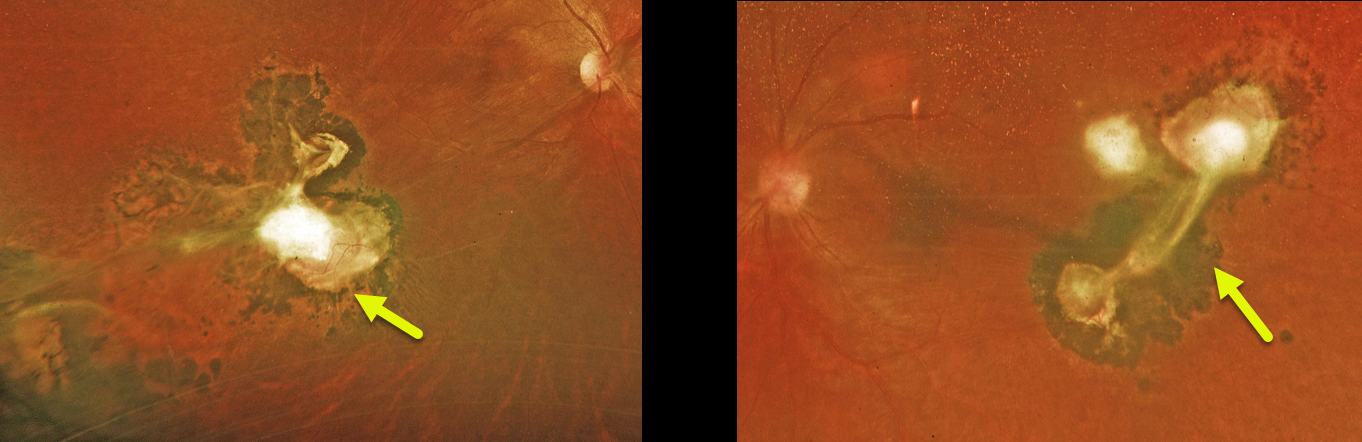
TOXOCARA
Matt Meredith
Originally posted on @retina.rocks October 23, 2024
This healthy 29YO female was referred for asymptomatic bilateral retinal findings. Vision was 20/40 OU.
Optos color RG imaging shows a large area of variably atrophic and fibrotic subretinal scarring inferotemporally OD, with a smaller area of scarring temporally OS. The scarring in her left eye has a more triangular appearance. Toxocara canis ELISA bloodwork was positive.
Learning Points:
Toxocariasis is a rare infection caused by the toxocara canis roundworm, which matures in the dog intestine. The eggs are passed in the stool, which can then be ingested when young children come into contact with infected sand or dirt and then touch their mouth. The ingested egg matures into a larva, which enters the systemic circulation from the intestine and can migrate into the eye. The death of the organism can cause variable findings, including endophthalmitis (typically a completely white, quiet eye), posterior granuloma, or peripheral granuloma.
Most commonly in our experience, they present as a triangular-shaped area of vitreous base organization/scarring, with a fold of retina extending more posteriorly. Since there is usually a single larva, the eye findings are almost always unilateral. Our case is therefore quite unusual in its bilaterality.
TOXOCARA
Will Gibson
Originally posted on @retina.rocks March 23, 2023
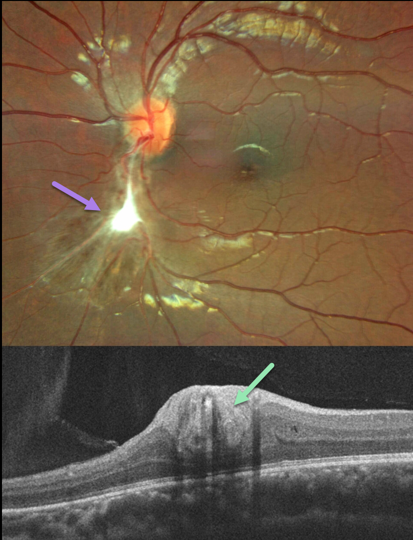
This healthy 13YO boy presented with a history of reduced vision in the left eye for 3 months. At the time of onset, he also reported having a red eye and headache.
Vision was 20/20 in his normal right eye and 20/70 in his left eye. Optos color imaging shows a triangular area of chorioretinal scarring extending inferiorly from the optic nerve. White fibrotic inner retinal scarring is noted more centrally.
OCT scanning through this region shows a thickened, featureless, hyperreflective inner retina.
After examination, further questioning revealed he was around multiple dogs at home, one of which had recalcitrant worms. Toxocara IgG antibody testing was surprisingly negative.
Learning Points:
Toxocariasis is a rare infection caused by the Toxocara canis roundworm, which matures in the dog intestine. The eggs are passed in stool, which can be ingested when young children come into contact with infected sand or dirt and then touch their mouths.
The ingested egg matures into a larva, which enters the systemic circulation from the intestine and can then migrate into the eye.
Death of the organism can cause variable findings, including endophthalmitis (typically a completely white and quiet eye), posterior granuloma, or peripheral granuloma. Most commonly in our experience, they present as a triangular-shaped area of vitreous base organization/scarring, with a fold of retina extending more posteriorly.
A positive test can help establish the diagnosis, although a false-negative result may occur, as sensitivity depends on the site of infection, the level of parasitic burden, and the timing of sample collection relative to exposure.
In our patient, despite the negative antibody testing, we still feel the history and clinical findings are most consistent with Toxocara. Other possible diagnoses include a combined hamartoma and posterior persistent fetal vasculature.
Regardless of the diagnosis, we expect the macular appearance to remain stable going forward and not require any treatment.
TOXOCARA
Originally posted on @retina.rocks June 8, 2021
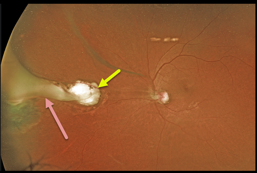
This 63YO female was referred for these asymptomatic unilateral fundoscopic changes. Optos imaging shows a midperipheral atrophic chorioretinal scar. Associated with this scar is a whitish fold of retina that extends anteriorly as a broader area of traction and scarring in the vitreous base.
A positive ELISA test can help to establish the diagnosis. Since our patient was asymptomatic, we did not pursue any further workup and recommended observation.
Learning Points:
Toxocariasis is a rare infection caused by the Toxocara canis roundworm, which matures in the dog intestine. The eggs are passed in stool, which can be ingested when young children come into contact with infected sand or dirt and then touch their mouths.
The ingested egg matures into a larva, which enters the systemic circulation from the intestine and can then migrate into the eye.
Death of the organism can cause variable findings, including endophthalmitis (typically a completely white and quiet eye), posterior granuloma, or peripheral granuloma.
Most commonly, in our experience, they present later in life as a triangular-shaped area of vitreous base organization/scarring, with a fold of retina extending more posteriorly.


