

Toxoplasmosis
<< Back to Cases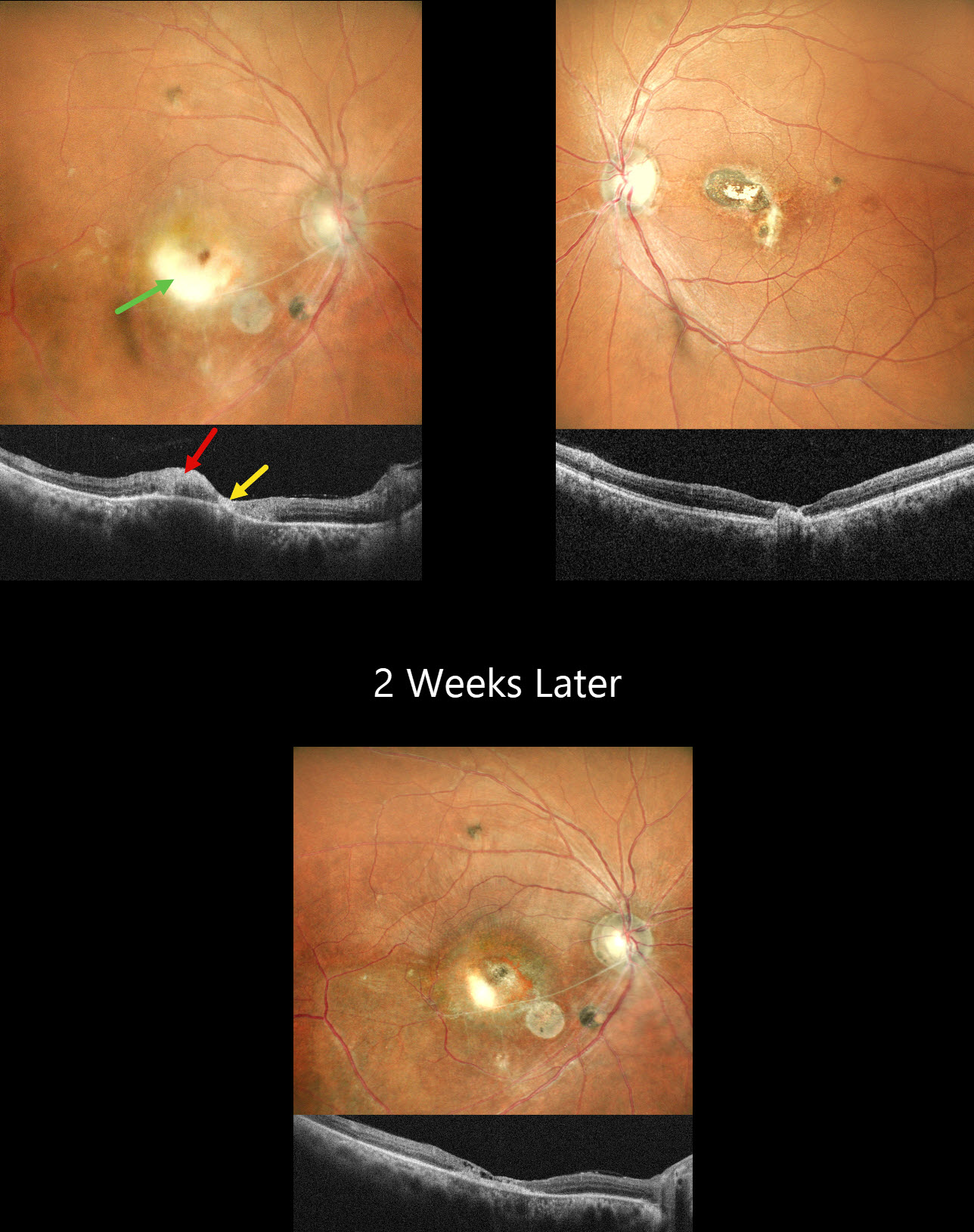
TOXOPLASMOSIS
Shraddha Raj Shrivastava, Akansha Sharma and Manish Nagpal
Originally posted on @retina.rocks March 31, 2026
This healthy 45YO male presented with a 3-week history of decreased vision in his right eye. He reported a similar episode in the left eye 10 years earlier, with poor vision since. Vision was 20/120 OD and 20/200 OS.
Pseudocolor SLO imaging OD shows active white retinitis adjacent to inactive nasal scarring. The vessels appear sclerosed in the inferonasal macula. OCT shows hyperreflectivity within the area of active retinitis with atrophy within the nasal scarring. The left eye has inactive nasal macular scarring. He was diagnosed with recurrent toxoplasmosis and started on a 6-week course of oral Bactrim DS. Oral steroids were also started 3 days later. When he returned 2 weeks later, he was unchanged at 20/120 with resolution of the active retinitis.
Learning Points:
Toxoplasmosis, the most common cause of infectious chorioretinitis in humans, is caused by the protozoan parasite, Toxoplasma gondii. It is usually transmitted through infected felines or by eating raw meat. Most infections are acquired, although it can also be transmitted congenitally. The body’s immune system is unable to completely kill the organism, which often lies dormant within a chorioretinal scar for years. When the immune balance favors the encysted organism, active chorioretinitis develops. Focal retinitis usually develops adjacent to a pigmented retinochoroidal scar. Overlying retinal arterial vasculitis is not uncommon.
The inflammation usually resolves spontaneously within 6 weeks. If the optic nerve or macula are threatened, as in our patient, a six-week course of Bactrim DS is recommended (Soheilian et al, Ophthalmology 2004;112:1876-1882). Oral prednisone can be added as well to help with severe inflammation.
TOXOPLASMOSIS
Tejaswita Verma and Manish Nagpal
Originally posted on @retina.rocks March 19, 2025
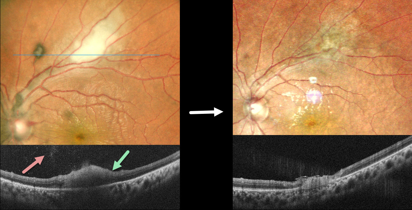
This 26YO male presented with 6 days of blurred vision in his left eye. Vision was 20/20 in his normal OD and 20/40 OS. The left cornea was edematous with a moderate nongranulomatous anterior uveitis and moderate vitreous cells. The intraocular pressure (IOP) was 47 mm HG.
Pseudocolor SLO imaging shows active white retinitis just superior to the macula. OCT scanning shows full-thickness hyperreflective retinal thickening with moderate overlying vitreous cells. He was diagnosed with toxoplasmosis panuveitis and started on a 6-week course of oral Bactrim DS. Oral steroids were also started 3 days later. The elevated IOP was aggressively treated with topical drops and oral acetazolamide. Six weeks later, vision improved to 20/30, and the IOP was 12. The uveitis and retinitis resolved, leaving behind inactive chorioretinal scarring.
Learning Points:
Toxoplasmosis, the most common cause of infectious chorioretinitis in humans, is caused by the protozoan parasite, Toxoplasma gondii. It is usually transmitted through infected felines or by eating raw meat. Most infections are acquired, although they can also be transmitted congenitally. The body’s immune system is unable to completely kill the organism, which often lies dormant within a chorioretinal scar for years. When the immune balance favors the encysted organism, active chorioretinitis develops. Focal retinitis usually develops adjacent to a pigmented retinochoroidal scar. Overlying retinal arterial vasculitis is not uncommon.
The inflammation usually resolves spontaneously within 6 weeks. If the optic nerve or macula is threatened, as in our patient, a six-week course of Bactrim DS is recommended (Soheilian et al, Ophthalmology 2004;112:1876-1882). Oral prednisone can be added as well to help with severe inflammation.
TOXOPLASMOSIS WITH RETINAL CHOROIDAL ANASTOMOSIS
Originally posted on @retina.rocks October 21, 2024
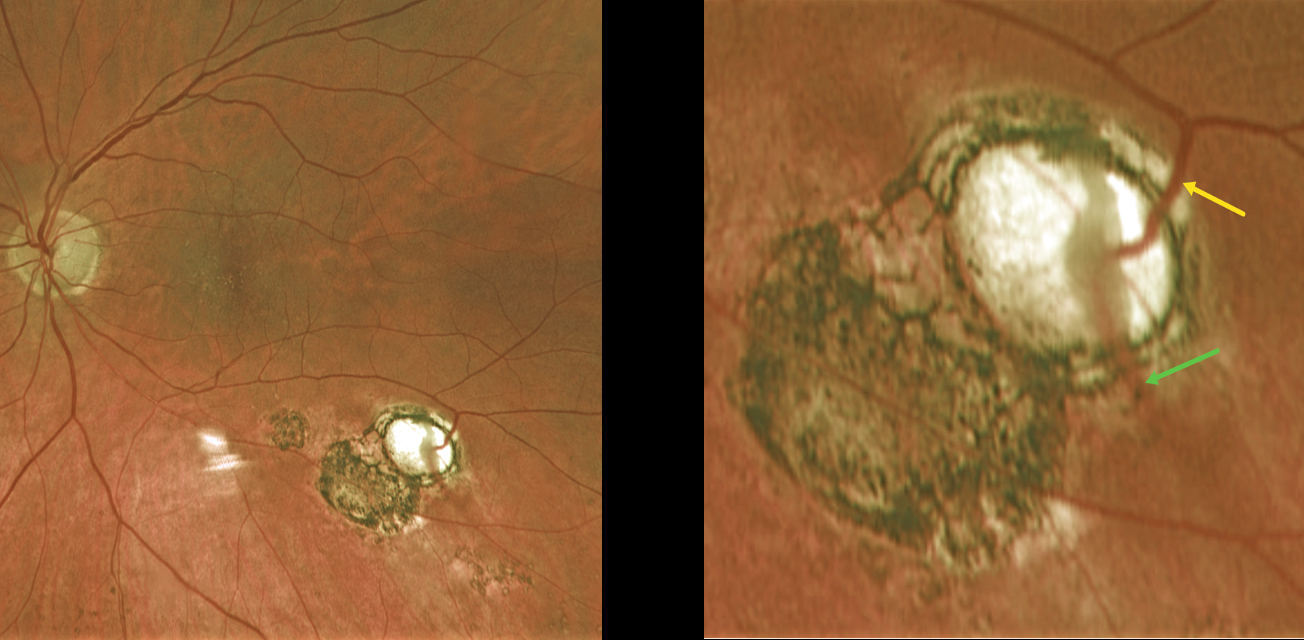
This 60YO female was referred for asymptomatic scarring in her left eye. Vision was 20/50 OD and 20/40 OS due to early bilateral cataracts. Small macular drusen were noted bilaterally.
Optos color RG imaging of her left shows multifocal variably pigmented scars below the inferotemporal arcade. A magnified inferotemporal view shows a retinal vein diving into an atrophic scar and directly connecting to a choroidal vein.
Learning Points:
Under normal conditions the retinal and choroidal circulations remain separate and distinct. Rarely, the two circulations directly connect with each other through a chorioretinal anastomosis. These can occur in chorioretinal scars (typically from inactive toxoplasmosis lesions as in this patient), in macular telangiectasia, and in disciform scars. We felt our patient’s findings most consistent with inactive toxoplasmosis scarring.
TOXOPLASMOSIS
Originally posted on @retina.rocks August 24, 2023
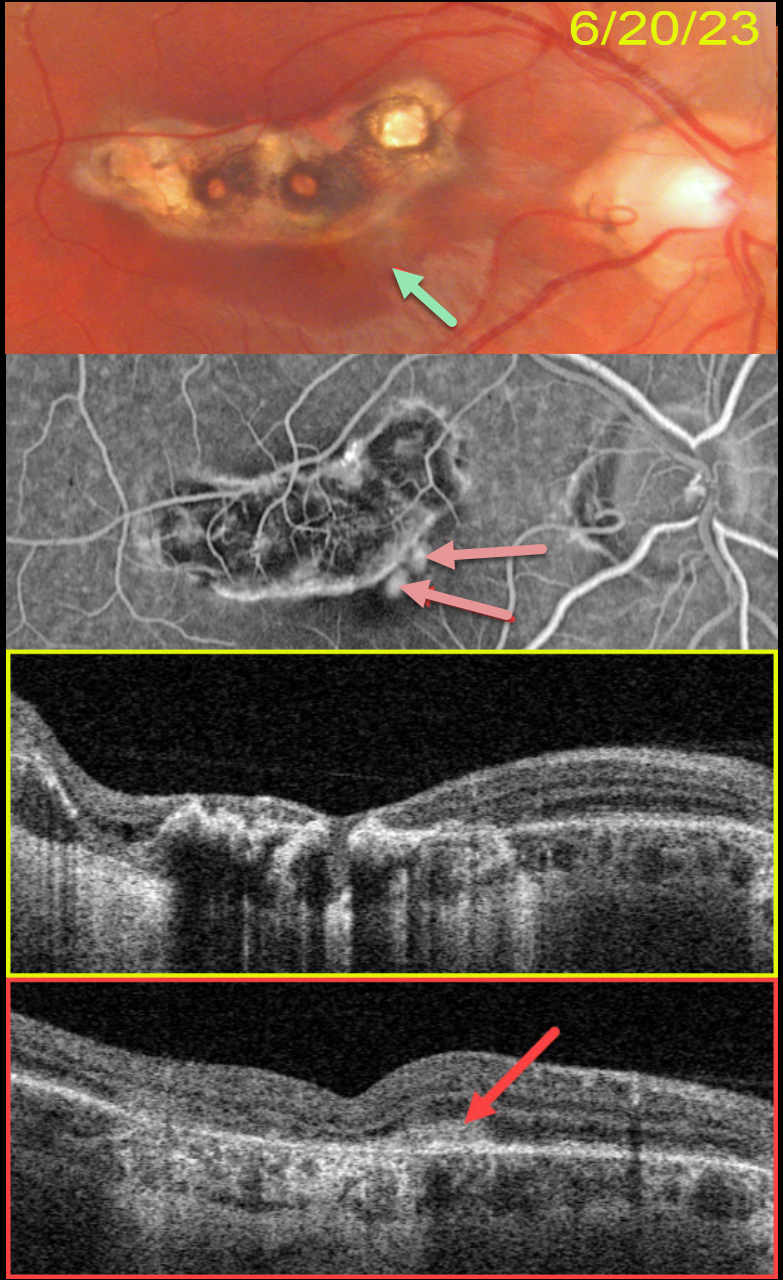
This 22YO male presented with several days of central blurred vision in his left eye. There was no prior ocular or medical history. Vision was 20/25 in his normal right eye and 20/25 in his left eye.
Color photography shows a variably pigmented comma-shaped colobomatous scar in the superior macula. A tiny depigmented scar is noted along its inferonasal edge. OCT scanning through this lesion showed no fluid (not shown).
He returned about 2 months later, complaining of increasing vision loss in his eye. Vision had decreased to 20/400. A faint area of subretinal pigment is now noted.
Optos fluorescein angiography shows leaking macular neovascularization (MNV) within this region. Swept-source OCT confirms a type 2 MNV (located above the RPE).
Intravitreal Avastin was injected.
Learning Points:
Virtually any disorder that affects the RPE-Bruch membrane layer can cause MNV, the most common being age-related macular degeneration, ocular histoplasmosis, idiopathic high myopia (lacquer cracks), trauma (choroidal rupture), and angioid streaks.
MYSTERY CASE
Asma Samsudeen and Ashish Sharma
Originally posted on @retina.rocks April 7, 2023
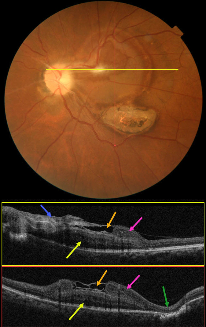
This 61YO male presented with 16 days of blurred vision and floaters in his left eye. There was no prior ocular history, and the past medical history was normal. Vision was 20/30 in his normal right eye and 20/200 in his left eye.
Color imaging shows an unusual circular area of subretinal yellow-orange pigmentary changes extending temporally from the optic nerve throughout the central and superior macula. A contracted localized area of premacular fibrosis is noted in the superonasal fovea. A flat area of variably pigmented chorioretinal scarring is found inferotemporally.
OCT B-scans show a constellation of interesting findings. The nasal epimacular fibrosis extends temporally, giving the appearance of either an ILM drape or local separation over the macular center. More temporally disorganized hyperreflective tissue descends and seems to merge into definite retinal inner tissue. There is complete outer retinal loss overlying the inferotemporal area of scarring.
The deeper retina is occupied by a mound-like area of alternating hyper- and hyporeflective bands. It is unclear whether this represents thickened outer retina or an unusual scar, as it appears distinct and anterior to the well-visualized ellipsoid zone below.
We believe this constellation of findings most likely represents changes following toxoplasmosis chorioretinitis, but we also can’t rule out an independent combined hamartoma. Or maybe we’re dealing with another diagnosis. What do you think?
TOXOPLASMOSIS
Originally posted on @retina.rocks December 19, 2022
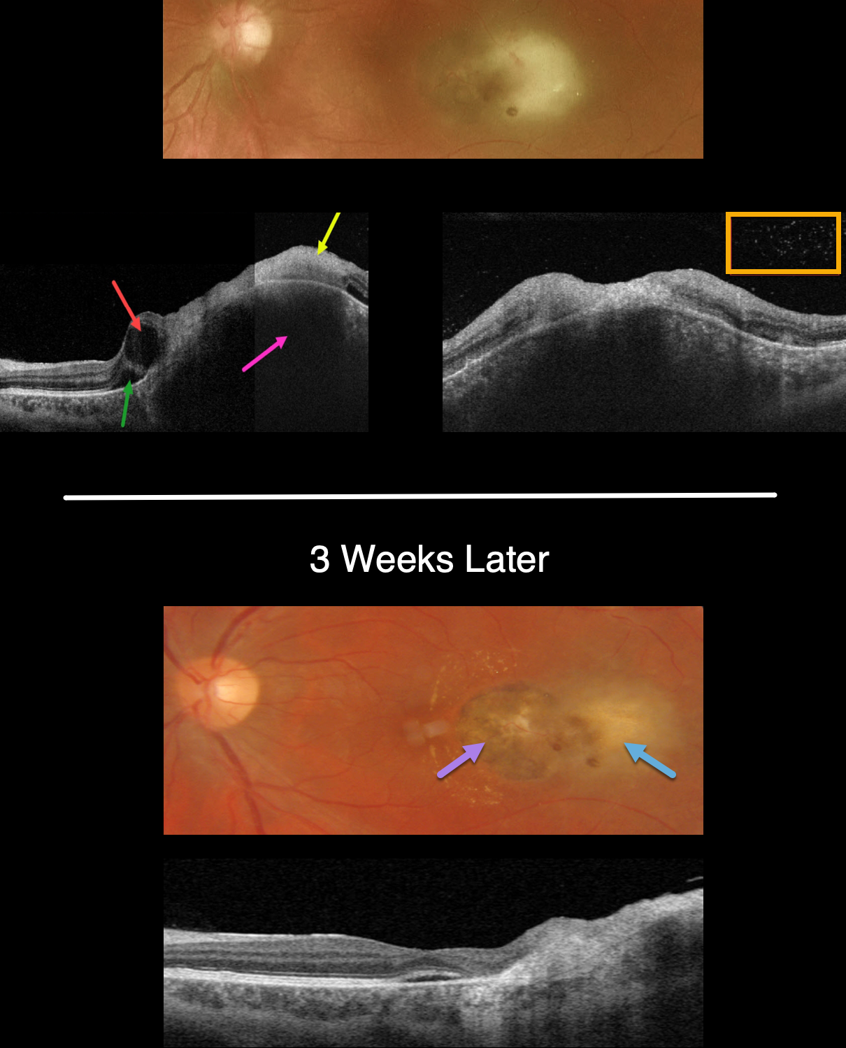
This 34YO male presented with recent vision loss of counting fingers vision in his left eye. He gave a history of prior ocular toxoplasmosis.
Optos color imaging shows opaque and white retina temporal to the fovea. Swept-source OCT shows the clinical retinitis involving full-thickness retina and is markedly hyperreflective. The underlying choroid is markedly thickened. The temporal fovea has a large cyst with a small amount of subretinal fluid. Overlying vitreous cells are noted.
The patient was started on a six-week course of oral Bactrim-DS BID. Laboratory testing for toxoplasmosis showed high IgG and negative IgM titers. ACE, lysozyme and QuantiFERON TB were negative.
The chorioretinal inflammation improved at each visit. When last seen 3 weeks into therapy, the likely pre-existing temporal pigmented toxoplasmosis scar is seen with resolving temporal retinitis. A few dots of retinal blood are seen, and faint lipid is noted nasally.
OCT scanning shows resolved vitreous cells and markedly decreased choroidal thickening.
Learning Points:
Toxoplasmosis, the most common cause of infectious chorioretinitis in humans, is caused by the protozoan parasite, Toxoplasma gondii.
It is most commonly transmitted through infected felines or by eating raw meat. The majority of infections are acquired, although it can also be transmitted congenitally.
The body’s immune system is unable to completely kill the organism, which often lies dormant within a chorioretinal scar for years. When the immune balance favors the encysted organism, active chorioretinitis develops. This usually resolves spontaneously within 6 weeks.
If the optic nerve or macular are threatened, a six-week course of Bactrim DS is recommended (see Ophthalmology 2004;112:1876-1882). Oral prednisone can be added as well to help with severe inflammation.
TOXOPLASMOSIS WITH RETINAL CHOROIDAL ANASTOMOSIS
Originally posted on @retina.rocks September 20, 2022
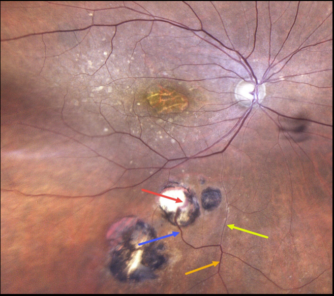
This 86YO female has a history of atrophic age-related macular degeneration with vision of 20/400 OD and 20/60 OS. Optos imaging of her right eye shows foveal atrophy with surrounding mixed drusen.
Multifocal inactive toxoplasmosis scars are noted inferiorly. An inferior retinal vein bifurcates, with a markedly attenuated branch continuing on its path towards the optic nerve. The other branch has a more normal caliber, dives into one of the toxoplasmosis scars, and drains into the choroid.
Learning Points:
Under normal conditions, the retinal and choroidal circulations remain separate and distinct. Rarely, the two circulations connect directly through a chorioretinal anastomosis.
These can occur in chorioretinal scars (typically from inactive toxoplasmosis lesions, as in this patient), in macular telangiectasia, and in disciform scars.
TOXOPLASMOSIS
Originally posted on @retina.rocks May 16, 2022
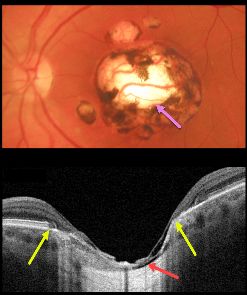
This 48YO male gave a history of lifelong poor vision in his left eye. Vision was 20/40 OD and 20/200 OS.
Triton color imaging shows a large, round, variably pigmented, excavated macular scar with a few smaller surrounding scars.
Swept-source OCT reveals a thinned atrophic retina and RPE overlying a shallow coloboma. Within the coloboma, the choroid is virtually absent, and the sclera is excavated. The yellow arrows mark the hyper-reflective plane of the surrounding intact retina and RPE. The right fundus was normal.
Learning Points:
For a great discussion on how to differentiate these infectious macular colobomatous lesions (congenital toxoplasmosis, Zika, and cytomegalovirus) from inherited dystrophies like North Carolina Macular Dystrophy (NCMD), see Kumar and Mahalingam AJO 2019;200:47-56.
TOXOPLASMOSIS
Originally posted on @retina.rocks May 4, 2022
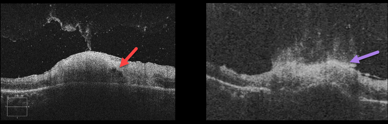
This is a great case of active toxoplasmosis.
The OCT B-scans show full-thickness retinitis with a volcanic-like eruption of the inflammatory process extending into the attached vitreous.
Learning Points:
Toxoplasmosis, the most common cause of infectious chorioretinitis in humans, is caused by the protozoan parasite, Toxoplasma gondii.
It is most commonly transmitted through infected felines or by eating raw meat. The majority of infections are acquired, although they can also be transmitted congenitally.
The body’s immune system is unable to completely kill the organism, which often lies dormant within a chorioretinal scar for years. When the immune balance favors the encysted organism, active chorioretinitis develops. This usually resolves spontaneously within 6 weeks.
If the optic nerve or macula is threatened, a six-week course of Bactrim DS is recommended (see Ophthalmology 2004;112:1876-1882).
Oral prednisone can be added as well to help with severe inflammation.
TOXOPLASMOSIS
Originally posted on @retina.rocks March 11, 2022
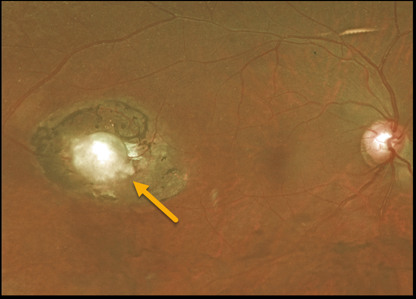
This 55YO female presented with 20/30 vision in her right eye and an asymptomatic colobomatous scar in her distal temporal macula. The left eye was normal.
We felt this lesion most likely represented an inactive toxoplasmosis scar.
Learning Points:
For a great discussion on how to differentiate these infectious macular colobomatous lesions (congenital toxoplasmosis, Zika, and cytomegalovirus) from inherited dystrophies, see Kumar and Mahalingam, AJO 2019;200;47-56.
TOXOPLASMOSIS
Originally posted on @retina.rocks August 12, 2021
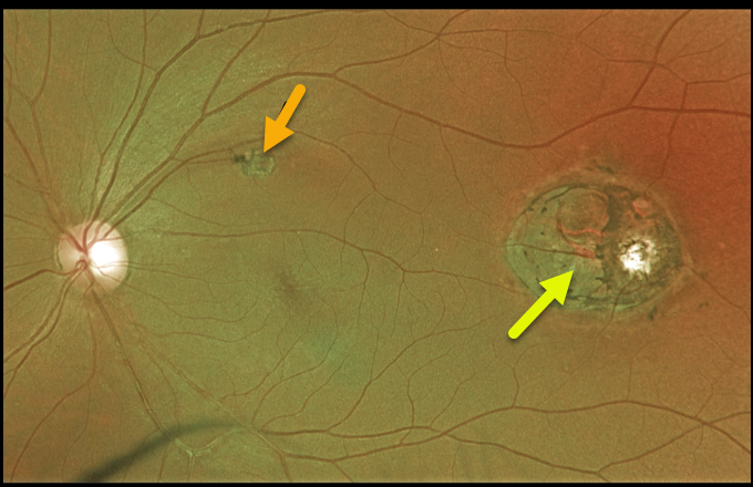
This 44YO male presented with 20/25 vision and a presumed inactive toxoplasmosis scar in the distal left macula. Although we do not have an OCT through this scar, it likely represents a colobomatous lesion.
This lesion somewhat resembles torpedo maculopathy, but the additional scar in the superior macula more suggests a prior inflammatory event.
Learning Points:
For a great discussion on how to differentiate these infectious macular colobomatous lesions (congenital toxoplasmosis, Zika, and cytomegalovirus) from inherited dystrophies like North Carolina Macular Dystrophy (NCMD), see Kumar and Mahalingam AJO 2019;200;47-56.
TOXOPLASMOSIS
Originally posted on @retina.rocks March 26, 2021
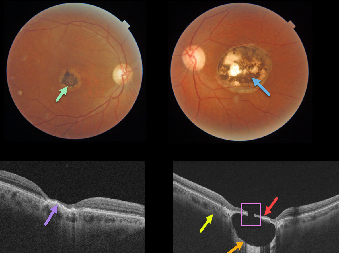
This 57YO male gave a history of lifelong poor vision in his left eye. Vision was 20/40 OD and 20/200 OS.
A small hyperpigmented foveal scar is noted in his right eye with neurosensory retinal atrophy and irregular elevation of the RPE.
A larger, round, variably pigmented, excavated macular scar is noted in his left eye. Triton swept-source OCT reveals a thinned atrophic retina and intact RPE suspended over an optically empty hyporeflective excavated colobomatous type lesion. The plane of the intact surrounding sclera is indicated by the yellow arrow. A small full-thickness retinal and RPE defect is noted centrally.
Learning Points:
For a great discussion on how to differentiate these infectious macular colobomatous lesions (congenital toxoplasmosis, Zika, and cytomegalovirus) from inherited dystrophies like North Carolina Macular Dystrophy (NCMD), see Kumar and Mahalingam AJO 2019;200;47-56.
In case you’re thinking our patient has NCMD, also see the first OCT description of congenital toxoplasmosis macular lesions (Garg et al, Retina 2009;29;631-637). Their Figures 4 and 6 look eerily similar to our patient’s right and left eyes!
TOXOPLASMOSIS
Originally posted on @retina.rocks January 12, 2021
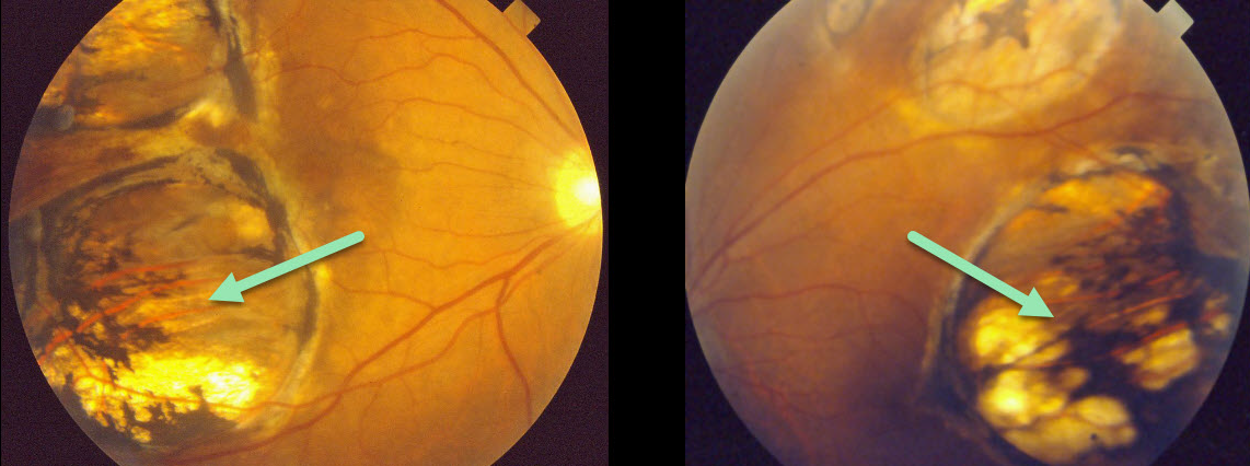
This patient presented in 1990 with what we assumed was bilateral inactive colobomatous congenital toxoplasmosis scarring.
Learning Points:
Choroidal colobomas are most commonly due to failure of the optic vesicle and choroidal fissure to close during fetal development. Multifocal lesions, as seen in our patient, can be caused by congenital toxoplasmosis and Zika.
For a comprehensive review of ocular coloboma, see Onwochei et al Surv Ophthalmol 2000;45:175-194.
TOXOPLASMOSIS
Originally posted on @retina.rocks November 16, 2020
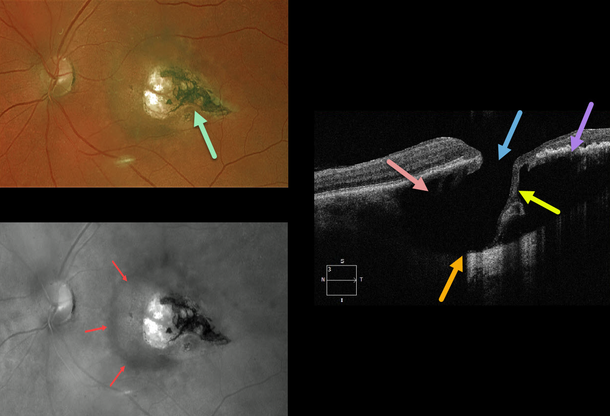
This 67YO female was referred with 20/30 vision and asymptomatic retinal findings. There was a tear-drop-shaped, variably pigmented chorioretinal scar in the temporal macula. The associated increased choroidal pigmentation is best seen in the red-channel image.
The OCT shows some unexpected findings. There is either a coloboma or posterior staphyloma causing marked posterior displacement of the sclero-choroidal junction. Temporally thinned retina dives posteriorly with an adjacent full-thickness retinal defect.
Temporally, the increased pigmentation of the scarring likely makes it difficult to see more posterior choroidal detail due to shadowing. Nasally, the faint, increased choroidal pigmentation also causes shadowing.
What do you think this is? Our best guess is a choroidal cavitation or an atypical colobomatous toxoplasmosis scar.
Learning Points:
For a great discussion on how to differentiate these infectious macular colobomatous lesions (congenital toxoplasmosis, Zika, and cytomegalovirus) from inherited dystrophies like North Carolina Macular Dystrophy (NCMD), see Kumar and Mahalingam AJO 2019;200:47-56. Also see the first OCT description of congenital toxoplasmosis macular lesions (Garg et al, Retina 2009;29;631-637).
TOXOPLASMOSIS
Originally posted on @retina.rocks November 13, 2020
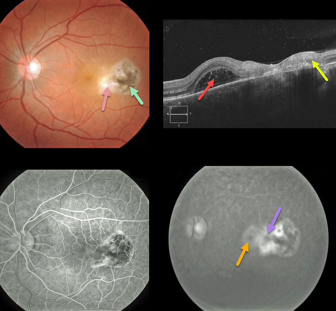
This patient presented with blurred vision from an area of active retinitis nasal to a prior toxoplasmosis scar.
An OCT B-scan shows full-thickness retinal necrosis and scarring temporally, with a foveal bacillary detachment. Tiny vitreous cells are scattered above the retina.
Fluorescein angiography shows staining of the inflamed retina and pooling within the bacillary detachment.
This patient was successfully treated with a 6-week course of oral Bactrim DS.
Learning Points:
Toxoplasmosis, the most common cause of infectious chorioretinitis in humans, is caused by the protozoan parasite, Toxoplasma gondii. It is most commonly transmitted through infected felines or by eating raw meat.
The majority of infections are acquired, although they can also be transmitted congenitally. The body’s immune system is unable to completely kill the organism, which often lies dormant within a chorioretinal scar for years.
When the immune balance favors the encysted organism, active chorioretinitis develops. This usually resolves spontaneously within 6 weeks.
If the optic nerve or macula is threatened, a six-week course of Bactrim DS is recommended (see Ophthalmology 2004;112;1876-1882). Oral prednisone can be added as well to help with severe inflammation.
TOXOPLASMOSIS
Originally posted on @retina.rocks June 23, 2020
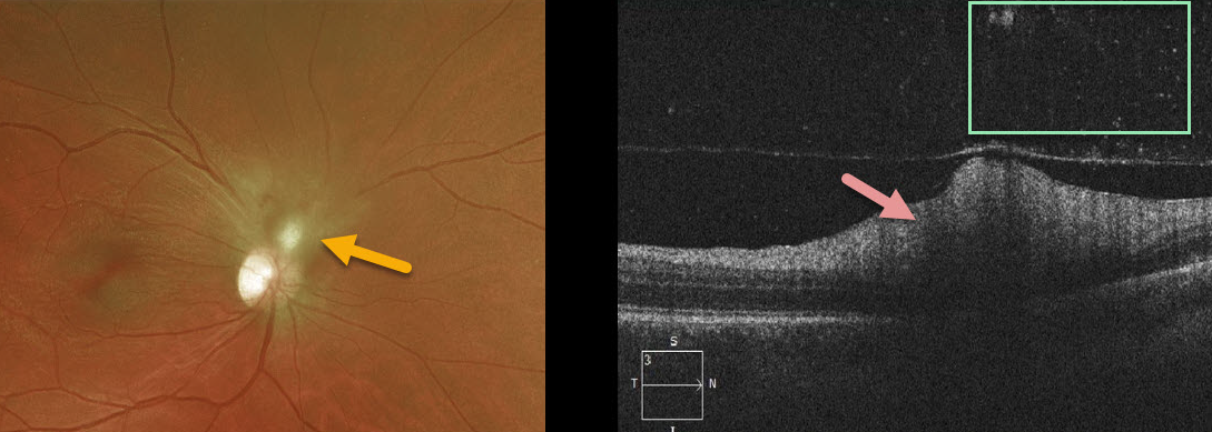
Our patient had active toxoplasmosis inflammation directly adjacent to the optic nerve and was placed on Bactrim double-strength (DS) and prednisone.
The OCT line scans show hyperreflective retinitis with overlying vitreous cells.
Learning Points:
Toxoplasmosis, the most common cause of infectious chorioretinitis in humans, is caused by the protozoan parasite, Toxoplasma gondii. It is most commonly transmitted through infected felines or by eating raw meat.
The majority of infections are acquired, although they can also be transmitted congenitally. The body’s immune system is unable to completely kill the organism, which often lies dormant within a chorioretinal scar for years.
When the immune balance favors the encysted organism, active chorioretinitis develops. This usually resolves spontaneously within 6 weeks.
If the optic nerve or macula is threatened, a six-week course of Bactrim DS is recommended (see Ophthalmology 2004;112;1876-1882). Oral prednisone can be added as well to help with severe inflammation.


