

Diabetic Retinopathy Tractional Retinal Detachment (TRD)
<< Back to Cases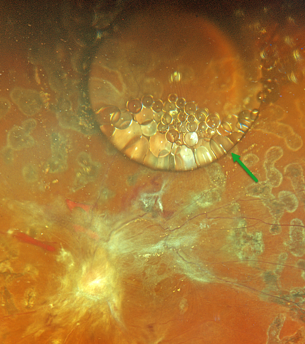
SILICONE FISH EGGS
Originally posted on @retina.rocks July 21, 2026
This 43YO male underwent a vitrectomy elsewhere for a diabetic traction retinal detachment about 5 months earlier. Vision was 20/400.
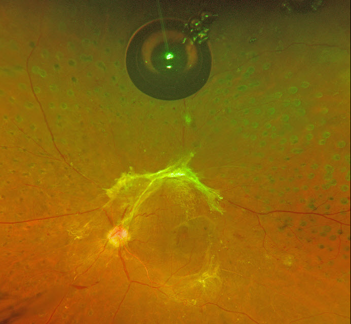
Optos color RGB imaging shows a large, round silicone oil droplet in the superior midperiphery. This bubble acts like a convex lens, magnifying the details of the underlying retina. Suspended within and along the edges of this droplet are numerous fish egg droplets. Residual fibrotic membranes extend from the nerve into the macula and along the superotemporal arcade. Repeat vitrectomy was scheduled.
TRACTIONAL RETINAL DETACHMENT WITH PSEUDO-GIANT MACULAR HOLE
The European VitreoRetinal Society (EVRS) and Shishir Verghese
Originally posted on @retina.rocks January 16, 2026
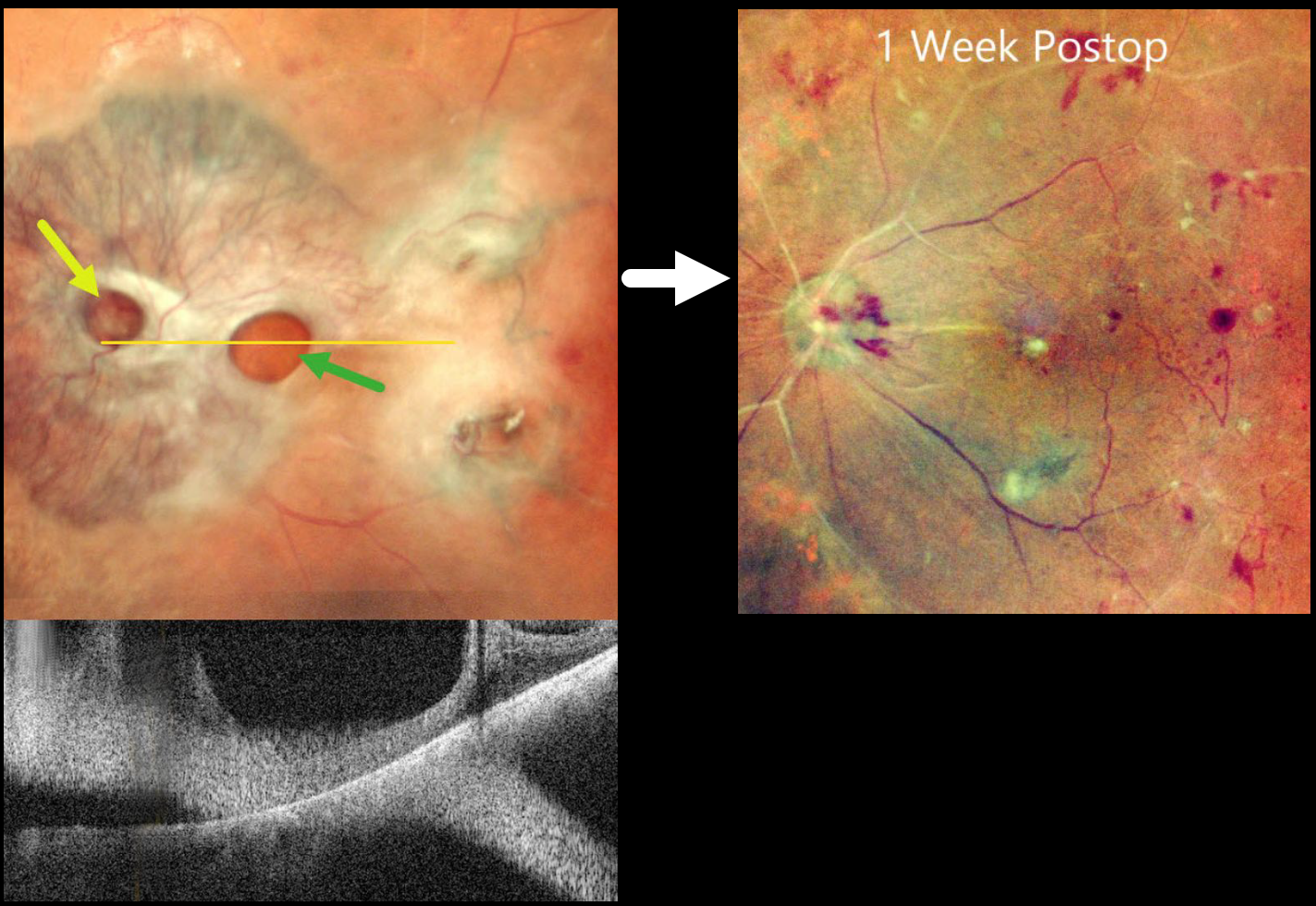
This 65YO diabetic female presented with counting fingers bilaterally from proliferative diabetic retinopathy with macular tractional retinal detachments.
Preoperative pseudocolor SLO imaging of her left eye shows a complex fibrovascular membrane covering the posterior pole. Pseudo giant-macular holes are noted overlying the disc and macula. OCT scanning confirms the lack of a true macular hole. One week following vitrectomy, membrane peeling, and air injection, vision improved to 20/200, and the retina was completely attached with no residual traction.
Learning Points:
A giant macular hole, also reported in Alport syndrome (Shah and Weinberg Ophthalmic Genetics 2010;31(2):93-97), is an exceedingly rare traumatic event (Hernandez-Da Mota Case Rep Ophthalmol 2011;2:283-286). The unusual anatomy of our patient’s fibrovascular diabetic membrane simulated a giant macular hole.
COMBINED TRACTIONAL-RHEGMATOGENOUS RETINAL DETACHMENT
The European VitreoRetina Society (EVRS) and Shishir Verghese
Originally posted on @retina.rocks September 19, 2025
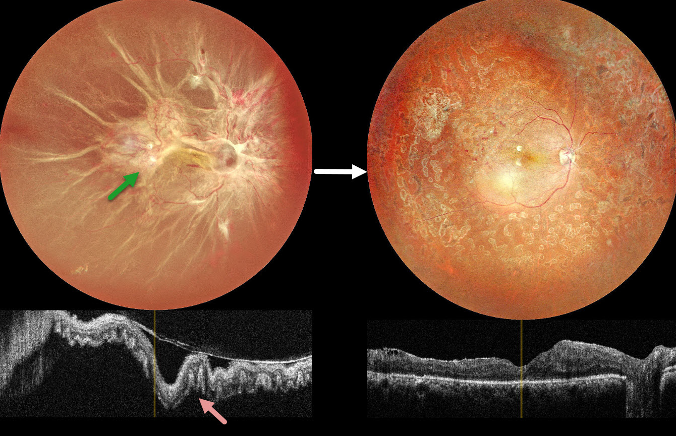
This 40YO female presented with vision loss in her right eye. There was a 15-year history of type 1 diabetes. Vision was hand motion OD.
Color photography shows a total combined traction-rhegmatogenous retinal detachment (RRD). There is a complex pattern of fibrovascular tissue covering the posterior pole with radiating retinal folds extending peripherally. OCT scanning shows a variably undulated detached retina with outer retinal corrugations.
Five days following an intravitreal anti-VEGF injection, she underwent pars plana vitrectomy with membrane peeling, endolaser, and silicone oil injection. Three months later, the silicone oil was removed. Postoperatively, vision improved to 20/80 with complete retinal reattachment.
Learning Points:
Outer retinal corrugations, also known as hydration outer retinal folds, are one of the key findings that help differentiate a RRD from exudative and traction retinal detachments. The cause of these folds remains uncertain but may result from the hydration of glycosaminoglycans in the interphotoreceptor matrix (Dalvin et al, Retinal Cases & Brief Reports 2020;14:355-359). This OCT biomarker confirms the rhegmatogenous component of our patient’s detachment.
COMBINED TRACTIONAL-RHEGMATOGENOUS RETINAL DETACHMENT WITH MACULAR HOLE
Sucheta Kulkarani, Anita Gaikwad and Anjana Mirajkar
Originally posted on @retina.rocks July 24, 2025
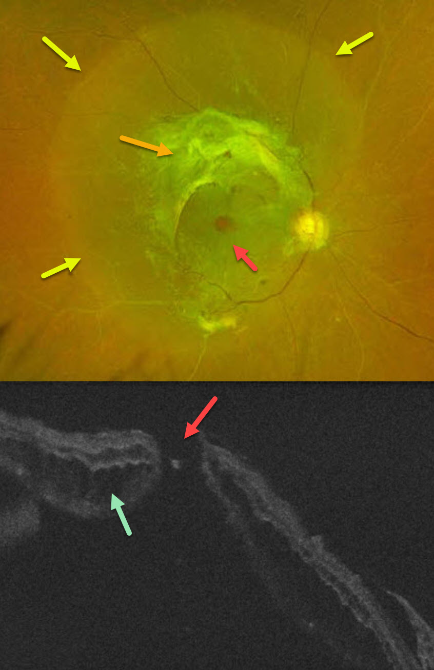
This 49YO male with type 2 diabetes noticed decreased vision in his right eye for 4 months. A single anti-VEGF injection was given elsewhere in his right eye around the time of his vision loss. Vision was 20/400 OD and 20/30 OS.
Optos color RG imaging OD shows a combined tractional-rhegmatogenous retinal detachment from dense fibrovascular proliferation and a full-thickness macular hole. OCT shows diffuse cystic edema within the detached retina, predominantly in the outer nuclear layer, and a macular hole.
Learning Points:
Tractional macular holes as a cause of combined tractional-rhegmatogenous diabetic retinal detachments are a rare event in our experience. Unfortunately, our patient was lost to follow-up immediately. We expect him to lose central vision in his left eye soon if he does not seek care.
TRACTIONAL RETINAL DETACHMENT
Originally posted on @retina.rocks January 29, 2025
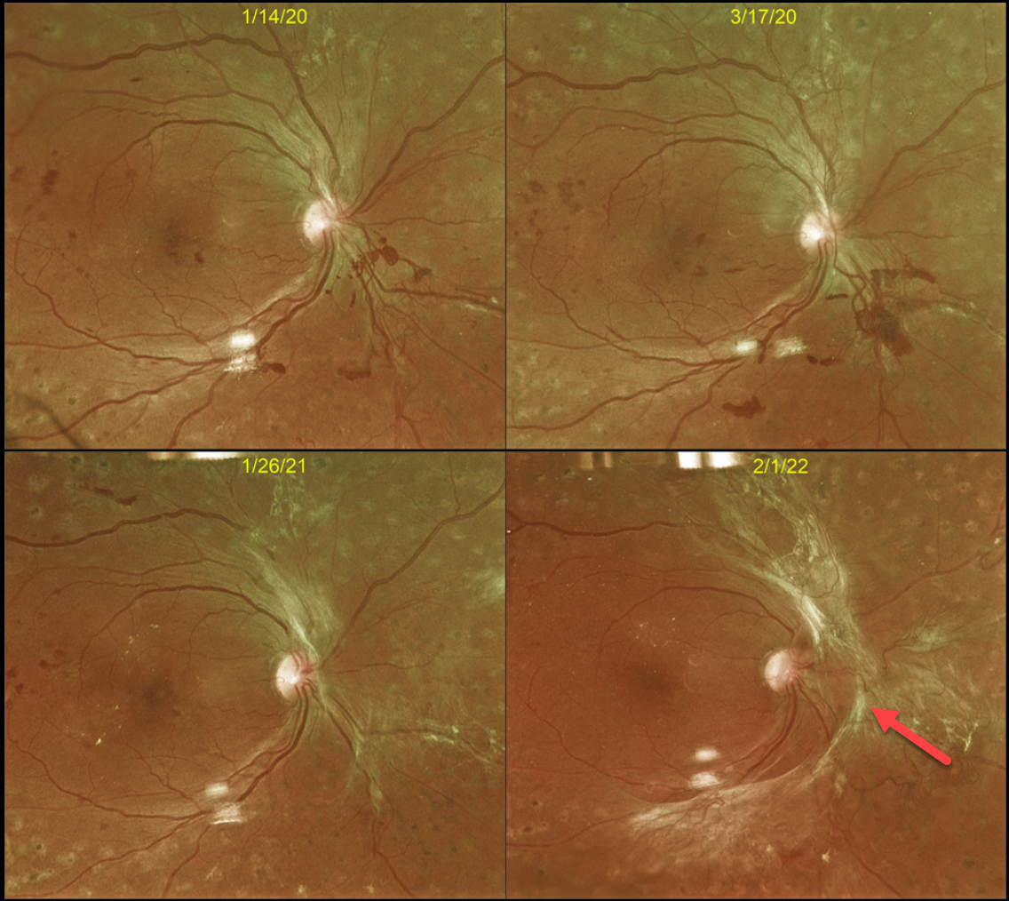
This 43YO male with type 2 diabetes initially responded beautifully in 2016 to full panretinal photocoagulation (PRP) for proliferative diabetic retinopathy (PDR). However, he presented on 3/17/20 with some new preretinal blood (Optos color RG image), and fill-in PRP was applied.
Over the course of 2 years, progressive fibrotic neovascularization with extramacular traction developed. Despite the traction, vision remained at 20/40, and careful observation was recommended. We were hesitant to add further laser or anti-VEGF therapy for fear of causing increased traction and ‘crunch’ (Tan et al, Surv Ophthalmology 2021;66:926-932). When last examined on 4/3/24, the retinal findings remained unchanged, and vision was 20/30 (not shown).
Although PRP is often a ‘one and done’ procedure, this is not always the case (Gonzalez et al, Ophthalmology 2021;128:1448-1457). Our patient is a perfect example of why these patients require lifelong monitoring even if their disease appears quiescent.
POSTOP DIABETIC TRD WITH INNER RETINAL FOLDS
Originally posted on @retina.rocks September 5, 2024
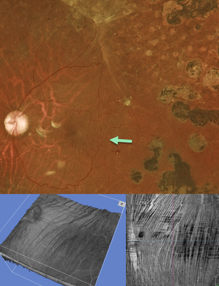
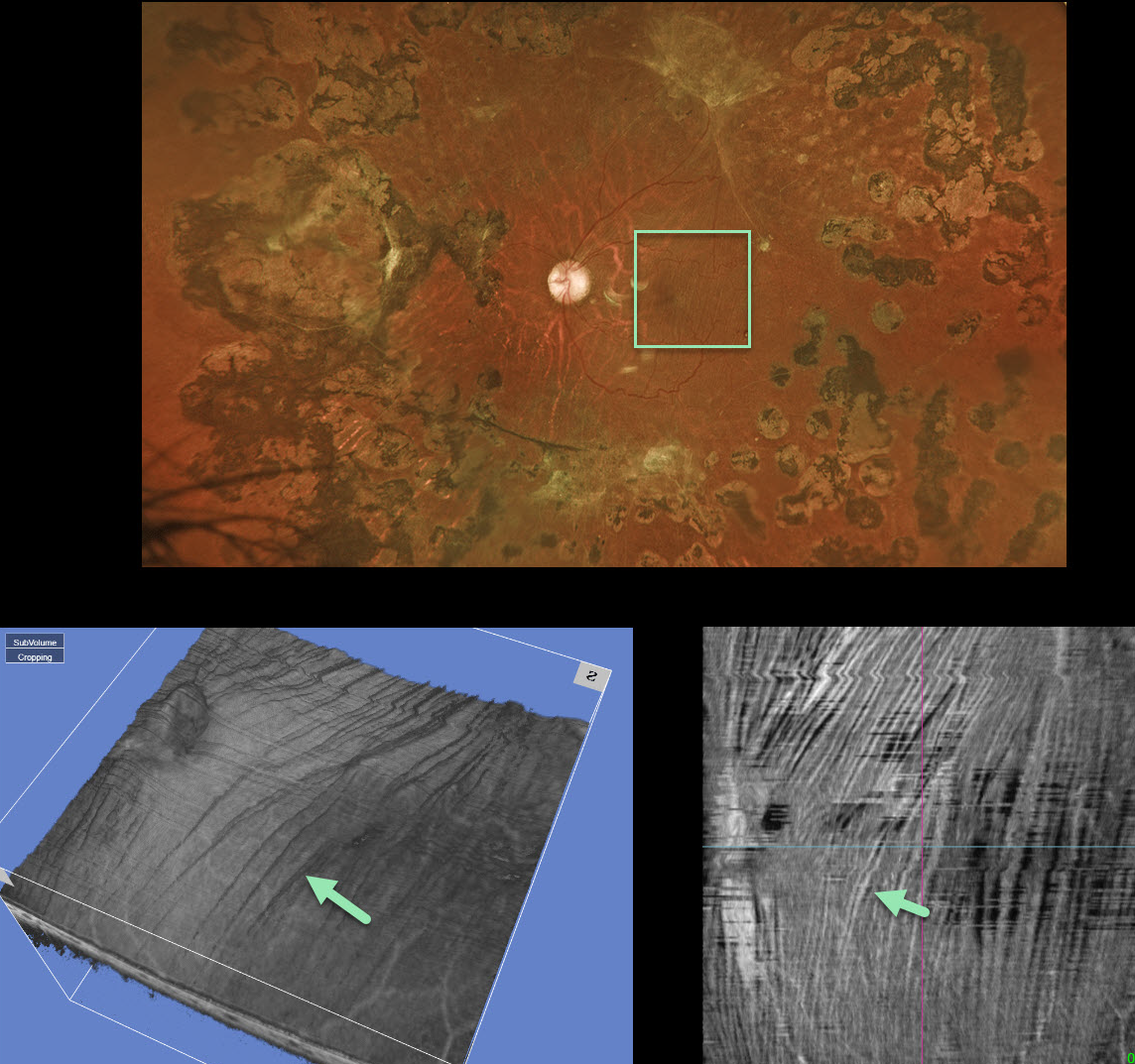
This 35YO diabetic male underwent prior vitrectomy surgery for a diabetic traction retinal detachment (TRD) five years earlier. Vision was stable at 20/40.
Optos color RG imaging shows fine macular striae extending superiorly towards the area of prior traction. Triton swept-source OCT 3D reconstruction and inner retinal en face show differing perspectives of these inner retinal folds.
Inner retinal folds are common following vitrectomy for diabetic TRD. They often fail to completely resolve, and vision can be quite good despite the persisting findings.
LOCALIZED TRACTIONAL RETINAL DETACHMENT
Ali Lamin and Ritu Chaturvedi
Originally posted on @retina.rocks July 4, 2024
This 55YO female with type 2 diabetes presented for a routine follow-up without visual complaint. Vision was 20/30 bilaterally.
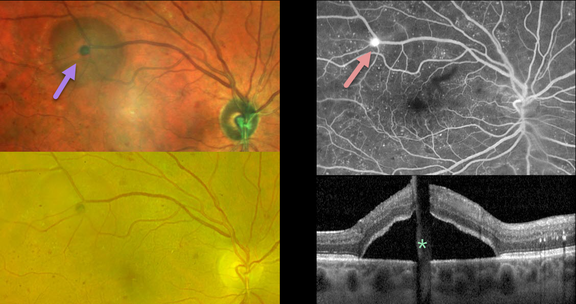
MultiColor imaging of her right eye shows a localized traction retinal detachment (TRD) in the distal superotemporal macula. The TRD is much less obvious with Optos color RG imaging.
The source of the traction is a small area of retinal neovascularization that leaks on fluorescein angiography. This localized traction is also confirmed on OCT. Although vitrectomy was not recommended given the localized nature of the TRD, panretinal photocoagulation was performed.
Learning Points:
Although pretty color images that match what we see on biomicroscopy are most intuitive, newer imaging modalities that ‘distort’ the normal fundus colors often contain very clinically relevant information. Standard Optos color RG images are generated from a red (635nm) and green (532nm) laser. Although these images have a greenish tint, they essentially contain two images in one: the RPE/neurosensory retina is best captured with the green channel, and the choroid is best seen with the red channel. A newer Optos true-color RGB unit has recently been introduced, which maintains the ability to view the separate RG channels while also generating a true-to-life color image.
Heidelberg Engineering’s Multicolor imaging module similarly utilizes a blue (488nm), green (515nm), and infrared (820nm) confocal laser to generate a fundus image. The blue laser best images the vitreoretinal interface and inner retina; the green laser images the mid retina; and the infrared laser images the outer retina, RPE, and choroid (Roy et al., Surv Ophthalmology 2024;69:378-402).
Combining these 3 wavelengths into a single file produces a pseudocolor image that, like the Optos, provides the clinician with more information than a standard color photo. As seen in our patient, Multicolor imaging best illustrates both the presence and extent of TRD (Gadde et al, Indian J Ophthalmol 2022;70:465-470).
TRACTIONAL RETINAL DETACHMENT
Originally posted on @retina.rocks June 10, 2024
This 42YO male with type 2 diabetes presented with 1 week of vision loss in his left eye. Vision was 20/40 OD and 20/100 OS.
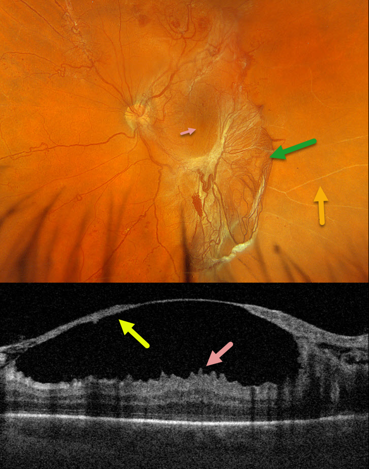
Optos color RGB imaging shows a large patch of variably fibrosed retinal neovascularization extending from the temporal macula into the inferior midperiphery. Vertical macular striae are drawn towards the neovascularization inferiorly. Scattered neovascularization is seen elsewhere, along with nonperfused large retinal vessels temporally.
Triton swept-source OCT through the macular center shows a thickened detached posterior hyaloid that extends from the optic nerve into the temporal macula. The macular striae noted on the Optos image correspond to inner macular folds on OCT. Vitrectomy surgery was scheduled.
COMBINED TRACTIONAL-RHEGMATOGENOUS RETINAL DETACHMENT
Originally posted on @retina.rocks March 27, 2024
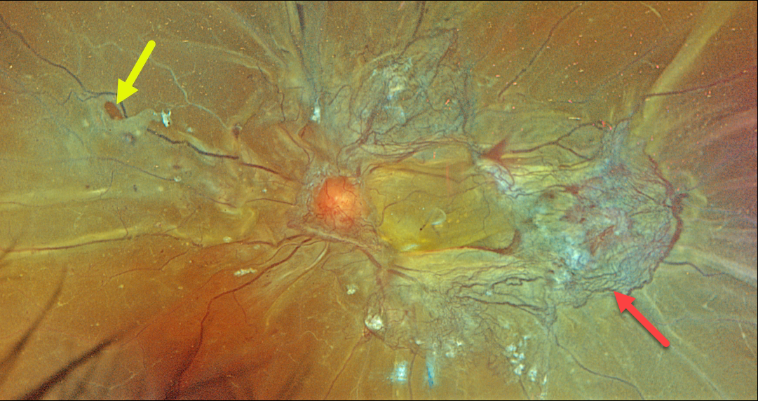
This 46YO female with type 2 diabetes presented with recent bilateral vision loss. Vision was 20/200 OD and hand motion OS.
Optos color RGB imaging of her left eye shows a combined tractional-rhegmatogenous total retinal detachment with fibrovascular tissue encircling the macula. Temporally radial outer retinal folds are dragged toward the more succulent, red neovascularization. A retinal tear is noted temporally.
Vitrectomy surgery was scheduled.
Proliferative diabetic retinopathy with disc and peripheral neovascularization was noted in her right eye (not shown). Panretinal photocoagulation was performed in her left eye and surgery was scheduled for her left eye.
MACULAR HOLE DUE TO DIABETIC TRACTION
Originally posted on @retina.rocks September 21, 2023
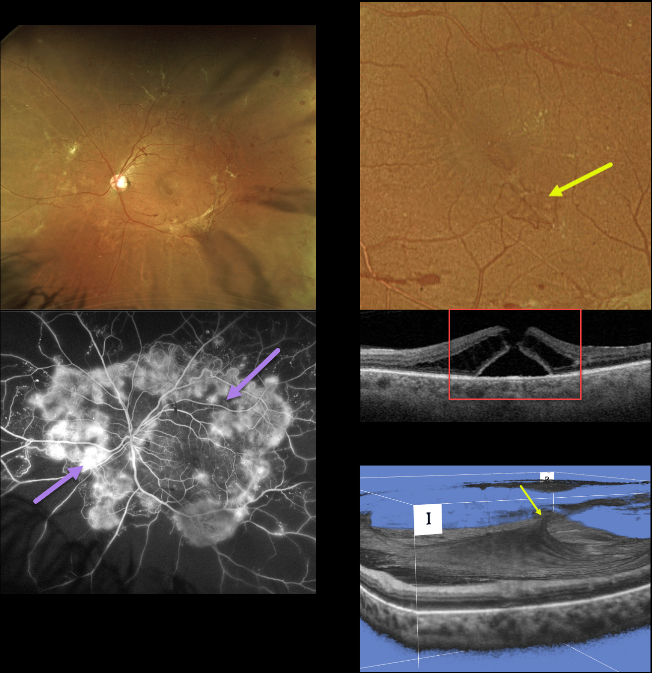
This 49YO female with type 2 diabetes presented with a several-year history of blurred vision in her left eye. Vision was 20/40 OD and 20/60 OS.
Optos RG imaging shows variably fibrosed retinal neovascularization encircling the posterior pole. Fluorescein angiography shows active leakage from these vessels.
An area of small neovascularization is present in the inferior temporal macula, which caused a tractional full-thickness macular hole. A 3D OCT view best shows this diabetic traction.
Vitrectomy surgery was scheduled for her left eye. Primary vitrectomy will be performed for her right eye as well once the left eye has stabilized.
Learning Points:
In 1988, Dr. Gass described his concept for the pathogenesis of idiopathic macular holes (Arch Ophthalmol 1988;106:629-639). His classification system, from stage 1 for an impending macular hole through stage 4 for a fully developed macular hole with complete posterior vitreous detachment, was based on his concept of localized, mostly tangential traction caused by the shrinking foveal vitreous cortex. This classic paper heralded modern macular hole surgery for a previously untreatable condition.
The advent of OCT technology, however, has shown that macular hole development is more often due to focal vitreomacular traction, as our case beautifully illustrates.
The current classification system, described by the International Vitreomacular Traction Study Group, emphasizes the characteristics that determine treatment choice and visual prognosis (Ophthalmology 2013;120:2611-2619). This includes size (small <= 250 microns, medium >250 microns and <= 400 microns, large >400 microns), presence or absence of VMT, and cause (primary from VMT or secondary).
Our patient emphasizes why this new classification system was implemented, since it provides information that guides our treatment. Our patient’s hole is secondary to diabetic traction, which obviously requires a very different vitrectomy procedure compared to a primary hole.
TRACTIONAL RETINAL DETACHMENT
Originally posted on @retina.rocks July 17, 2023
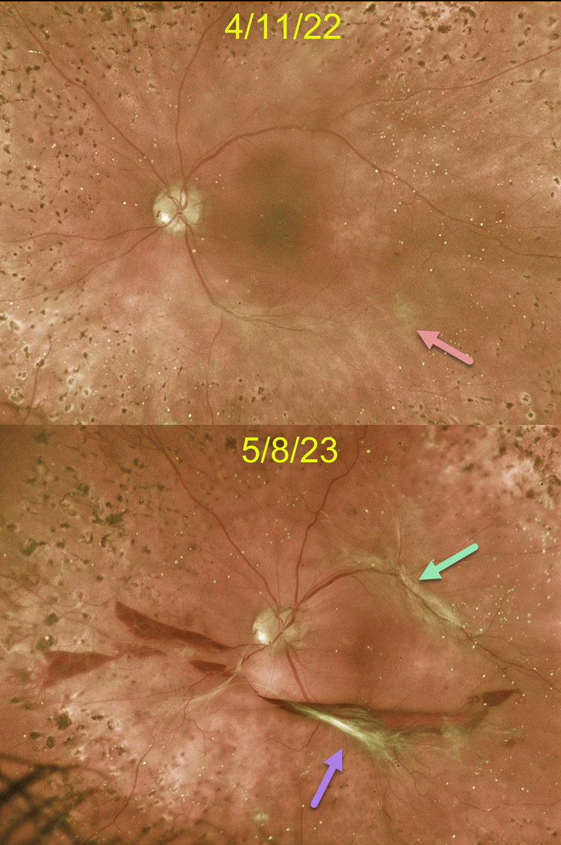
This 45YO female with type 1 diabetes responded beautifully to full panretinal photocoagulation (PRP) for her proliferative diabetic retinopathy (PDR). When examined 6 weeks following completion of her PRP, faint fibrotic neovascularization was noted along the inferotemporal arcade. Vision was 20/400 from residual central edema that was being treated with ongoing anti-VEGF therapy.
Unfortunately, she was lost to follow-up for over a year. Although vision remained at 20/400, increased traction was noted inferiorly, with new traction superiorly, resulting in an extrafoveal traction detachment. Symptomatic vitreous hemorrhage was also seen. Vitrectomy surgery was scheduled.
Learning Points:
Although PRP is often a ‘1 and done’ procedure, this is not always the case (Gonzalez et al, Ophthalmology 2021;128:1448-1457), and patients therefore need continued monitoring.
Although our patient showed involution of her PDR following PRP, increased fibrosis and traction still developed about a year later.
These findings would likely have been caught earlier if she had not been lost to follow-up, which is unfortunately quite common among patients with diabetes (Obeid et al., Ophthalmology 2018;125:1386-1392; Gao et al., Ophthalmology Retina 2019;3:230-236; Green et al, AJO 2020;216:18-27, etc.).
CRUNCH SYNDROME
Originally posted on @retina.rocks February 22, 2023
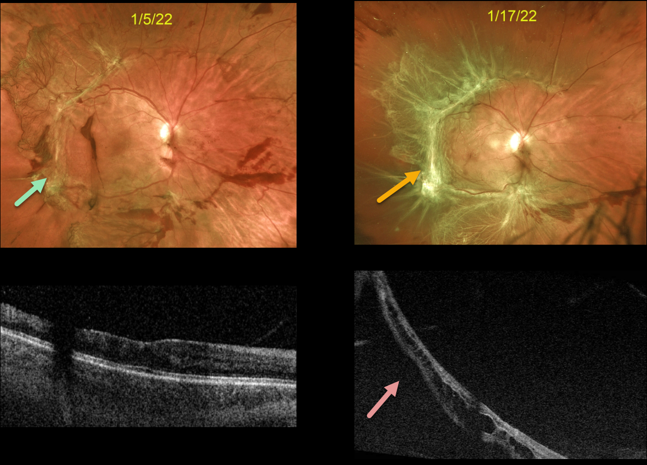
This 37YO female presented with a symptomatic diabetic traction retinal detachment (TRD) extending from the peripheral macula towards the mid-peripheries. Vision was 20/60. An intravitreal Avastin injection was given in preparation for vitrectomy within the week.
However, due to work-related reasons, the patient delayed the surgery and returned 2 weeks later, complaining of several days of increasing distortion and vision loss. Vision was 20/200. There was marked fibrosis of all neovascularization, and the macula was now detached. Vitrectomy was scheduled within the next several days.
Learning Points:
Intravitreal anti-VEGF is commonly used before diabetic vitrectomies to cause rapid involution of the vascularized preretinal tissue and control intraoperative bleeding during membrane dissection. These injections are given within a week of surgery since the neovascularization can contract with increased traction. This uncommon but known phenomenon is called the ‘crunch’ syndrome.
See Tan et al for a recent crunch review (Survey of Ophthalmology 2021;66:926-932).
TRACTIONAL RETINAL DETACHMENT
Originally posted on @retina.rocks December 12, 2022
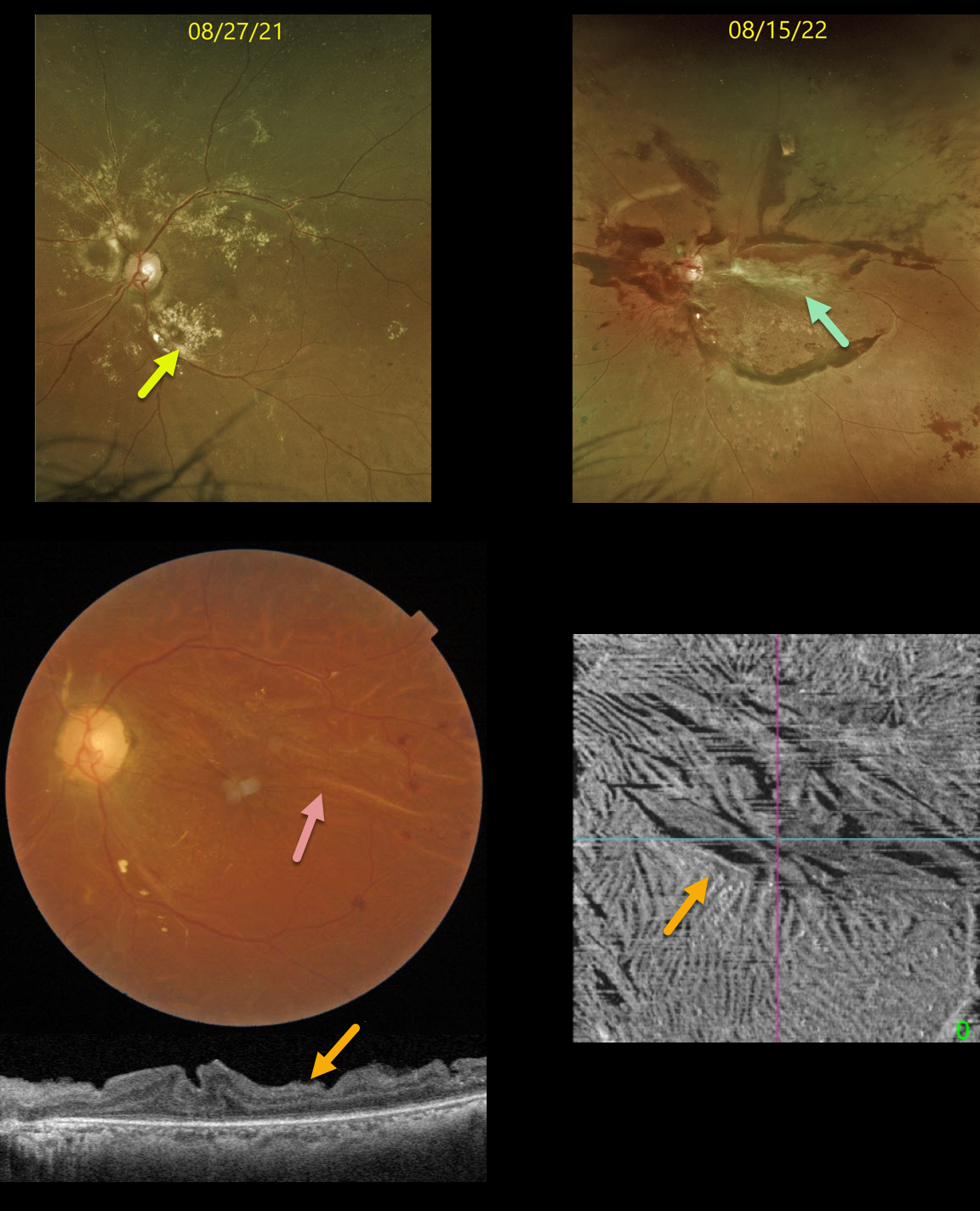
This 38YO diabetic female was initially seen on 8/27/21 with extensive dry macular lipid in her left eye and proliferative diabetic retinopathy. Vision was 20/40. Panretinal photocoagulation was recommended.
Unfortunately, she was lost to follow-up until she returned on 8/15/22 with 20/200 vision from a traction retinal detachment. The lipid spontaneously absorbed.
Vitrectomy surgery was performed, and three weeks later, the retina was completely attached with 20/70 vision.
Color imaging of the macula shows variable retinal folds centrally and superiorly.
OCT B-scan and en face of the inner retina variably show the dramatic inner retinal folds caused by the prior traction. We expect these to improve over time, but will probably never be completely resolved.
Learning Points:
Inner retinal folds, although usually less dramatic than in our patient, are fairly common following vitrectomy for diabetic TRD. They often fail to completely resolve, and vision can be quite good despite the OCT findings.
TRACTIONAL RETINAL DETACHMENT
Originally posted on @retina.rocks November 18, 2022
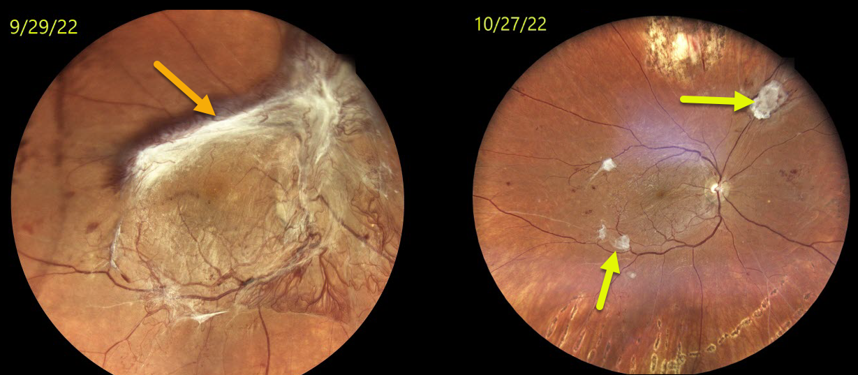
This 27YO female with type 1 diabetes presented with counting fingers vision OD after being lost to follow-up with a new tractional retinal detachment (TRD). Vision and retinal findings were unchanged OS.
Vitrectomy surgery was performed one week later.
Three weeks postop, vision improved to 20/40, and the retina was completely attached.
Residual fibrosed islands of neovascularization are seen in the distal temporal macula and superonasal midperiphery, representing the neovascular ingrowth sites onto the posterior hyaloid that created the TRD.
TRACTIONAL RETINAL DETACHMENT
Originally posted on @retina.rocks May 20, 2022
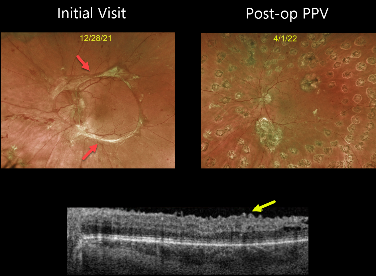
This 32YO female with type 1 diabetes presented with an 18-month history of vision loss in her left eye. Vision was counting fingers.
Optos imaging beautifully captures the pre-operative photos of the tractional retinal detachment, caused primarily by fibrosed retinal neovascularization extending in a circular pattern from the nerve along the arcades.
The second image shows the patient four months following vitrectomy. Swept-source OCT shows an irregular inner retinal contour, which is common following these vitrectomies.
Vision improved to 20/50. She is currently undergoing panretinal photocoagulation in her contralateral eye.
TRACTIONAL RETINAL DETACHMENT
Originally posted on @retina.rocks April 14, 2022
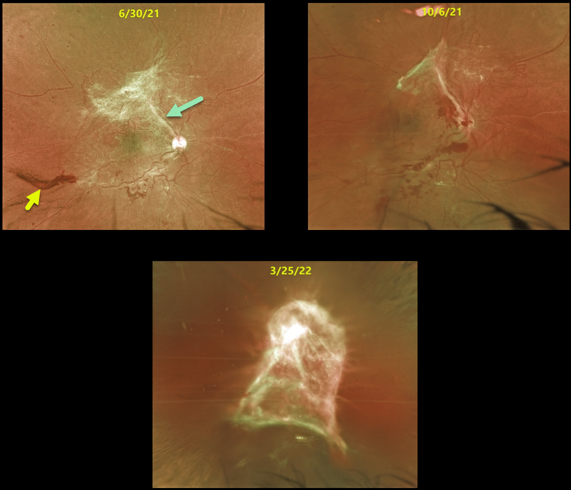
This 34YO female presented with hand motion vision from a progressive diabetic tractional retinal detachment with subhyaloid hemorrhage.
She was repeatedly noncompliant with appointment attendance and was lost to follow-up on numerous occasions.
The images show the progressive fibrosis and traction that developed over the course of a year. She will now require vitrectomy in hopes of recovering some vision in this eye.
TRACTIONAL RETINAL DETACHMENT
Originally posted on @retina.rocks January 3, 2022
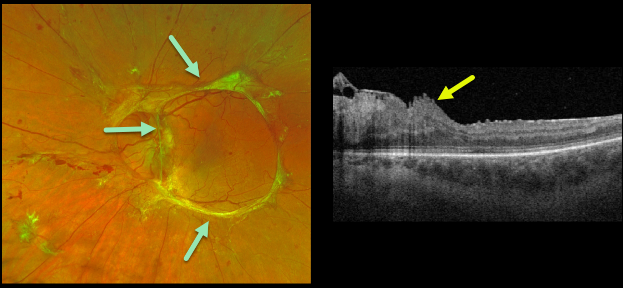
This 32YO female with type 1 diabetes presented with an 18-month history of vision loss in her left eye. Vision was counting fingers.
Optos imaging beautifully captures the traction retinal detachment from mostly fibrosed retinal neovascularization extending circularly from the nerve along the arcades.
Triton swept-source OCT surprisingly shows relatively mild macular distortion despite the severe vision loss, which is most likely due to vitreopapillary traction.
Intravitreal Avastin followed by vitrectomy will be performed.
Learning Points:
Tractional retinal detachment is one of the main complications of untreated or incompletely treated proliferative diabetic retinopathy.
TRACTIONAL RETINAL DETACHMENT
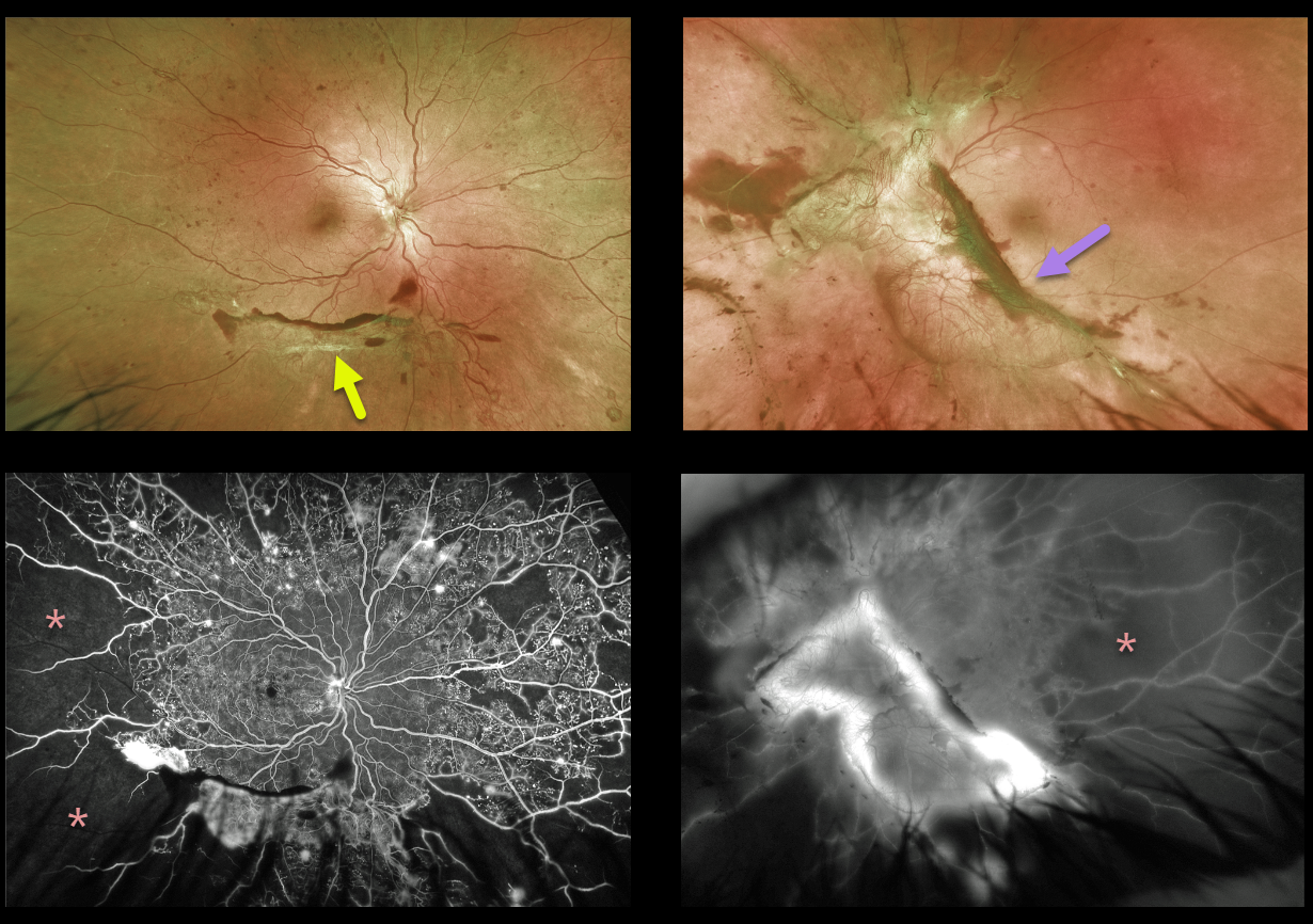
Originally posted on @retina.rocks July 19, 2021
This 34YO female presented with 20/20 vision OD and 20/80 vision OS. Optos imaging of her right eye shows inferior fibrotic neovascularization with marked peripheral ischemia, with more extensive fibrovascular proliferation and ischemia in her left eye.
The relatively good vision in her left eye is deceiving, with potentially blinding traction lurking just outside the fovea. Triton swept-source OCT imaging revealed a relatively normal, flat central macula (not shown here).
Panretinal photocoagulation was started in her right eye. She is scheduled for primary vitrectomy in her left eye.
Learning Points:
Tractional retinal detachments are one of the complications that can develop from proliferative diabetic retinopathy.
Although described as a specific manifestation in sickle cell disease (see Han, Retina 2021; 41;1361-1363), we think the left eye resembles a sea fan.
TRACTIONAL RETINAL DETACHMENT
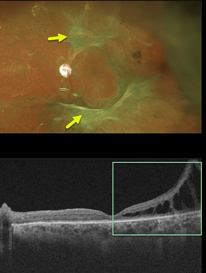
Originally posted on @retina.rocks October 22, 2020
For many years, we have been following this 55YO male with 20/50 vision and involuted proliferative diabetic retinopathy with a stable extrafoveal traction retinal detachment in his left eye.
His right eye was enucleated following several failed vitrectomy surgeries years ago, before he came to our practice.
Although completely stable funduscopically, OCT shows some subclinical, progressive tractional schisis near the temporal macular center.
After a lengthy discussion, the patient chose to avoid vitrectomy for now, given his total lack of symptoms and prior experience with his right eye. We continue to follow him extremely closely since surgery may be necessary in the near future.
Learning Points:
Tractional retinal detachment is one of the main complications of untreated or incompletely treated proliferative diabetic retinopathy.
TRACTIONAL RETINAL DETACHMENT
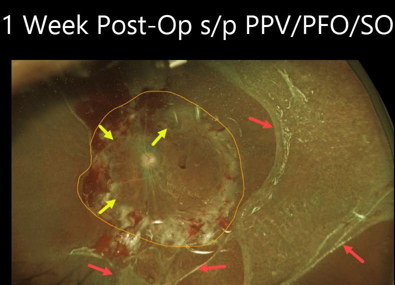
Originally posted on @retina.rocks July 13, 2020
This patient had hand motion vision in her left eye from a closed funnel, combined with diabetic tractional and rhegmatogenous retinal detachment.
Along with vitrectomy, a 360-degree retinectomy was made anterior to the macula. The posterior retina was then flattened with Perfluoron, followed by silicone oil. A large macular hole was also noted intraoperatively.
This image was taken 1 week postoperatively and shows the extent of the retinectomy. Although intraoperatively we felt that we were excising full-thickness retina, once the retina flattened, a remaining outer layer of retina was variably present temporally and inferiorly.
Although Perfluoron is completely removed during surgery, complex cases such as this often have residual subretinal bubbles.
Functionally blind before surgery, she was able to fully care for herself in her apartment and use her microwave and cell phone at 1 week.
Learning Points:
Sometimes during vitrectomy, the only way to partially flatten the retina is to do a retinectomy to excise the extremely thick, stiff, and scarred retina anterior to the macula.
RETINAL STRIAE FOLLOWNG VITRECTOMY FOR DIABETIC TRD
Originally posted on @retina.rocks May 21, 2020
This 35 YO male had a severe diabetic tractional retinal detachment with subsequent pars plana vitrectomy a few years prior.
Now, macular striae are apparent in the inner retina and are especially dramatic on en face imaging (image 4). Remarkably, this patient maintains 20/40 vision. It is quite common for these postoperative traction detachments to have residual retinal striae, which usually don’t adversely affect vision.
Learning Points:
En face OCT is extremely helpful in evaluating and managing retinal disease, offering a unique perspective for viewing pathology.
TRACTIONAL RETINAL DETACHMENT
Originally posted on @retina.rocks April 28, 2020
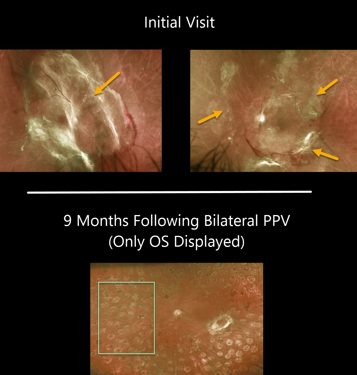
This patient presented with bilateral diabetic traction detachments with counting fingers OD and 20/400 OS. The fibrotic neovascularization is particularly severe in his right eye, obscuring the entire posterior pole.
He subsequently underwent bilateral pars plana vitrectomy. An image of the left eye is shown about 9 months later. All neovascular tissue was removed intraoperatively and endolaser was done. Vision remarkably improved to 20/60.
We were unable to get a photo of the right eye at that visit due to a recurrent vitreous hemorrhage. The blood subsequently resolved with an intravitreal Avastin injection, and vision was 20/200 OD at the most recent visit.
Learning Points:
Tractional retinal detachment is one of the main complications of untreated or incompletely treated proliferative diabetic retinopathy.
HUMMINGBIRD MACULOPATHY
Originally posted on @retina.rocks February 28, 2020
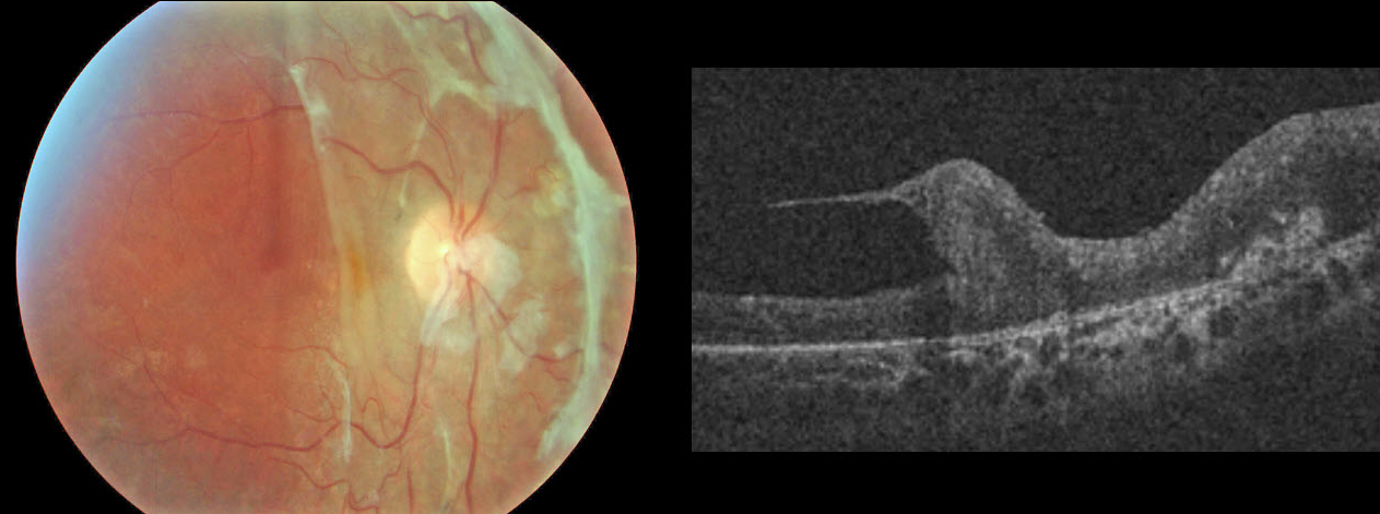
This rare condition was discovered after a diabetic TRD repair. Thankfully, no birds or retinas were harmed in this process.
TRACTIONAL RETINAL DETACHMENT
Originally posted on @retina.rocks February 20, 2020
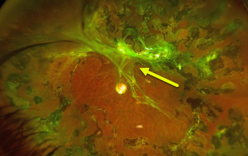
This patient underwent vitrectomy in 2013 for a severe table-top diabetic traction retinal detachment (TRD). We were able to free up all significant traction except superiorly and superotemporally, shown in the Optos image.
Despite this, the patient has been quite stable anatomically, maintaining 20/30 vision.
Learning Points:
Tractional retinal detachment is one of the main complications of untreated or incompletely treated proliferative diabetic retinopathy.
SILICONE OIL DROPLET
Originally posted on @retina.rocks January 2, 2020
This patient with proliferative diabetic retinopathy with tractional detachment was noted to have a large asymptomatic silicone oil bubble following multiple prior Avastin injections.
Learning Points:
The older insulin syringe needles provided by compounding pharmacies for the Avastin (bevacizumab) injections were lubricated with silicone.
Many eyes receiving intravitreal Avastin developed small vitreous silicone droplets, although the droplet is unusually large in this patient. Since then, these insulin syringe needles have been discontinued.


