

Trauma Miscellaneous
<< Back to Cases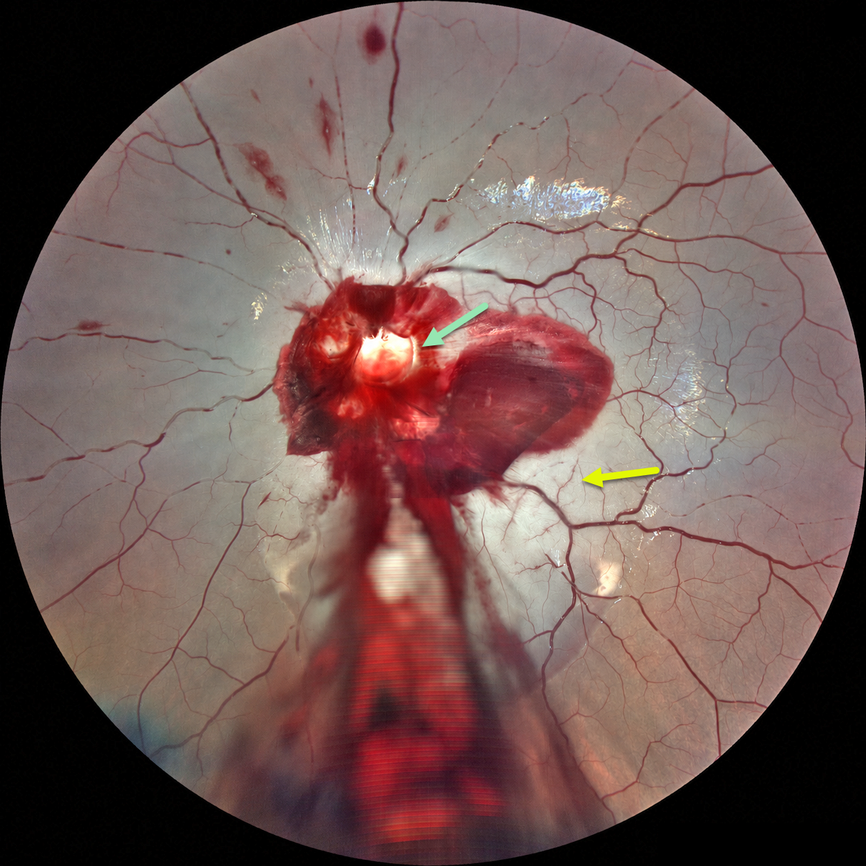
ACUTE OPTIC NERVE AVULSION WITH SECONDARY CRAO
Ayushi Gupta and Vishal Agrawal
Originally posted on @retina.rocks September 8, 2026
This 6YO boy developed sudden and complete loss of vision in his left eye immediately following blunt trauma. He was riding a bicycle as a pillion passenger when the bike stopped suddenly, throwing his face against the handlebar. Vision was no light perception. Anterior segments were normal.
Fundus photography shows preretinal blood surrounding the nerve, extending through the macula and into the inferior periphery. A pale-yellow cavity is noted where the nerve head should be. A fresh central retinal artery occlusion (CRAO) with a stagnant blood column is noted, along with some scattered inner retinal hemorrhages. There were no other physical or neurologic injuries.
Learning Points:
Traumatic optic nerve head avulsion is a rare and visually devastating traumatic event in which the optic nerve is forcibly separated from the globe at its junction with the sclera, typically resulting in immediate, severe, and irreversible vision loss. The mechanism involves sudden forceful rotation or anterior displacement of the globe following blunt periorbital trauma, including projectiles, falls, or finger-poke injuries, whereby the globe moves anteriorly or rotationally while the optic nerve remains relatively fixed, causing violent separation. Fundoscopic examination may reveal a partial or complete tear of the optic nerve head from the sclera (Foster et al, Arch Ophthalmol 1997;115:623-630). Avulsion can be partial or complete, and may be associated with a CRAO, as was noted in our patient.
CRAO FOLLOWING DOUBLE PERFORATION FROM PERIBULBAR INJECTION
Ayushi Gupta and Vishal Agrawal
Originally posted on @retina.rocks April 6, 2026
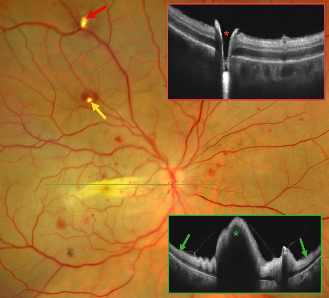
This 42YO female immediately lost vision in her right eye 4 days earlier following pterygium excision surgery performed under peribulbar anesthesia elsewhere. Vision in our office was light perception.
Fundus photography shows an opaque retina throughout the posterior pole consistent with an acute central retinal artery occlusion (CRAO). There is a horizontal retinal fold throughout the macula with scattered blot retinal hemorrhages. Entry and exit needle penetration sites are noted. OCT scan through the superior entry site confirms a full-thickness retinal break. Macular OCT shows diffuse inner retinal hyperreflectivity with elevation through the fold.
Learning Points:
Peribulbar anesthesia is considered to have a lower risk of inadvertent globe perforation, although this risk is not zero (Lim et al., J Clin Ophthalmol 2021;5:414-416). CRAO after such an injury is exceedingly rare and was reported by Lake et al. (J Cataract Refract Surg 2003;29:2234-2235), who felt the occlusion might have resulted from increased IOP due to intraocular anesthetic. A CRAO was also reported by Gyasi et al. following peribulbar anesthesia for pterygium excision, although they found no evidence of globe perforation (Ghana Medical Journal 2012;46:48-50).
TRAUMATIC RETINAL HOLES
Fatma Shakarchi and Ahmed Sallam
Originally posted on @retina.rocks October 6, 2025
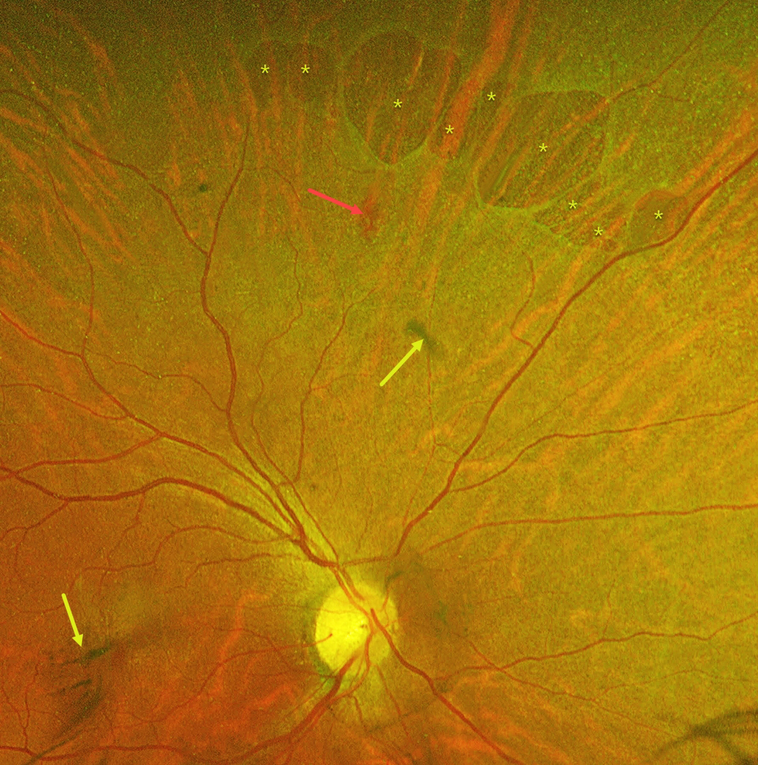
This 55YO female was referred with mildly decreased vision in her right eye two days following blunt ocular trauma. Vision was 20/40 OD and 20/20 in her normal OS.
Optos color RG imaging reveals numerous irregularly oval superonasal retinal holes, a retinal hemorrhage, and some clumps of vitreous blood. There was no vitreous detachment clinically.
Learning Points:
Retinal breaks following blunt trauma include retinal dialysis, horseshoe tears, and giant retinal tears. Breaks can also develop from concussive injury at the impact site or from a coup-contrecoup injury, so-called impact or necrotic breaks, which may be associated with varying degrees of commotio retinae acutely (Cox et al, Arch Ophthalmol 1966;76:678-685) or following its resolution (Longstaff et al, Br J Ophthalmol 1987;71:375-376).
In this case, the breaks identified 2 days after the injury were not associated with commotio. However, their appearance and the adjacent hemorrhage support an acute traumatic etiology. Therefore, prophylactic laser retinopexy was promptly applied around all breaks.
POST-TRAUMATIC ILM DETACHMENT
Nilesh Kumar
Originally posted on @retina.rocks September 2, 2025
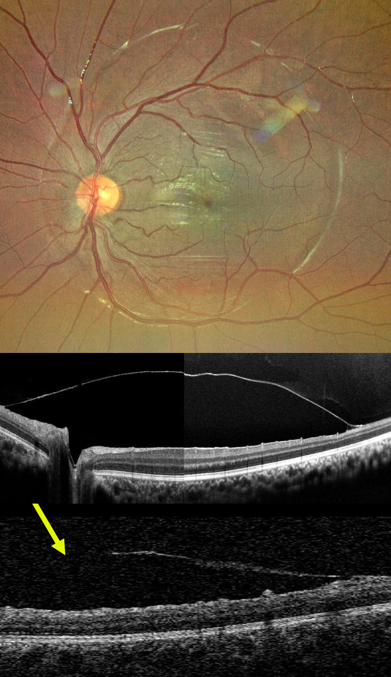
This 12YO boy came in for a routine eye examination. Although vision was 20/20 bilaterally, the right fundus showed a striking premacular sheen.
Ultra-widefield imaging reveals a round internal limiting membrane (ILM) elevation surrounding the posterior pole. On further questioning, he gave a history of blunt trauma to the right eye 2 years earlier that was treated elsewhere with the ND:YAG laser. The opening in the ILM is noticed inferiorly on OCT.
This case demonstrates a presumed chronic and stable sub-ILM detachment likely secondary to prior sub-ILM hemorrhage. Fortunately, he remains completely asymptomatic with perfect vision and no distortion.
A valuable reminder that retinal imaging can unearth forgotten chapters of ocular history, and that fundus examination with multimodal imaging can reveal prior stories.
TRAUMATIC MACULAR SCARRING AND MACULAR HOLE
Liubov Sitaruk and Ophthalmocoach
Originally posted on @retina.rocks June 10, 2025
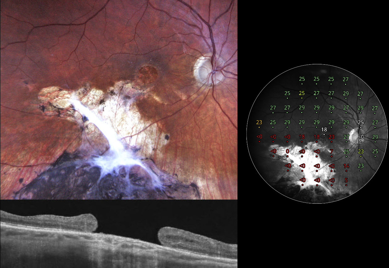
This 36YO Ukrainian male sustained a firearm-related facial injury during military combat in 2014 while in Eastern Ukraine. At the time, he was treated in a frontline hospital, where the left eye underwent primary enucleation due to a non-reconstructible globe rupture. His right eye sustained a penetrating injury with scleral rupture, which was primarily repaired. Over the following months, he underwent pars plana vitrectomy with endolaser and silicone oil due to complications, likely including retinal detachment and/or vitreous hemorrhage. Given the complexity of the trauma, the monocular status, and probable limited access to regular follow-up during wartime, silicone oil was likely retained for an extended period, potentially several years, until its removal at an undetermined later date. On our 2024 examination, he had no new visual complaints, and vision was 20/100.
Fundus photography shows atrophic inferior macular chorioretinal scarring, which becomes hyperpigmented as it extends into the inferior midperiphery. A large macular hole is noted funduscopically and on OCT. Macular perimetry shows marked depression within the area of scarring with preservation of some central sensitivity. Observation was recommended.
GIANT MACULAR HOLE WITH TOTAL RETINAL DETACHMENT
Anand Temkar and Manish Nagpal
Originally posted on @retina.rocks February 10, 2025
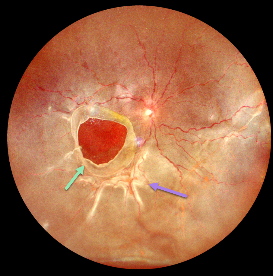
This 10YO boy presented with 1 week of vision loss in his left eye. The parents gave a vague history of trauma with a tennis ball 2 years earlier. Vision was counting fingers OD and 20/20 in his normal OS.
Ultrawidefield imaging shows a giant macular hole occupying nearly the entire macula. The hole’s edges are rolled, and proliferative vitreoretinopathy with star folds is present within the total rhegmatogenous retinal detachment (RRD).
Emergent vitrectomy surgery with silicone oil was scheduled.
Learning Points:
Blunt ocular trauma can result in numerous sequelae, including commotio retina, retinal breaks and detachment, choroidal rupture, and macular hole. A giant macular hole, also reported in Alport syndrome (Shah and Weinberg Ophthalmic Genetics 2010;31(2):93-97), is an exceedingly rare traumatic event (Hernandez-Da Mota Case Rep Ophthalmol 2011;2:283-286).
Traumatic macular holes are thought to result from a vitreous coup contrecoup injury or a jet stream of anterior chamber aqueous humor shooting through Cloquet canal into the premacular bursa.
TRAUMATIC MACULAR HOLE
Originally posted on @retina.rocks December 18, 2024
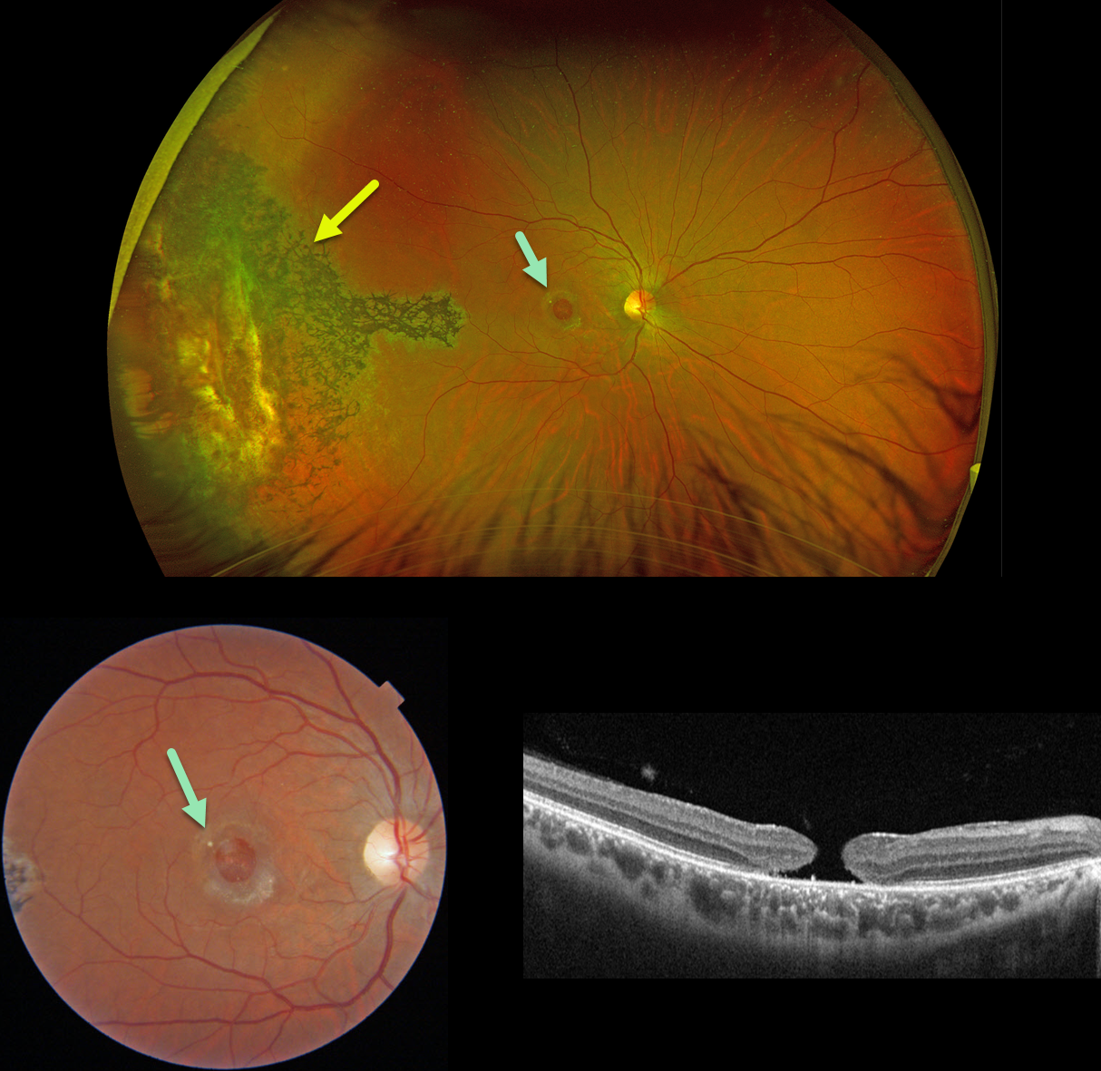
This 31YO male suffered a paint gun injury to his right eye when he was 12YO. Vision has since been stable and poor in this eye. Vision was 20/200.
Optos color RG imaging shows temporal peripheral scarring with intraretinal pigment migration. A large macular hole with surrounding pigmentary changes is noted on Triton color imaging and swept-source OCT.
Learning Points:
Since fresh traumatic holes can spontaneously flatten, especially in pediatric cases (Liu et al, AJO 2020;210:174-183), an initial period of observation is recommended before considering vitrectomy. Given the size and chronicity of our patient’s hole, surgery was not considered.
Photoreceptor loss from numerous causes, including blunt trauma, retinal detachment, macular telangiectasia, and retinitis pigmentosa (RP), can lead to intraretinal RPE migration, a shared phenotype. Unilateral RP is exceedingly rare and requires normal electrophysiologic testing with long-term observation to ensure that delayed findings are present in the fellow eye.
TRAUMATIC MACULAR HOLE
Barbara Parolini, Veronika Matello, Giulia Freschi, and Roberta Penzani
Originally posted on @retina.rocks April 9, 2024
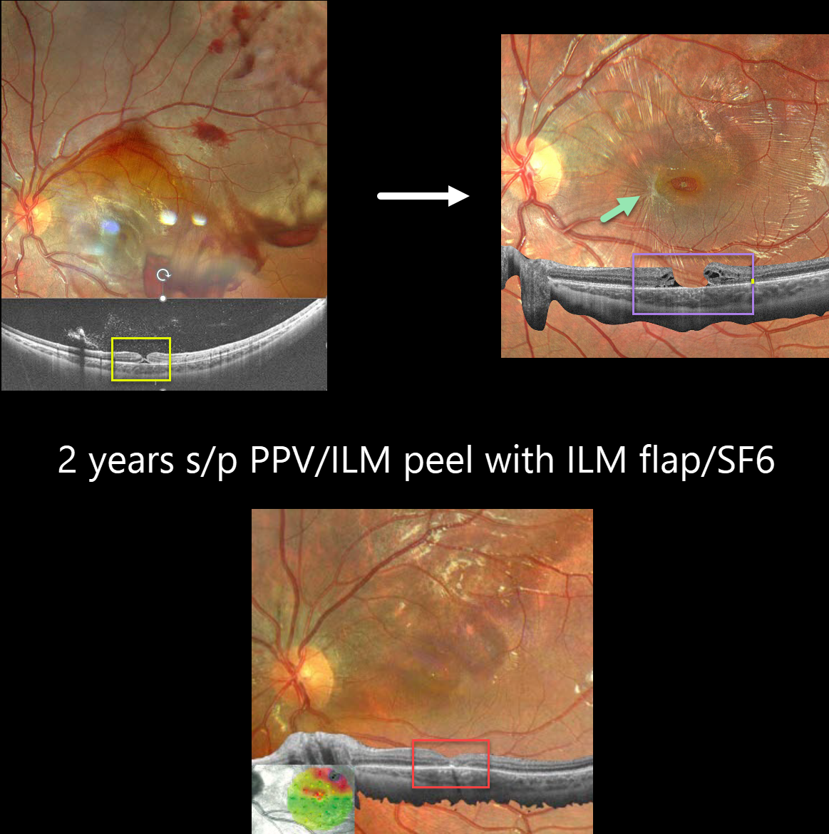
This 10YO boy was referred for vision loss immediately after getting hit in his left eye by a soccer ball 7 days earlier. Vision was 20/70.
Fundus photography shows scattered intra- and preretinal hemorrhages with a large confluent area of commotio retina superiorly and temporally.
Widefield OCT shows a partial-thickness macular hole with the external retina intact but elevated. Three months later, vision was 20/100, and an irregular, full-thickness macular hole with surrounding macular pucker was seen.
25-gauge vitrectomy with ILM peeling, ILM flap, SF6 gas tamponade, and 3 days face-down positioning was performed. Two months later, vision improved to 20/40 with complete closure of the hole. Two years later, vision was 20/25.
Learning Points:
This patient illustrates several sequelae of blunt trauma, including retinal/preretinal blood, commotio retinae, and macular hole formation. Since fresh traumatic holes can spontaneously flatten, especially in pediatric cases (Liu et al, AJO 2020;210:174-183), an initial period of observation is recommended before considering vitrectomy.
In our patient’s case, the hole did not resolve, and surgery provided good anatomic and long-lasting visual outcomes.
TRAUMATIC UNILATERAL WIPE-OUT
Originally posted on @retina.rocks May 4, 2023
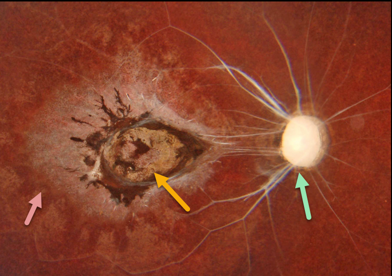
This healthy 10YO boy presented with a 5-year history of blindness in his right eye. His mother said that he injured his eye after falling while playing at school.
Color imaging of his right eye shows a white nerve and retinal vessels, a variably pigmented macular scar, and a moth-eaten appearance to the RPE outside the macula. The left eye was normal.
Learning Points:
The differential diagnosis for unilateral optic nerve atrophy, chorioretinal scarring, and narrowing/sclerosis of the major retinal vessels is relatively small.
The term ‘unilateral wipe-out syndrome’ was coined by Dr. Gass (Ophthalmology 1978;85:521-545) in describing end-stage diffuse unilateral subacute neuroretinitis (DUSN).
Other causes include trauma, ophthalmic artery occlusion, and post-inflammatory (i.e., acute retinal necrosis syndrome, ARNS). Despite the history, it is hard to imagine how a 5-year-old child falling at a playground could cause this type of injury.
AIRBAG TRAUMA
Will Gibson
Originally posted on @retina.rocks August 25, 2022
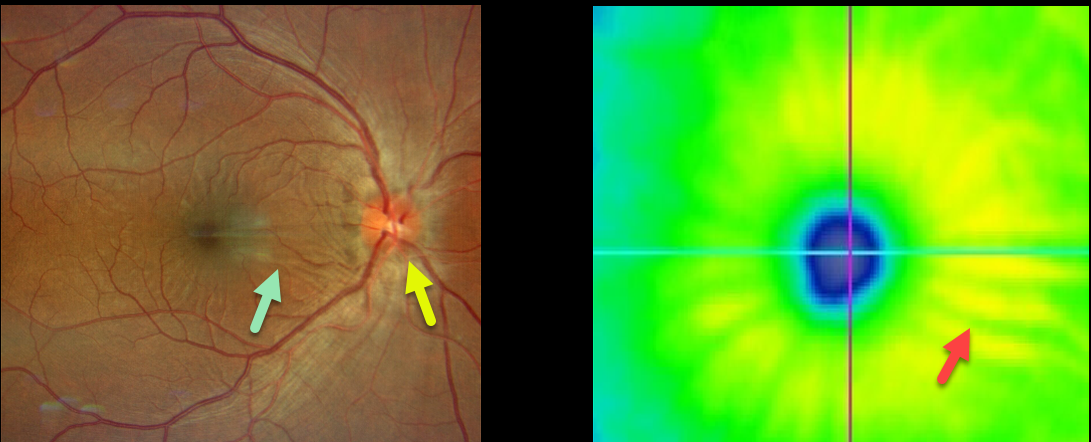
This 35YO male presented with a unilateral swollen nerve and retinal striae following blunt trauma from airbag deployment Vision was 20/25. The intraocular pressure was 15, and there were no signs of open-globe injury.
Optos imaging shows retinal folds radiating from the macular center. These striae are also evident on the OCT thickness map.
He was started on oral prednisone, and 10 days later, the ocular findings had improved (images not available).
TRAUMATIC PSEUDO-RP
Originally posted on @retina.rocks May 31, 2022
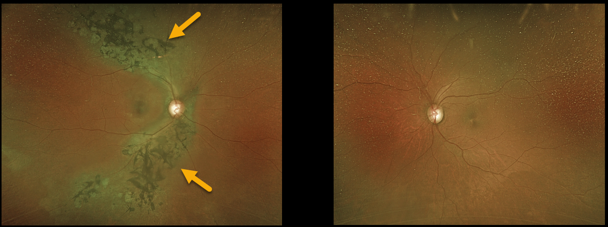
A 67YO female was referred for asymptomatic retinal findings in her right eye. She gave a history of unspecified head trauma as a child.
Optos imaging shows chorioretinal scarring with intraretinal pigment migration, extending superotemporally and inferotemporally from the optic nerve. The left fundus was completely normal.
Learning Points:
Photoreceptor loss from numerous causes, including blunt trauma, retinal detachment, macular telangiectasia, and retinitis pigmentosa (RP), can allow for intraretinal RPE migration with this shared phenotype.
Unilateral RP is exceedingly rare and requires normal electrophysiologic findings and long-term observation to ensure delayed findings in the fellow eye.
HENLE LAYER HEMORRHAGE
Originally posted on @retina.rocks April 18, 2022
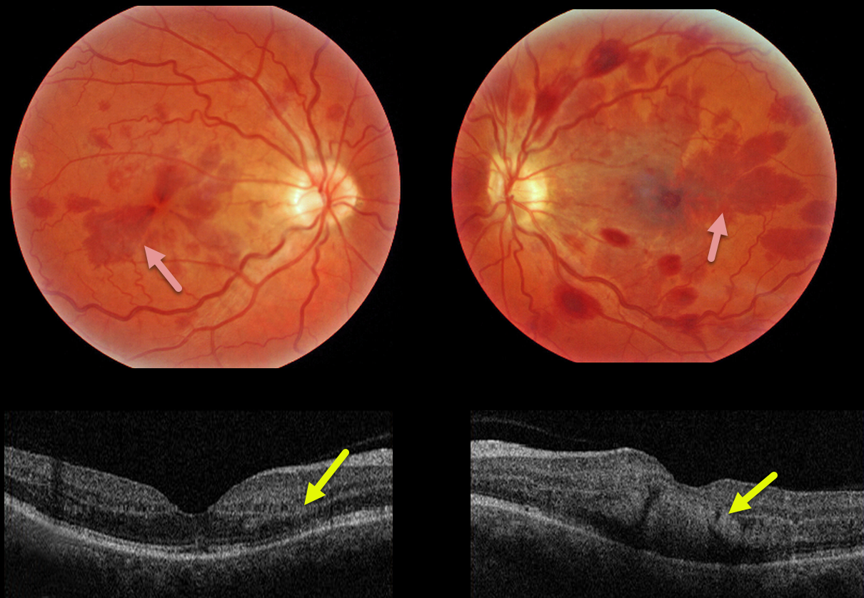
This 47YO female presented with bilateral vision loss. She had a seizure 10 days prior, losing consciousness and hitting her head. Upon awakening, she had reduced vision in her right eye and complete central loss in her left. Vision was 20/60 OD and counting fingers at 3 feet OS. Head CT scanning was normal.
Triton color imaging shows deep radiating wedge-shaped retinal hemorrhages centered in each macula. Swept-source OCT shows that these hemorrhages are located primarily in the outer plexiform layer (OPL).
Learning Points:
Radiating deep macular hemorrhages are seen in multiple conditions, including Valsalva (likely this case) and trauma. Their unique radiating appearance is caused by blood accumulating in Henle’s layer.
Kon Graversen et al described hemorrhagic unilateral retinopathy, an idiopathic disorder with a similar appearance (Retina 2014;34:483-489).
OCULAR PERFORATION FROM SUBTENON INJECTION
Originally posted on @retina.rocks September 28, 2021
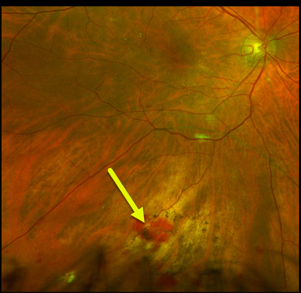
This 73YOF was referred for persistent bilateral symptomatic pseudophakic cystoid macular edema (CME) despite a course of topical therapy. Vision was 20/70 OU.
During the triamcinolone subtenon injection, given through the inferior conjunctival cul-de-sac, the patient complained of severe pain, and the red reflex turned white. We immediately removed the needle. Triamcinolone was dispersed throughout the vitreous cavity; no retinal detachment or break could be identified, and the intraocular pressure was remarkably normal.
When examined two days later, vision was hand motion due to residual triamcinolone, and the retina remained attached. One week later, the examination was unchanged, and she requested that the material be removed. At vitrectomy, the penetration was noted in the inferior midperiphery without open breaks or detachment. Three weeks following vitrectomy, vision was 20/40, with faint residual blood and chorioretinal scarring.
Not knowing how to proceed immediately after the initial injection, the patient allowed us to perform a subtenon injection in the left eye, which was thankfully completely uneventful. Vision returned to 20/40 within a few weeks with resolved CME.
MACULAR HOLE
Originally posted on @retina.rocks August 24, 2021
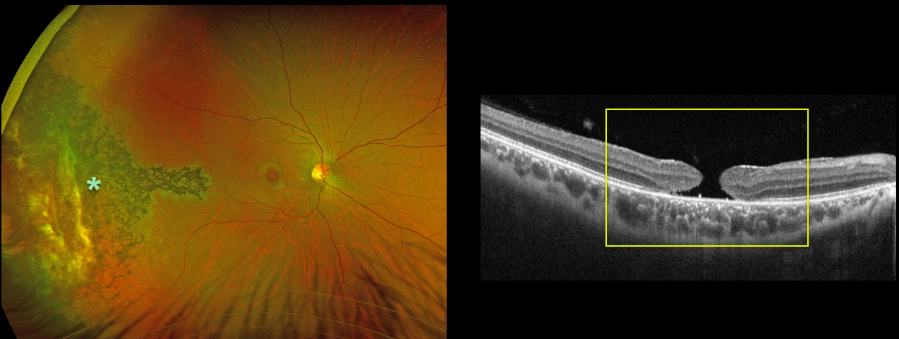
This 31YO male was hit in his right eye by a paintball when he was 12 years old. His vision was 20/200 due to a large, chronic 1100-micron full-thickness macular hole.
Optos imaging shows chorioretinal scarring with intraretinal pigment migration extending from the distal macula into the temporal periphery, underlying the likely impact site.
Learning Points:
Traumatic macular holes are more common in younger patients, often from recreational or sports injury, and tend to be more eccentric in shape.
The mechanism could be sudden vitreous separation or stretching of the posterior pole resulting from anteroposterior ocular compression. The holes can spontaneously close.
Unfortunately for our patient, the hole remained open, and at this point, we saw little possible surgical benefit given its chronicity, size, and secondary pigmentary changes.
SUPRACHOROIDAL HEMORRHAGE
Originally posted on @retina.rocks August 20, 2021
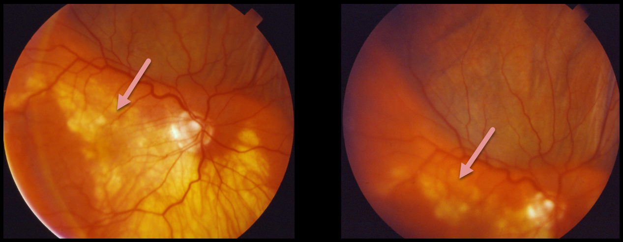
This patient presented with a pigmented, well-defined, and irregularly lumpy superior subretinal mass due to a suprachoroidal hemorrhage. This simulated a uveal melanoma.
Unfortunately, we don’t have any further information or images for this patient. However, we suspect that the blood spontaneously resolved with normalization of the fundus appearance.
Learning Points:
Patients presenting with suprachoroidal hemorrhage will usually have a history of recent intraocular surgery, trauma, or Valsalva. A clinical clue to this being a hemorrhage and not a melanoma includes the often somewhat lumpy and corrugated appearance, as in this patient.
See Chu and Green for a good review of suprachoroidal hemorrhage (Surv Ophthalmol 1999;43:471-486).
TRAUMA
Originally posted on @retina.rocks October 5, 2020
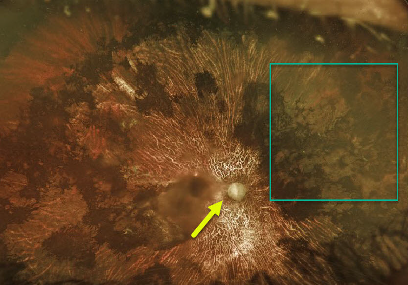
There is extensive chorioretinal scarring with optic atrophy in this patient who suffered severe blunt trauma 10 years earlier following a heroin overdose.
Vision is now counting fingers (CF), but thankfully, the other eye is normal with 20/20 acuity.
Note that the areas of intraretinal pigment migration mimic the bone spicules found with RP.
Learning Points:
Near total unilateral chorioretinal scarring has a relatively small differential diagnosis, including severe trauma, diffuse unilateral subacute neuroretinitis (DUSN), acute retinal necrosis syndrome (ARNS), and Saturday night retinopathy.
TRAUMA
Originally posted on @retina.rocks September 29, 2020
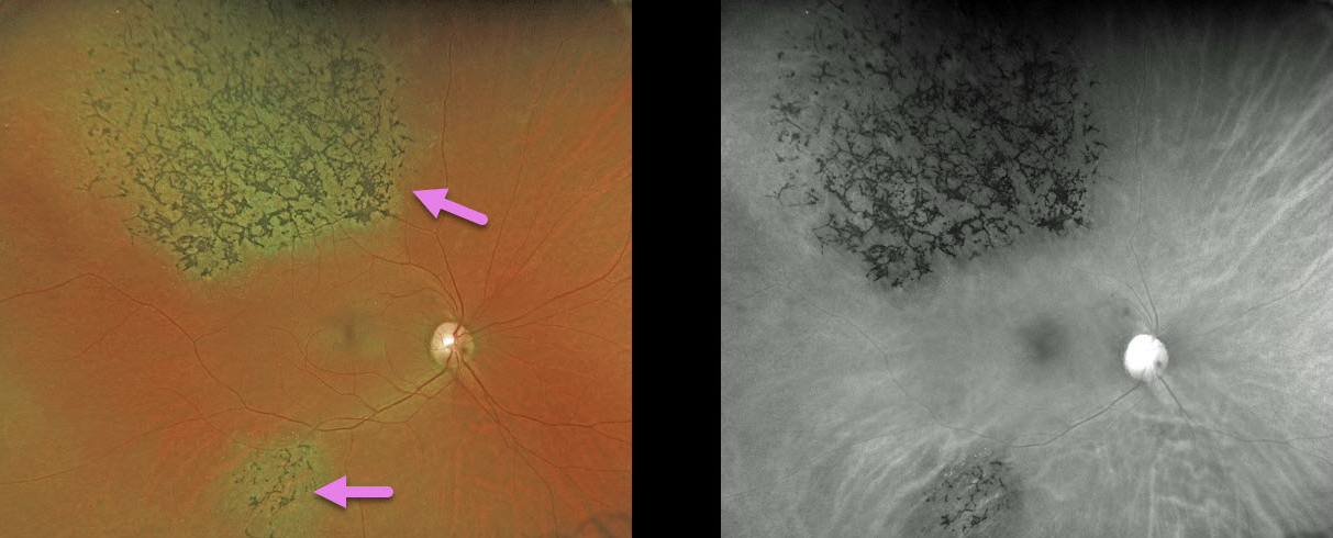
A 49YO female was involved in a motor vehicle accident 30 years ago.
Her retina showed signs of pigmentary retinopathy from the ocular injury, which could be mistaken for retinitis pigmentosa (RP). Unlike typical RP, our patient had multifocal, sectoral, unilateral findings.
Learning Point:
Intact photoreceptors inhibit RPE migration. Photoreceptor loss from numerous causes, including blunt trauma, retinal detachment, macular telangiectasia, and retinitis pigmentosa, can allow intraretinal RPE migration with this shared phenotype.
TRAUMA
Originally posted on @retina.rocks August 7, 2020
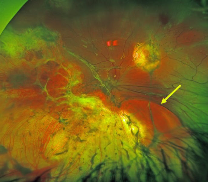
This patient was shot several times in the head and remarkably survived.
He underwent numerous retinal procedures at an outside hospital for a retinal detachment, and currently has light perception (LP) vision.
Examination revealed extensive chorioretinal scarring with a mostly detached retina under silicone oil.
A large retinal break, possibly from an intraoperative retinectomy, was seen nasally.
Learning Points:
Traumatic retinal detachments can occur from blunt trauma or from perforating injury to the eye.
TRAUMA
Originally posted on @retina.rocks March 26, 2020
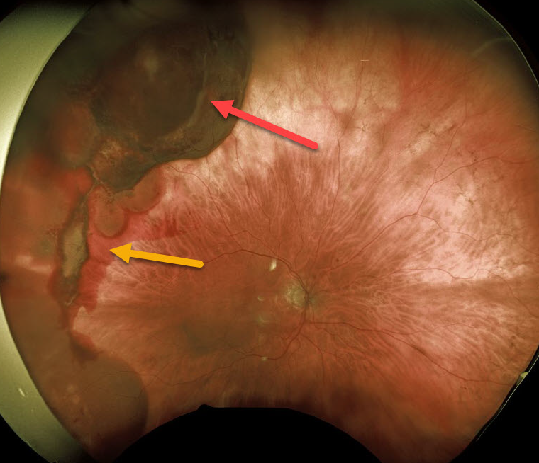
This patient presented with unilateral subretinal and suprachoroidal hemorrhages from a fall 1 month earlier causing blunt facial trauma.
Vision was normal and she was asymptomatic. She is being followed without treatment.
Learning Points:
There will likely be at least one choroidal rupture visible as the overlying blood clears over time. Subretinal blood following blunt trauma virtually always indicates the presence of an underlying choroidal rupture.


