

Tuberculosis
<< Back to Cases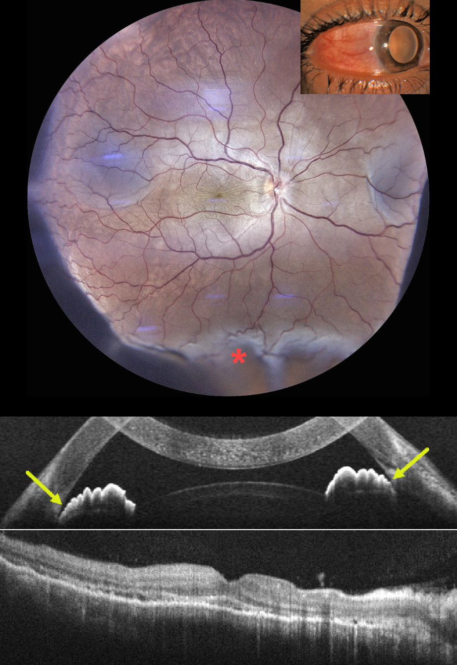
TUBERCULOUS POSTERIOR SCLERITIS
Aniruddh Soni
Originally posted on @retina.rocks July 31, 2026
This previously healthy 35YO male presented with 1 week of pain, redness, and decreased vision in his right eye. He had been treated by an outside doctor for conjunctivitis. Vision 20/200 OD and 20/20 in his normal OS. Slit lamp examination showed temporal scleral injection. Intraocular pressure was 23 mm Hg.
Color photography shows radiating chorioretinal folds throughout the macula with an inferior exudative retinal detachment. OCT scanning of the macula shows chorioretinal folds, with anterior chamber OCT showing a narrow-to-closed angle. B-scan ultrasonography showed subtenon’s fluid (not shown).
Following a 3-week course of tapering oral steroids and glaucoma drops, vision improved to 20/40 with reduced chorioretinal folds and complete resolution of the exudative detachment. Rheumatologic workup was negative, but Mantoux skin testing and QuantiFERON-TB GOLD were positive, with a subsequent diagnosis of latent tuberculosis (TB). Antituberculous therapy was recommended, but the patient refused treatment.
Learning Points:
Tuberculous posterior scleritis is a rare manifestation of ocular TB (Agarwal and Majumder, Indian J Ophthalmol 2019;67:1362-1365), which itself occurs in approximately 1.5% of patients with confirmed systemic TB. Ocular findings include optic disc edema, choroiditis, and sclerochoroidal thickening with widening of the sub-Tenon space. It may occur via direct hematogenous dissemination of Mycobacterium tuberculosis or through a hypersensitivity-mediated immune response to the organism and can develop even in the absence of clinically active pulmonary disease.
Diagnosis, as in our patient, is largely presumptive, requiring a high index of suspicion supported by bloodwork, chest imaging to assess pulmonary involvement, and exclusion of mimics such as sarcoidosis, syphilis, and metastatic disease.
TUBERCULAR SERPIGINOUS-LIKE CHOROIDITIS
Aniruddh Soni
Originally posted on @retina.rocks July 6, 2026
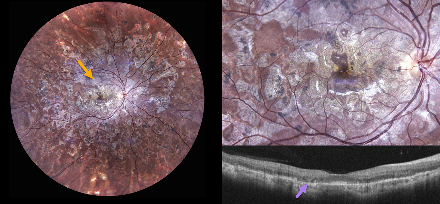
A 30YO female presented with a 1.5-year history of bilateral floaters and blurred vision. She was diagnosed elsewhere with ‘choroiditis’ and treated with oral steroids and azathioprine, with 2 recurrences. When seen in our office, vision was 20/30 OU.
Color photography OD shows extensive, variably confluent, and meandering chorioretinal scarring extending from the disc and macula to the retinal periphery. The foveal center is just spared. OCT scanning shows variable outer retinal and RPE atrophy. Identical findings were noted in her left eye (not shown).
Tuberculin skin testing and QuantiFERON-TB Gold were positive. She was referred to pulmonary and infectious disease specialists, was diagnosed with latent TB, and started on anti-tuberculosis treatment (ATT) with tapering oral steroids.
Learning Points:
Although TB most commonly presents with pulmonary involvement, extrapulmonary sites can involve the gastrointestinal, skin, cardiovascular, genitourinary, and central nervous systems, as well as the eyes. Ocular involvement, like syphilis, can mimic virtually any type of uveitis, including anterior, intermediate, posterior, and panuveitis, retinitis, and retinal vasculitis, neuroretinitis, optic neuropathy, choroidal granuloma, choroiditis, and scleritis.
Tubercular serpiginous-like choroiditis mimics serpiginous but affects younger patients with more multifocal and peripheral recurrences, foveal sparing, vitritis, and progression if untreated (Agarwal et al, AJO 2020;220:160-169). Although the exact mechanism is not yet clear, a direct or indirect infectious trigger by the Mycobacterium tuberculosis is thought to cause choroiditis. Its origin is probably immunogenic, as it appears to respond to corticosteroids and other immunosuppressants. Our patient was a bit unusual in that he required systemic steroids, ATT, and adalimumab to finally control his disease.
TUBERCULAR RETINITIS AND VASCULITIS
The European VitreoRetinal Society (EVRS), Saarang Hansraj and Sanjiv Hansraj
Originally posted on @retina.rocks May 8, 2026
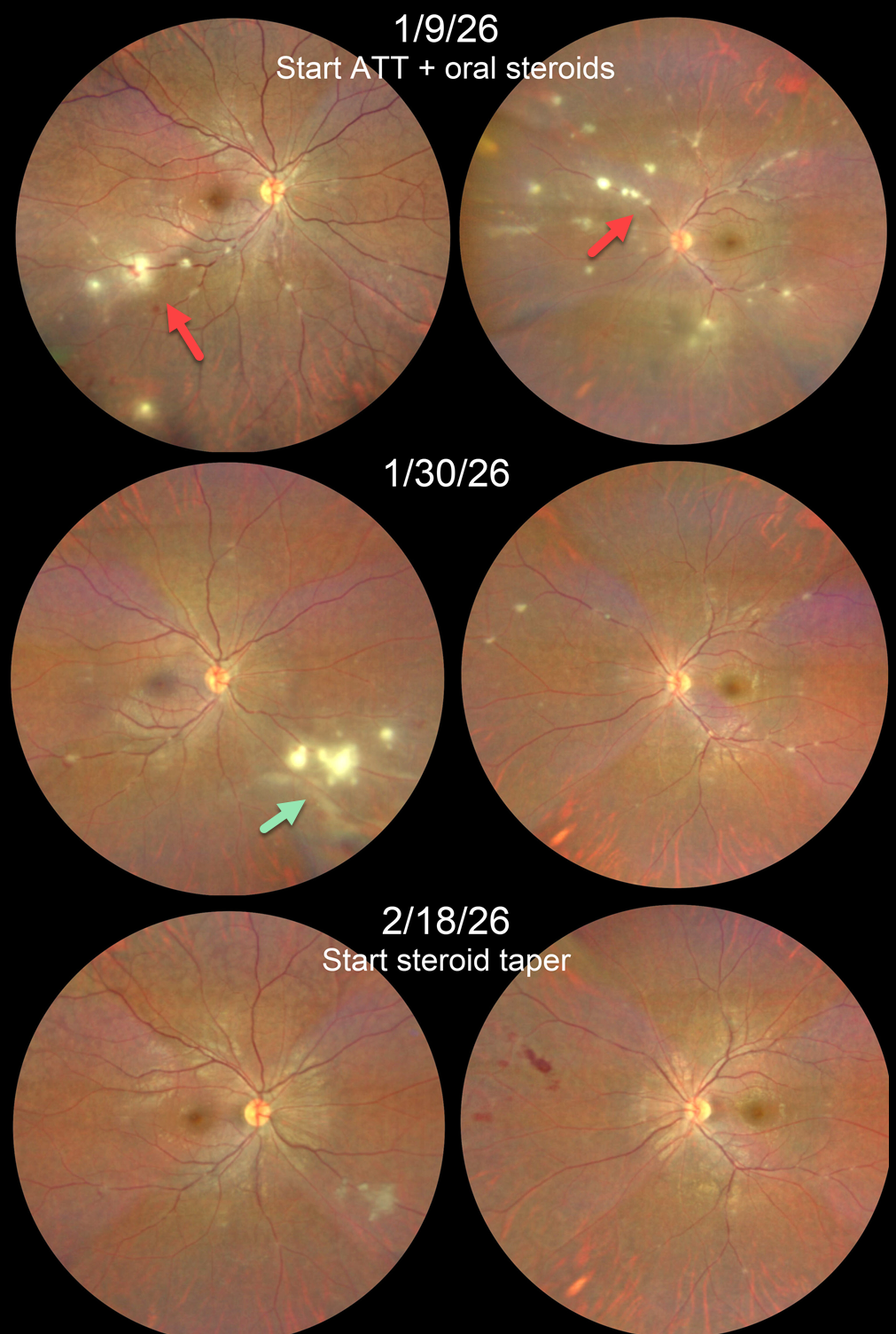
This 22YO male presented with a 1-week history of blurred vision in his left eye. His sister had been undergoing treatment for pulmonary tuberculosis 1 year ago. She also had a history of Takayasu arteritis requiring renal artery embolization and was on multiple immunosuppressants. Vision was 20/20 OD and 20/25 OS.
Color photography shows multifocal areas of retinitis/phlebitis bilaterally. Chest CT revealed paratracheal and subcarinal lymph node calcification, and Mantoux skin testing was strongly positive. Anti-tubercular therapy (ATT) and oral steroids were begun.
He returned 3 weeks later with 20/20 OU. Both eyes were markedly improved except for a new area of active inflammation inferonasally OD. Two weeks later, the retinitis/phlebitis was inactive bilaterally, although some new retinal hemorrhages were noted in the left nasal midperiphery. He was then asked to taper his steroids.
Learning Points:
Although TB most commonly presents with pulmonary involvement, extrapulmonary sites can include the gastrointestinal, skin, cardiovascular, genitourinary, and central nervous systems, as well as the eyes. Ocular involvement, like syphilis, can mimic virtually any type of uveitis, including anterior, intermediate, posterior, and panuveitis, retinitis and retinal vasculitis, neuroretinitis, optic neuropathy, choroidal granuloma, choroiditis, and scleritis.
Tubercular retinitis and vasculitis are prevalent in endemic countries such as India. Laboratory investigations should validate/rule out, not determine, the diagnosis. It is important to closely follow these patients after starting treatment. Despite initial improvement in our patient’s retinitis and vasculitis, a new area developed that needed an additional 2 weeks of therapy before we could start steroid tapering.
TUBERCULAR SERPIGINOUS-LIKE CHOROIDITIS
Akansha Sharma
Originally posted on @retina.rocks March 9, 2026
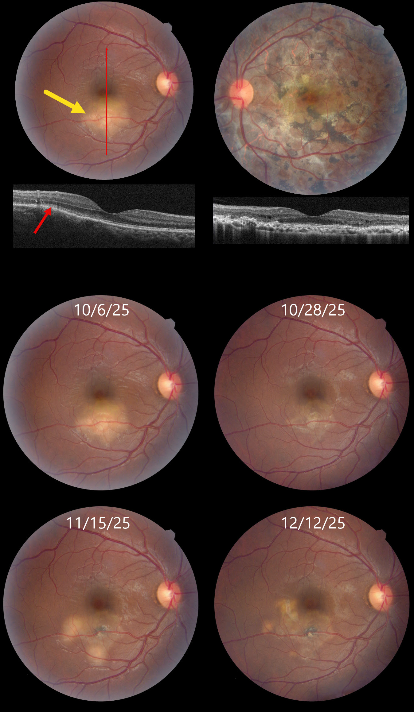
This 32YO male presented on 10/6/25 with a 6-day history of decreased vision in his right eye. About 2 years earlier, he was diagnosed elsewhere with tubercular serpiginous-like choroiditis in his left eye. Extensive workup at the time, including a chest X-ray, was negative except for a positive QuantiFERON TB Gold result. He was lost to follow-up without treatment for his latent tuberculosis (TB). Vision in our office was 20/30 OU.
Color photography shows a deep, creamy-white retinal inflammatory lesion in the inferior macula. OCT shows outer retinal hyperreflectivity with loss of the outer retinal band details. Extensive scarring is noted throughout the left posterior pole, with a small island of preserved foveal tissue.
Tapering systemic corticosteroid therapy was begun, followed by anti-tuberculosis treatment. Over the ensuing 2 months, he experienced waxing and waning inflammation necessitating intravitreal Ozurdex, oral steroids, and adalimumab while vision remained stable at 20/30. We continue to follow him closely.
Learning Points:
Although TB most commonly presents with pulmonary involvement, extrapulmonary sites can include the gastrointestinal, skin, cardiovascular, genitourinary, and central nervous systems, as well as the eyes. Ocular involvement, like syphilis, can mimic virtually any type of uveitis, including anterior, intermediate, posterior, and panuveitis, retinitis, and retinal vasculitis, neuroretinitis, optic neuropathy, choroidal granuloma, choroiditis, and scleritis.
Tubercular serpiginous-like choroiditis mimics serpiginous but affects younger patients with more multifocal and peripheral recurrences and progression if untreated (Agarwal et al, AJO 2020;220:160-169). Although the exact mechanism is not yet clear, a direct or indirect infectious trigger by the Mycobacterium tuberculosis is thought to cause choroiditis. Its origin is probably immunogenic since it seems to respond to corticosteroids and other immunosuppressants. Our patient was a bit unusual in that he required systemic steroids, ATT, and adalimumab to finally control his disease.
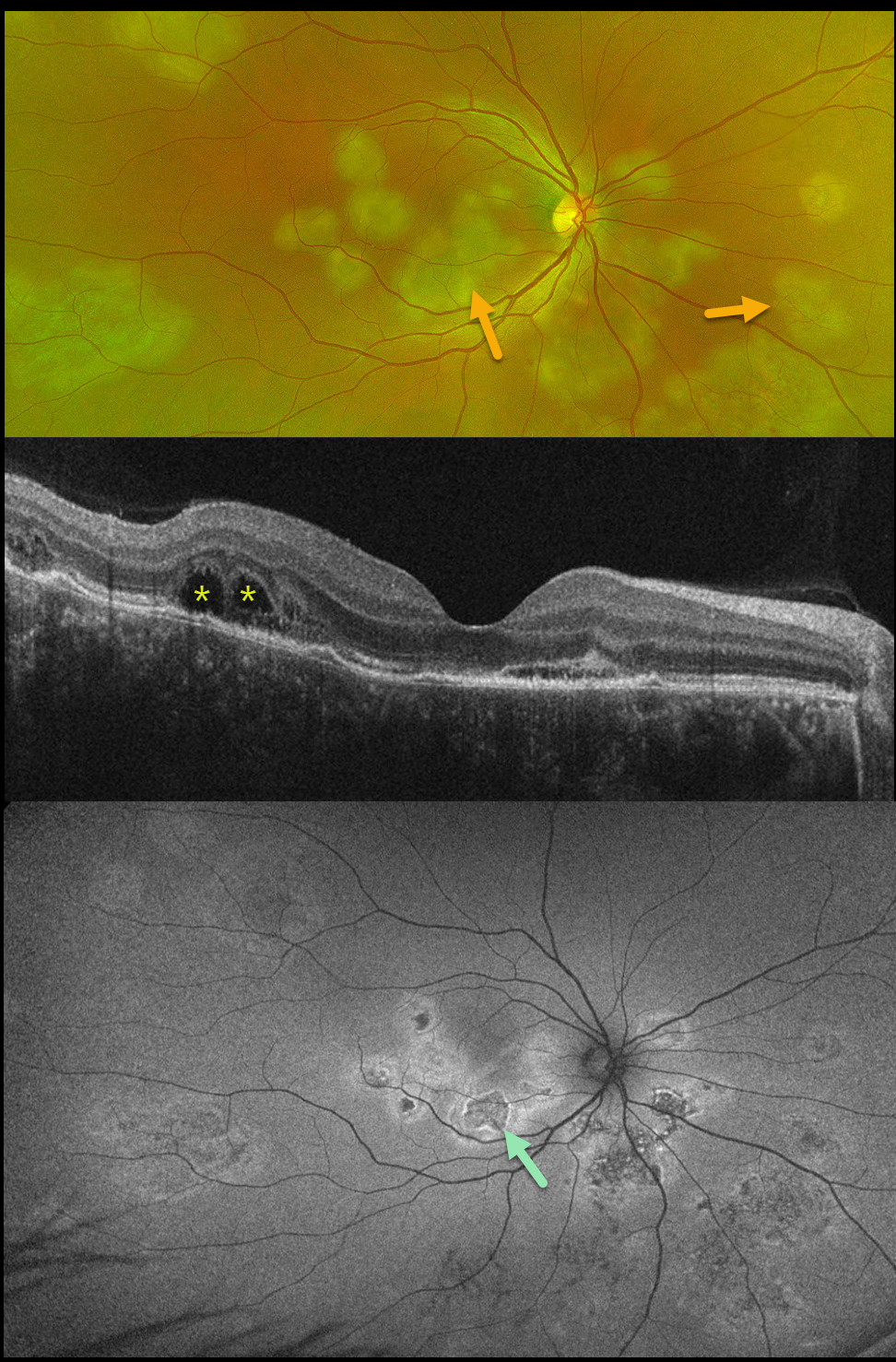
TUBERCULAR SERPIGINOUS-LIKE CHOROIDITIS
The European VitreoRetinal Society (EVRS) and Vaibhav Sethi
Originally posted on @retina.rocks January 2, 2026
This previously healthy 41YO male presented with 4 days of blurred vision in his right eye. Vision was 20/80 OD and 20/20 in his normal OS.
Optos color RG imaging shows multifocal subretinal creamy white placoid lesions in the macula, peripapillary retina, and midperiphery. OCT scanning shows variable disruption of the outer retinal bands, as well as bacillary layer detachments (BALAD) temporally. The placoid lesions on fundus autofluorescence (FAF) show variable central hypo-FAF with more hyper-FAF borders.
Mantoux skin testing was positive at 24mm, and chest CT revealed left lower lobe ground glass opacities with hilar adenopathy. He was started on anti-tubercular therapy and oral prednisone.
Although tuberculosis most commonly presents with pulmonary involvement, extrapulmonary sites can include the gastrointestinal, skin, cardiovascular, genitourinary, and central nervous systems, as well as the eyes. Ocular involvement, like syphilis, can mimic virtually any type of uveitis, including anterior, intermediate, posterior, and panuveitis, retinitis, and retinal vasculitis, neuroretinitis, optic neuropathy, choroidal granuloma, choroiditis, and scleritis.
Learning Points:
Tubercular serpiginous-like choroiditis mimics serpiginous but affects younger patients with more multifocal and peripheral recurrences and progression if untreated (Agarwal et al, AJO 2020;220:160-169). Although the exact mechanism is not yet clear, a direct or indirect infectious trigger by the Mycobacterium tuberculosis is thought to cause choroiditis. Its origin is probably immunogenic since it seems to respond to corticosteroids and other immunosuppressants.
BALAD appears as a unique dome-shaped collection of intraretinal fluid from photoreceptor splitting. These detachments can be observed in an increasing number of conditions, most classically Vogt-Koyanagi-Harada disease (Cicinelli et al, Ophthalmology Retina 2020;4:454-456).
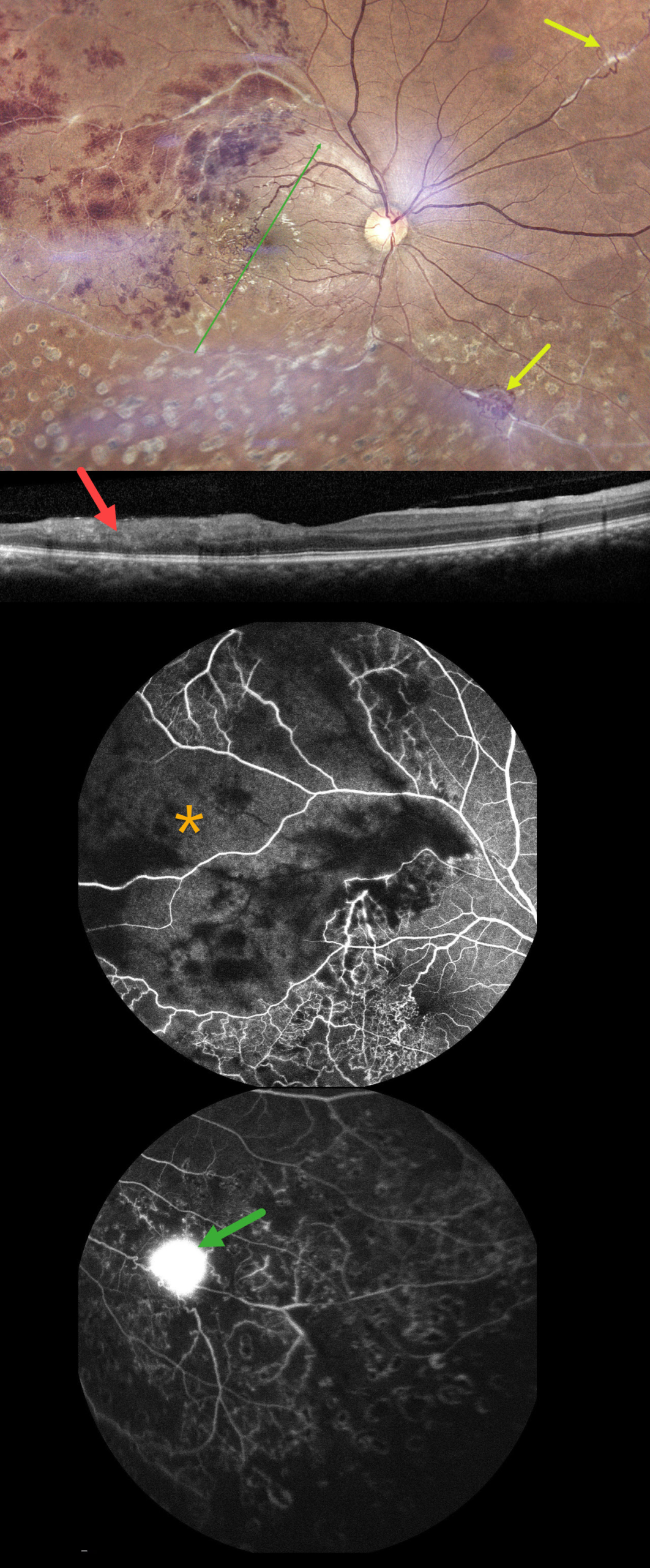
TUBERCULAR ISCHEMIC VASCULITIS
The European VitreoRetina Society (EVRS) and Abhishek Upadhyaya
Originally posted on @retina.rocks November 28, 2025
This 40YO male presented with 10 days of vision loss in his right eye. There was a history of prior laser treatment to this eye elsewhere. Vision was 20/40 OD and 20/20 in his normal OS.
Fundus photography shows superotemporal retinal hemorrhages consistent with a branch retinal vein occlusion (BRVO), with a possible additional BRVO inferotemporally. Collateral vessels are noted in the temporal fovea with scatter laser scarring in the inferior hemiretina. Skip vasculitis can be seen in the superotemporal quadrant along with venous sheathing in other areas as well. There are a few areas of fibrosed peripheral neovascularization (NV). OCT scanning shows variable temporal macular thinning with disorganization of retinal inner layers (DRIL). Fluorescein angiography shows profound ischemia within the superotemporal BRVO, along with vascular leakage and peripheral ischemia. Leakage from the Inferonasal NVE can be seen in the late stages.
Systemic workup was consistent with Ocular Tuberculosis (TB), including a positive Mantoux skin test (12mm), positive Quantiferon TB testing, and calcified lung nodules on contrast-enhanced computed tomography. He was started on oral steroids and antitubercular therapy, and scatter laser was planned for the areas of angiographic ischemia.
Learning Points:
The differential for occlusive peripheral retinal vasculitis (Huvard et al, Ophthalmology Retina 2022;6:43-48) includes TB, rheumatologic disorders/systemic vasculitides (granulomatosis with polyangiitis, Bechet’s disease, systemic lupus, etc.), idiopathic retinal vasculitis and neuroretinitis, and sarcoidosis. Historically, Eales disease has been used to describe a variety of heterogeneous disorders, including tuberculous vasculitis, and, due to its ambiguity, we prefer not to use this term.
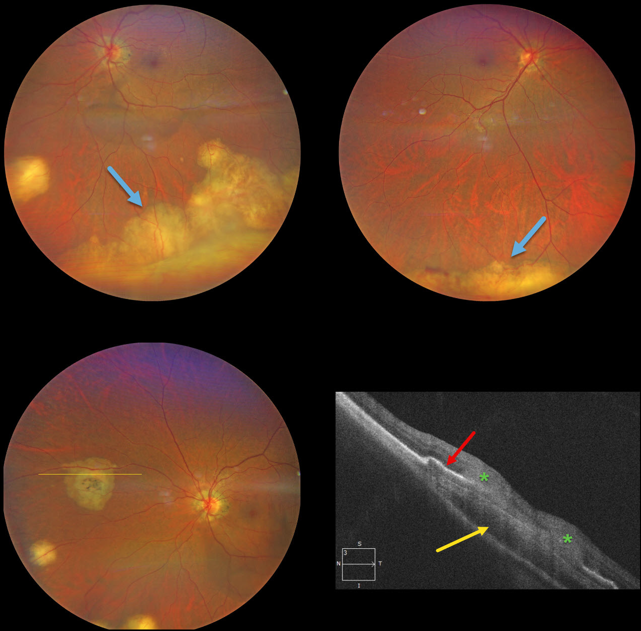
MULTIFOCAL TUBERCULOSIS CHOROIDAL GRANULOMAS
Will Gibson
Originally posted on @retina.rocks July 3, 2025
This previously healthy 49YO Indian female was visiting the United States and presented with new floaters in her left eye. Vision was 20/25 OD and 20/40 OS. There was no anterior uveitis, and the vitreous was clear bilaterally.
Color photography shows multifocal bilateral amelanotic choroidal lesions, some isolated and others confluent in the inferior periphery. OCT scanning through a nasal lesion OS shows a relatively hyporeflective choroidal lesion, irregular overlying RPE elevation, and probable infiltration of the overlying retina.
QuantiFERON-TB testing was positive with a negative chest X-ray. The patient was referred to the infectious disease department and started on anti-tuberculosis treatment (ATT).
Learning Points:
Although TB most commonly presents with pulmonary involvement, extrapulmonary sites can include the gastrointestinal, skin, cardiovascular, genitourinary, and central nervous systems, as well as the eyes. Ocular involvement, like syphilis, can mimic virtually any type of uveitis, including anterior, intermediate, posterior, and panuveitis, retinitis and retinal vasculitis, neuroretinitis, optic neuropathy, choroidal granuloma (our patient), choroiditis, and scleritis. For an excellent review of intraocular tuberculosis, see Bupta et al, Survey Ophthalmology 2007;52:561-587.
Tubercular granulomas can be distinguished from sarcoid lesions by being solitary, yellow, larger, and vascularized (Agarwal et al, AJO 2021;226:42-55). Our patient is therefore somewhat unusual with bilateral multifocal lesions. Sarcoid workup was negative.
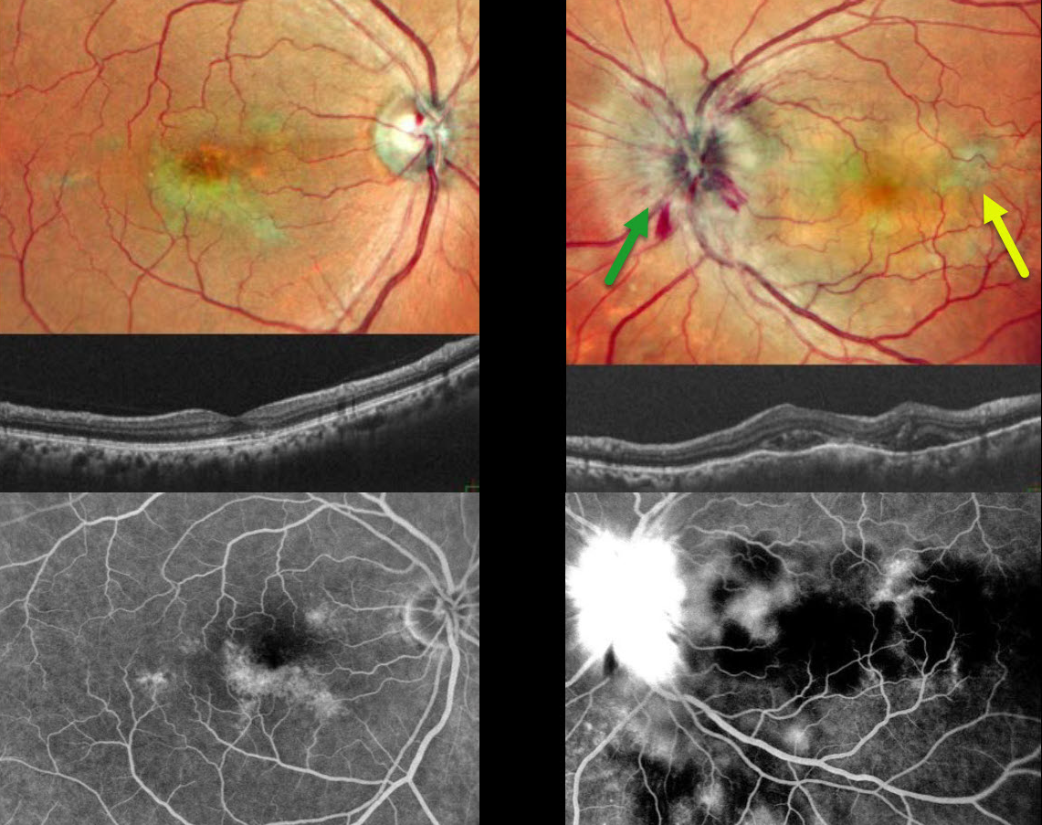
TUBERCULAR SERPIGINOUS-LIKE CHOROIDITIS
Originally posted on @retina.rocks March 11, 2025
This 54YO male presented with 2 weeks of vision loss in his left eye. He was diagnosed elsewhere with papilledema, and brain MRI scanning was normal. Vision was 20/20 in his asymptomatic OD and counting fingers OS. Pseudocolor SLO imaging of the OS shows marked disc swelling with radiating hemorrhages in the nerve fiber layer. Some deep, hypopigmented retinal lesions are also seen.
OCT scanning shows irregular choroidal thickening vs choroidal folds with overlying pockets of variably reflective subretinal fluid. The right eye shows multifocal serpiginous-like areas of chorioretinal scarring with variable outer retinal thinning on OCT.
On fluorescein angiography, the macular lesions stain OD. The left nerve shows profound leakage, and the choroidal lesions show variable blockage and staining.
We suspected tubercular serpiginous-like choroiditis OD with acute tubercular papillitis and choroiditis OS. Chest X-ray showed a mass pressing on the trachea. Chest CT confirmed the mass, and subsequent biopsy revealed granulomatous thyroiditis. His Mantoux skin test was positive. We advised emergent Infectious disease consultation regarding starting anti-tubercular therapy. Unfortunately, he was immediately lost to follow-up.
Learning Points:
Although tuberculosis most commonly presents with pulmonary involvement, extrapulmonary sites can include the gastrointestinal, skin, cardiovascular, genitourinary, and central nervous systems, as well as the eyes. Ocular involvement, like syphilis, can mimic virtually any type of uveitis, including anterior, intermediate, posterior, and panuveitis, retinitis, and retinal vasculitis, neuroretinitis, optic neuropathy, choroidal granuloma, choroiditis, and scleritis.
Tubercular serpiginous-like choroiditis mimics serpiginous but affects younger patients with more multifocal and peripheral recurrences and progression if untreated (Agarwal et al, AJO 2020;220:160-169). Although the exact mechanism is not yet clear, a direct or indirect infectious trigger by the Mycobacterium tuberculosis is thought to cause the choroiditis. Its origin is probably immunogenic since it seems to respond to corticosteroids and other immunosuppressants.
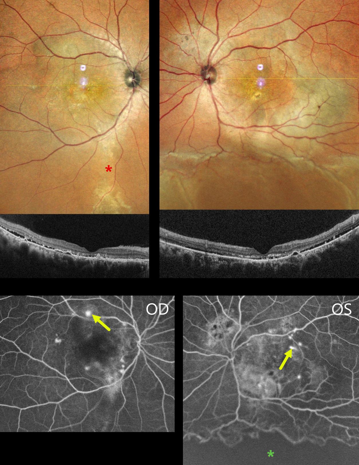
CENTRAL SEROUS CHORIORETINOPATHY WITH EXUDATIVE RD AND COMCOMITANT TB
Anand Temkar, Navneet Mehrotra, and Manish Nagpal
Originally posted on @retina.rocks November 7, 2024
This 39YO male presented with 4 months of bilateral vision loss. He was diagnosed elsewhere with chronic idiopathic central serous chorioretinopathy (ICSC) and was placed on azathioprine and spironolactone 3 months earlier. There was no history of steroid use or injections. Vision was 20/120 OD and 20/200 OS.
MultiColor imaging shows macular pigmentary changes that extend in a gutter inferiorly OD. Diffuse macular pigmentary changes are noted in his left eye that communicate with an inferior exudative retinal detachment. OCT scanning shows shallow subretinal fluid with hyperreflective shed outer segments, outer retinal hyperreflective foci, and RPE detachments. Fluorescein angiography shows bilateral multifocal subretinal blot leakage. A broad area of ischemia is present overlying the inferior detachment of the OS.
Mantoux skin testing (15x18mm) and QuantiFERON-TB Gold testing were positive. Chest CT showed fibrotic strands in the right lung. He was referred for systemic antituberculosis treatment. A week later, his local ophthalmologist reported that his vision and retinal findings improved, but he was then lost to follow-up.
Learning Points:
Our patient presented with severe bilateral idiopathic central serous chorioretinopathy (ISCS) with multifocal leaks in each eye and a secondary exudative detachment in his left eye. There are rare case reports of TB occurring in patients with ICSC, and these are likely coincidental findings (Khan et al, BMJ Case Rep 2017:bcr-2016-216471).
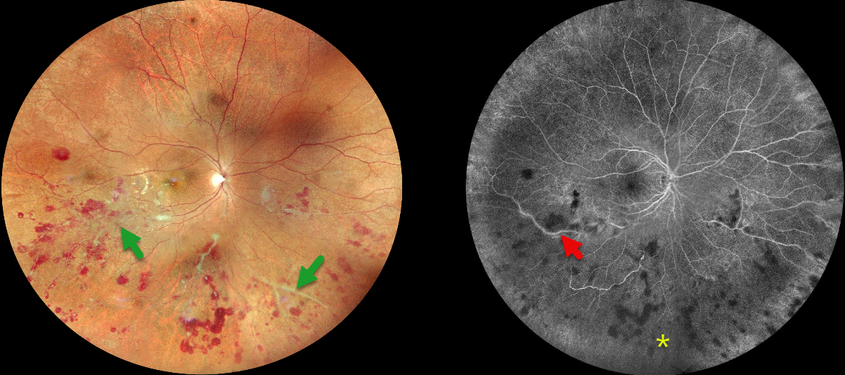
TUBERCULOSIS VASCULITIS
Tejaswita Verma, Navneet Mehrotra and Manish Nagpal
Originally posted on @retina.rocks September 18, 2024
This 27YO male presented with 10 days of floaters and photophobia in his right eye. He was tentatively diagnosed with tuberculosis (TB) several days earlier and sought a second opinion with us. Recent Mantoux skin testing was markedly positive (20 mm induration), and QuantiFERON-TB Gold was positive as well. Vision was 20/20 bilaterally.
Fundus photography shows inferior and nasal vascular sheathing, inner and deep retinal hemorrhages in the inferior hemisphere, and focal areas of whitish inner retinal ischemia in the inferior macula. Fluorescein angiography shows variable midperipheral ischemia with focal vascular staining. The left eye was normal.
Chest CT was supportive of active pulmonary TB, and the patient was started on antitubercular treatment (ATT) by the pulmonary team on the day of our exam.
When he returned four days later, his vision was 20/40 OD. We recommended an intravitreal anti-VEGF injection followed by scatter laser, but unfortunately, he was then lost to follow-up.
Learning Points:
The differential for occlusive peripheral retinal vasculitis (Huvard et al, Ophthalmology Retina 2022;6:43-48) includes TB, rheumatologic disorders / systemic vasculitides (granulomatosis with polyangiitis, Bechet’s disease, systemic lupus, etc), idiopathic retinal vasculitis and neuroretinitis, and sarcoidosis. Historically, Eales’ disease has been used for a variety of heterogeneous disorders, including tuberculous vasculitis, and due to its ambiguity, we prefer not to use this term.
The diagnosis of tubercular vasculitis is often presumptive and difficult to diagnose without isolation of TB from ocular tissue or fluids. However, as with our patient, it is a reasonable diagnosis in the proper clinical setting, including in those from endemic areas and with a systemic diagnosis of TB (Agrawal et al, 2017 37:112-117). Systemic ATT, along with local ocular treatment for neovascular complications (anti-VEGF injections, scatter laser photocoagulation), is recommended.
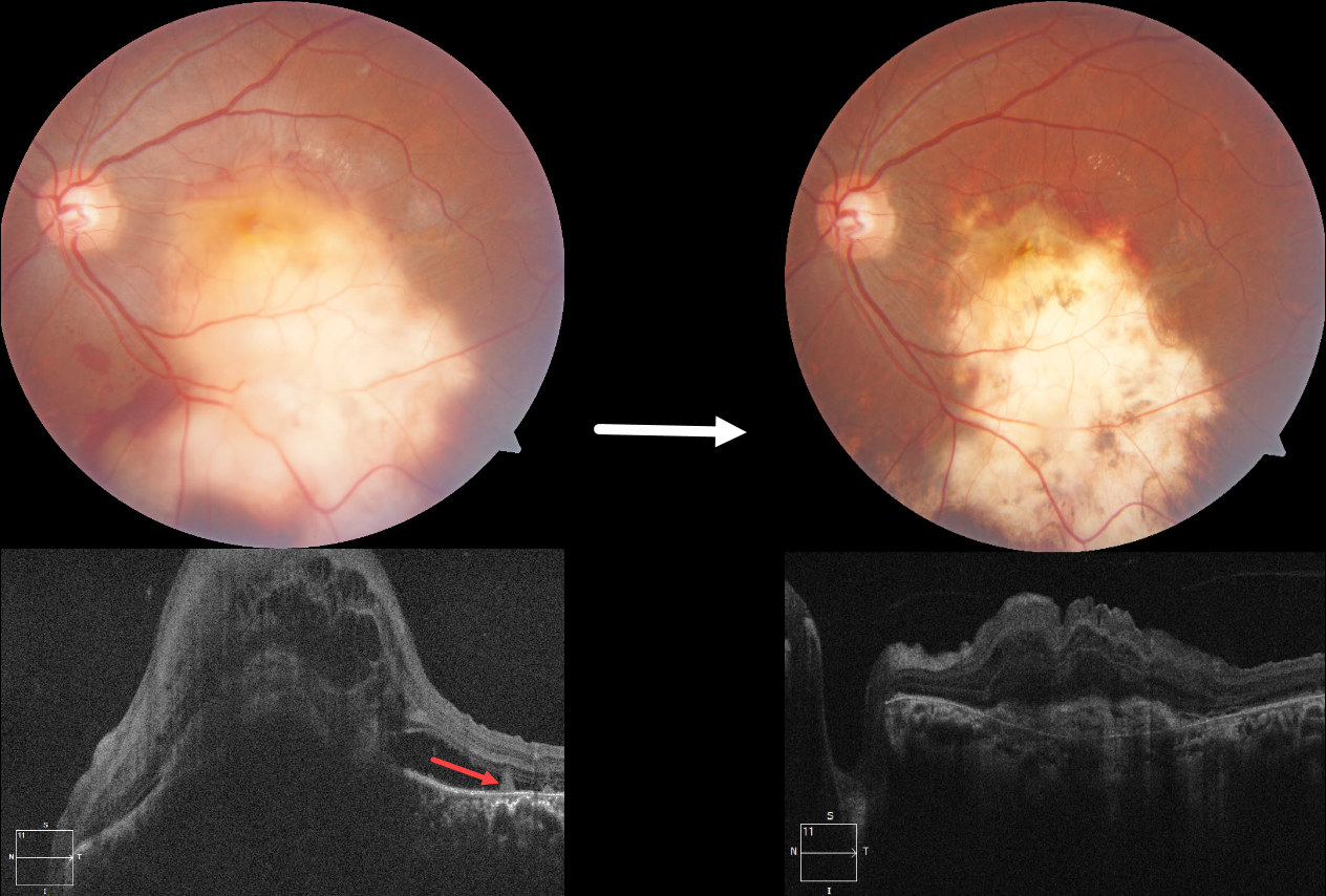
TUBERCULOSIS CHOROIDAL GRANULOMA
Divya Nair
Originally posted on @retina.rocks July 22, 2024
This 36YO male presented with 10 days of vision loss in his left eye. One month earlier, he was diagnosed with disseminated tuberculosis (TB), peritonitis, and pleural effusion, and was placed on systemic antitubercular therapy (ATT). Vision was 20/20 in his normal right eye and counting fingers in his left eye.
Fundus photography shows a large elevated yellowish submacular lesion that extends into the inferior midperiphery. Subretinal blood is noted along its superior and inferior margins. Faint lipid is present superotemporally, and some retinal striae are seen temporally. Spectral domain OCT shows a large dome-shaped hyporeflective choroidal lesion with overlying cystic retinal thickening, subretinal fluid, and subretinal hyperreflective material. A few hyperreflective pyramidal lesions are noted above the RPE temporally.
We started him on oral corticosteroids. In view of the vascularized lesion with subretinal blood, an intravitreal injection of Avastin was also given. Six weeks later, there is a relatively flat scar with near complete resolution of the blood. The choroidal lesion has resolved with an overlying hyperreflective subretinal scar and decreased retinal thickening. Unfortunately, vision remained at counting fingers.
Learning Points:
Although TB most commonly presents with pulmonary involvement, extrapulmonary sites can include the gastrointestinal, skin, cardiovascular, genitourinary, and central nervous systems, including the eyes. Ocular involvement, like syphilis, can mimic virtually any type of uveitis, including anterior, intermediate, posterior, and panuveitis, retinitis and retinal vasculitis, neuroretinitis, optic neuropathy, choroidal granuloma, choroiditis, and scleritis.
For an excellent review of intraocular tuberculosis, see Bupta et al, Survey Ophthalmology 2007;52:561-587.
Tubercular granulomas can be distinguished from sarcoid lesions by their solitary nature, yellow color, larger size, and vascularization (Agarwal et al., AJO 2021;226:42-55). All these features were found in our patient. Vascularization is thought to arise from localized hypoxia, leading to increased levels of vascular endothelial growth factor. Anti-VEGF injections and systemic corticosteroids, in addition to ATT, are helpful in hastening granuloma resolution and reducing secondary exudation (Lee et al, Graefe’s 2022;260:1641-1650).
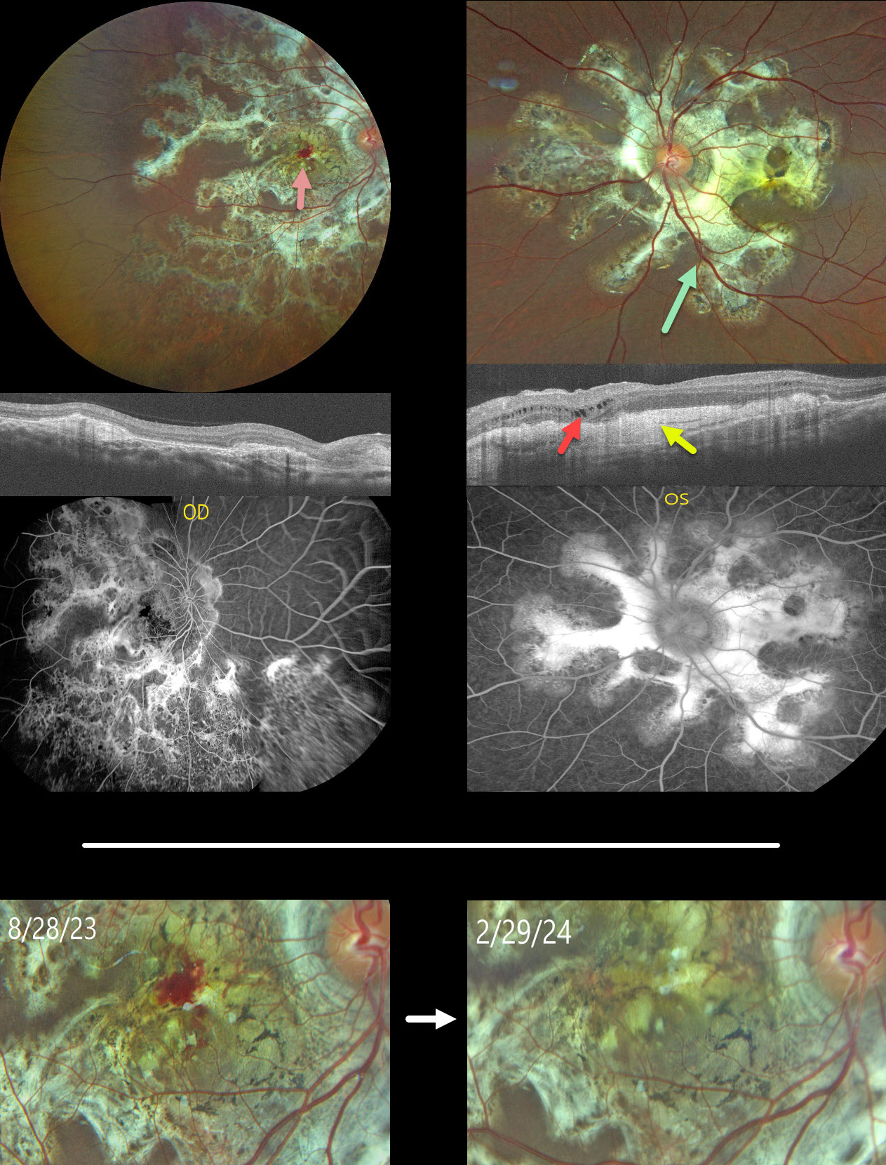
TUBERCULAR SERPIGINOUS-LIKE CHORIORETINITIS
Ogugua Okonkwo, Adekunle Olubola Hassan, Ayodele Harriman, Ogochukwu Sibeudu, and Idris Akintayo Oyekunle
Originally posted on @retina.rocks May 1, 2024
This healthy 13YO male presented with a 5-month history of bilateral vision loss. Vision was 20/200 OU.
Fundus photography shows bilateral subretinal, variably fibrotic peripapillary scarring that extends outwards with finger-like projections. A foveal hemorrhage is noted in the right macula. More mottled pigmented scarring extends into the right inferior periphery.
OCT scanning shows variable bilateral outer retinal atrophy and hyperreflective subretinal fibrosis with mild outer nuclear cysts in the nasal left macula. This scarring stains angiographically.
We were hesitant to start anti-VEGF therapy in a young boy and felt that the macular neovascularization causing the right macular blood might be inflammatory. He was therefore placed on a rapidly tapering course of oral prednisone.
At the 6-month follow-up, vision remained stable at 20/200 in this eye, with resolution of the blood.
Mantoux skin testing was negative. We have made multiple attempts to have the patient get QuantiFERON-TB Gold testing, but to date have been unsuccessful.
Learning Points:
Serpiginous choroiditis is an idiopathic, usually bilateral chorioretinal inflammatory condition, most commonly found in middle-aged men (Khanamiri and Rao, Surv Ophthalmology 2013;58:203-232). Recurrent areas of inflammation develop along the edge of a previous scar, producing scarring that meanders from the optic nerve outwards. Vision is good unless scarring or macular neovascularization extends through the macular center.
Tubercular serpiginous-like choroiditis mimics serpiginous but affects younger patients with more multifocal and peripheral recurrences and progression if untreated (Agarwal et al, AJO 2020;220:160-169). Although the exact mechanism is not yet clear, a direct or indirect infectious trigger by the Mycobacterium tuberculosis is thought to cause the choroiditis. We believe our patient has this diagnosis and will continue to confirm it with further testing.
TUBERCULOUS NEURORETINITIS
Keith Slayden
Originally posted on @retina.rocks April 2, 2024
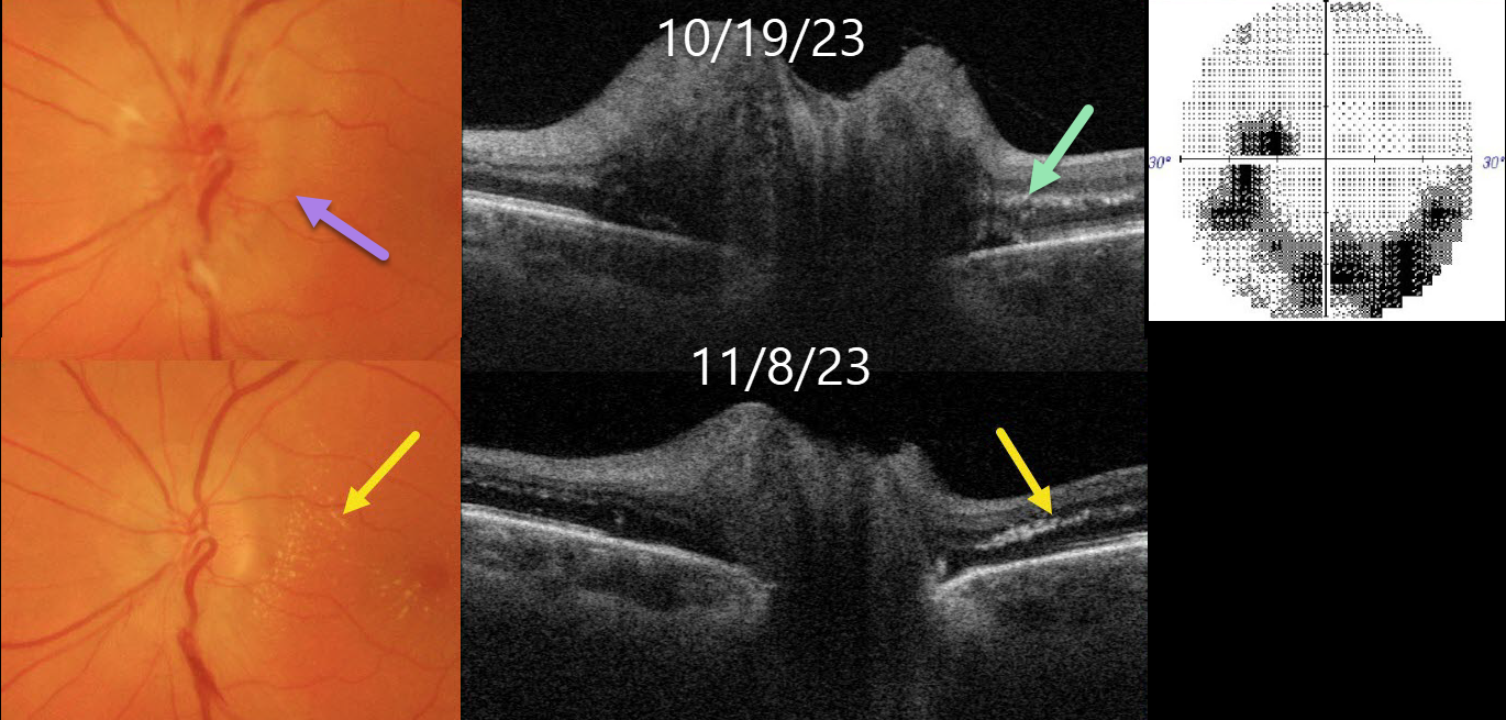
This healthy 56YO female presented with 1-2 weeks of blurred vision in her left eye. Vision was 20/25 in her normal OD and 20/100 OS.
Color imaging shows a diffusely swollen left nerve. Optic nerve Triton swept-source OCT B-scan confirms the diffuse retinal nerve fiber layer edema. A few dots of subclinical hyperreflective lipid are noted in the outer plexiform layer. 30-2 visual field testing shows an inferior unilateral arcuate scotoma extending from the blind spot.
Extensive blood work and chest X-ray were negative except for a positive QuantiFERON-TB Gold. Infectious disease consultation led to the diagnosis of latent tuberculosis, and she was started on systemic anti-tuberculous therapy.
Over the following several weeks, as the optic nerve edema resolved, the papillomacular outer plexiform layer lipid became more prominent.
Learning Points:
The diagnosis for a unilateral swollen nerve is extensive. However, it becomes much smaller when associated with acute inner retinal ischemia (not seen in our case) and later lipid exudation within Henle’s layer (neuroretinitis), including cat scratch (Bartonella), Lyme disease, and syphilis. When no underlying cause is found, the entity is called Leber’s idiopathic stellate neuroretinitis.
Tuberculosis is a rare cause of neuroretinitis (Stechschulte et al, J Neuroophthalmology 1999;19:201-204). It is unclear whether our patient’s findings are idiopathic or related to her latent tuberculosis.
TUBERCULAR SERPIGINOUS-LIKE CHORIORETINITIS
Asma Samsudeen and Ashish Sharma
Originally posted on @retina.rocks January 18, 2024
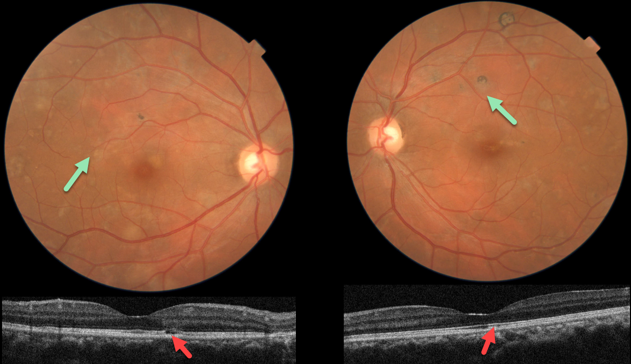
This 38YO male presented with floaters in each eye. He denied any past medical history. Vision was 20/20 OU.
Fundus photos show multifocal patches of variably pigmented chorioretinal scarring throughout each posterior pole.
OCT scanning in each eye shows variable outer retinal atrophy and ellipsoid disruption. Unfortunately, fundus autofluorescence and fluorescein angiography are not available.
Subsequent workup was consistent for active tuberculosis, including a positive QuantiFERON-TB Gold and chest x-ray findings. Systemic anti-tuberculous therapy was begun.
Learning Points:
Serpiginous choroiditis is an idiopathic, usually bilateral chorioretinal inflammatory condition most commonly found in middle-aged men (Khanamiri and Rao, Surv Ophthalmology 2013;58:203-232).
Recurrent areas of inflammation develop along the edge of a previous scar, producing scarring that meanders from the optic nerve outwards. Vision is good unless scarring or macular neovascularization extends through the macular center.
Tubercular serpiginous-like choroiditis mimics serpiginous, but affects younger patients with more multifocal and peripheral recurrences and progression if untreated (Agarwal et al, AJO 2020;220:160-169).
Although the exact mechanism remains unclear, a direct or indirect infectious trigger by Mycobacterium tuberculosis is believed to cause choroiditis.
TUBERCULOSIS CHOROIDAL GRANULOMA
Originally posted on @retina.rocks July 12, 2021
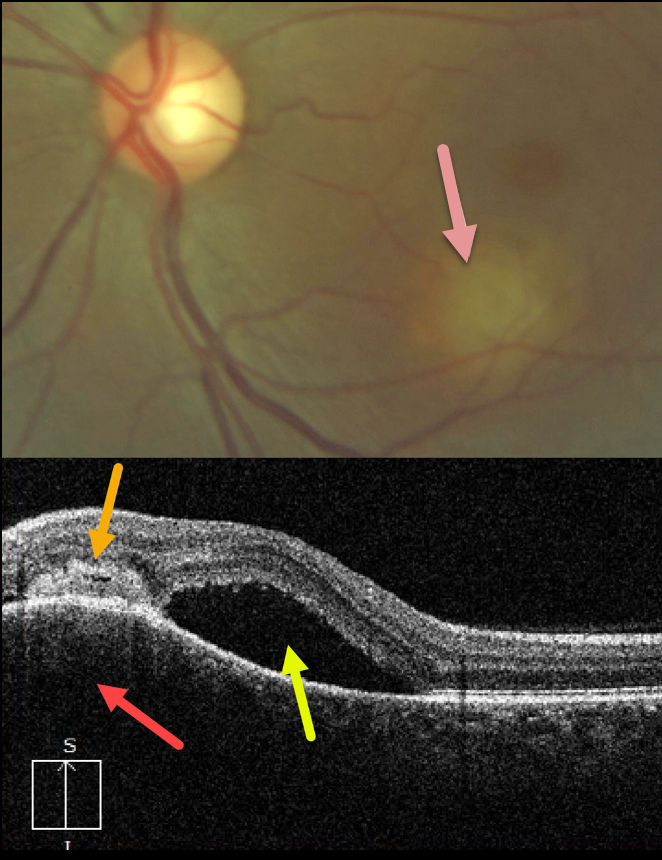
This 33YO female presented with this symptomatic lesion in her left macula. Vision was 20/25 in her normal right eye and 20/300 in her left eye.
A granulomatous lesion is noted in the inferior left macula. A vertical OCT B-scan through this lesion shows choroidal hyporeflective thickening, an overlying subretinal/intraretinal hyperreflective lesion, and superior subretinal fluid.
Laboratory testing was positive for QuantiFERON-TB Gold, and negative for FTA, CBC, and ACE. Unfortunately, despite numerous attempts to reach this patient, she was lost to follow-up.
Learning Points:
Tuberculosis (TB) is a systemic disease caused by Mycobacterium tuberculosis, characterized by the formation of caseating granulomas throughout the body. Although pulmonary involvement is the most common, extrapulmonary sites can involve the gastrointestinal, skin, cardiovascular, genitourinary, and central nervous systems, as well as the eyes.
Ocular involvement, similar to syphilis, can mimic virtually any type of uveitis, including anterior, intermediate, posterior, and panuveitis, retinitis and retinal vasculitis, neuroretinitis, optic neuropathy, and choroidal granuloma, choroiditis, and scleritis.
For an excellent review of intraocular tuberculosis, see Bupta et al, Survey Ophthalmology 2007;52:561-587.


