

Uveal Effusion
<< Back to Cases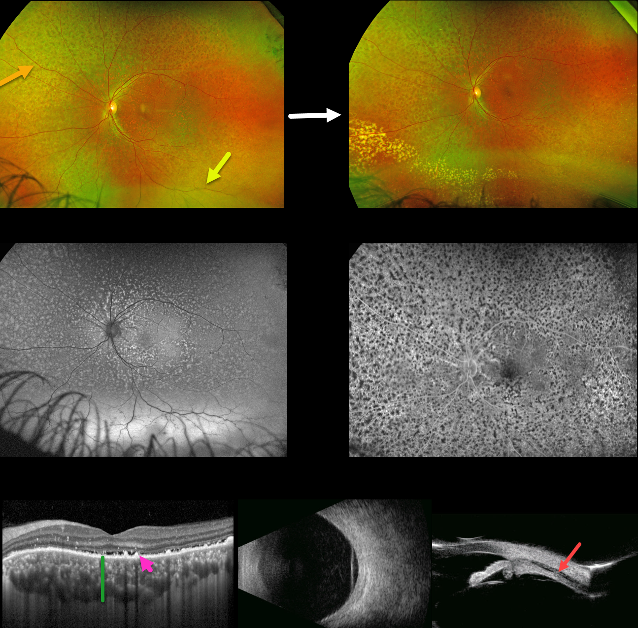
IDIOPATHIC UVEAL EFFUSION SYNDROME vs UNILATERAL BDUMP
Carl-Joe Mehanna
Originally posted on @retina.rocks July 17, 2024
This 48YO healthy male presented with two months of progressive loss of vision in his left eye. Vision was 20/50 OD due to a subcapsular cataract and 20/100 OS with a clear lens. Anterior segments were normal.
Optos color RG imaging of the left eye shows a diffuse orange-red leopard spot appearance to the fundus with an inferior exudative retinal detachment. Fundus autofluorescence (FAF) shows these lesions to be markedly hyper-FAF and block on fluorescein angiography.
Macular OCT shows a thickened choroid, shallow subretinal fluid, and multiple hyperreflective lipofuscin deposits on top of the RPE. B-scan ultrasonography shows a diffusely thickened choroid with overlying retinal detachment, and ultrasound biomicroscopy shows a ciliary body effusion.
Extensive medical workup, including bloodwork and brain MRI, was negative for malignancy or infectious processes (including syphilis, HIV, and TB). He was lost to follow-up for a year, when he presented with resolved subretinal but with new peripheral lipid. He has since been lost to follow-up.
Learning Points:
The idiopathic uveal effusion syndrome (non-nanophthalmic) is a rare entity presenting with idiopathic exudative choroidal, ciliary body, and shifting retinal detachment due to impaired transscleral suprachoroidal drainage caused by an abnormal thickened sclera (Elagouz et al, Surv Ophthalmology 2010;55:134-145). Chronic detachment can cause leopard-spot changes and vision loss.
The differential diagnosis includes leukemic infiltration, central nervous system lymphoma, and bilateral diffuse uveal melanocytic proliferation (BDUMP). The most likely cause of these red subretinal lesions is RPE-engorged, subretinal lipofuscin, which was noted on our patient’s OCT.
The five cardinal signs of idiopathic uveal effusion syndrome include 1) multifocal round/oval red patches at the level of the RPE, 2) early hypofluorescence of these patches, 3) multiple uveal melanocytic tumors with diffuse uveal thickening, 4) exudative retinal detachment, and 5) rapidly progressive cataract (Gass et al, Arch Ophthalmol 1990;108:427-533).
In our patient’s case, we feel the most likely diagnosis is idiopathic uveal effusion syndrome. However, we have seen similar findings in BDUMP, which can present unilaterally (Spaide, Retinal Cases & Brief Reports 2018;12:263-165). However, BDUMP is usually a paraneoplastic process, and our patient’s metastatic workup was negative.


