

Valsalva Retinopathy and Terson Syndrome
<< Back to Cases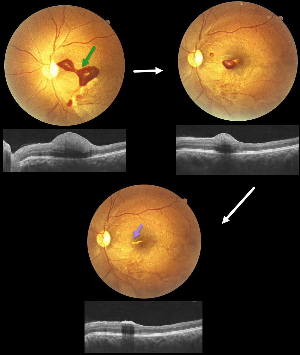
TERSON SYNDROME
Anand Temkar, Surendra Pal, Nagamani Gunasekar
Originally posted on @retina.rocks January 12, 2026
This 32YO male presented with 20 days of vision loss in his left eye. He underwent cardiovascular surgery 3 weeks earlier to remove a clot, followed 1 week later by a subarachnoid hemorrhage. Vision was 20/20 in his normal OD and 20/400 in his OS.
Fundus photography shows a subinternal limiting membrane (ILM) hemorrhage extending from the central macula, superonasally, towards the disc. This blood is hyperreflective on OCT. Observation was recommended.
One month later, vision improved to 20/70, with a marked decrease in sub-ILM blood. One month later, vision improved to 20/30, with near-complete resolution of the blood, except for a small amount of devitalized yellow blood extending nasally from the foveal center.
Learning Points:
Terson syndrome consists of intraocular hemorrhage (vitreous, subhyaloid, or retinal) associated with spontaneous or traumatic subarachnoid hemorrhage. Traditionally, the cause was attributed to elevated intracranial pressure (ICP), which caused cerebrospinal fluid effusion into the optic nerve sheath, compressing the central retinal vein and leading to subsequent rupture of retinal vessels. More recent evidence suggests glymphatic reflux, in which raised ICP causes subarachnoid blood to reflux through glymphatic channels into the globe (Kumaria et al, J Neurology 2022;269:1264-1271).
VALSALVA RETINOPATHY
The European VitreoRetina Society (EVRS) and Srishti Ramamurthy
Originally posted on @retina.rocks August 1, 2025
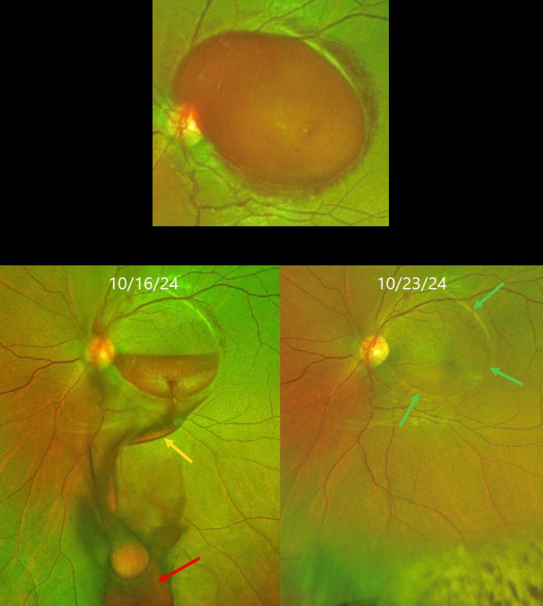
This 29YO pregnant female presented with sudden vision loss in her left eye immediately following a bout of severe vomiting. Vision was 20/20 in her normal OD and counting fingers OS.
Optos color RG imaging shows a large sub-internal limiting membrane (ILM) hemorrhage extending throughout the macula. Blood immediately drained into the subhyaloid and vitreous spaces following the YAG laser. One week later, the macular blood completely resolved. The margins of the prior elevated ILM are still visible, and some far inferior vitreous blood is noted. Vision was 20/20.
Learning Points:
Valsalva retinopathy is characterized by usually unilateral retinal and preretinal blood caused by rupture of the superficial retinal capillaries from suddenly raised intrathoracic or intra-abdominal pressure. Various causes include coughing, heavy lifting, and vomiting. These hemorrhages virtually always resolve without sequelae, although the preretinal blood in severe cases can be drained into the inferior vitreous by creating a small ILM opening using either a thermal or Nd:YAG laser (Durukan et al, Eye 2008;22:214-218).
The site chosen for laser delivery is ideally at the inferior margin of thick blood, well away from the fovea to avoid underlying retinal damage. For the Nd:YAG, the laser energy should begin at 4 mJ and be gradually titrated upward until an opening is created that allows the blood to drain inferiorly into the vitreous.
VALSALVA RETINOPATHY
Malvika Singh and Manish Nagpal
Originally posted on @retina.rocks June 9, 2025
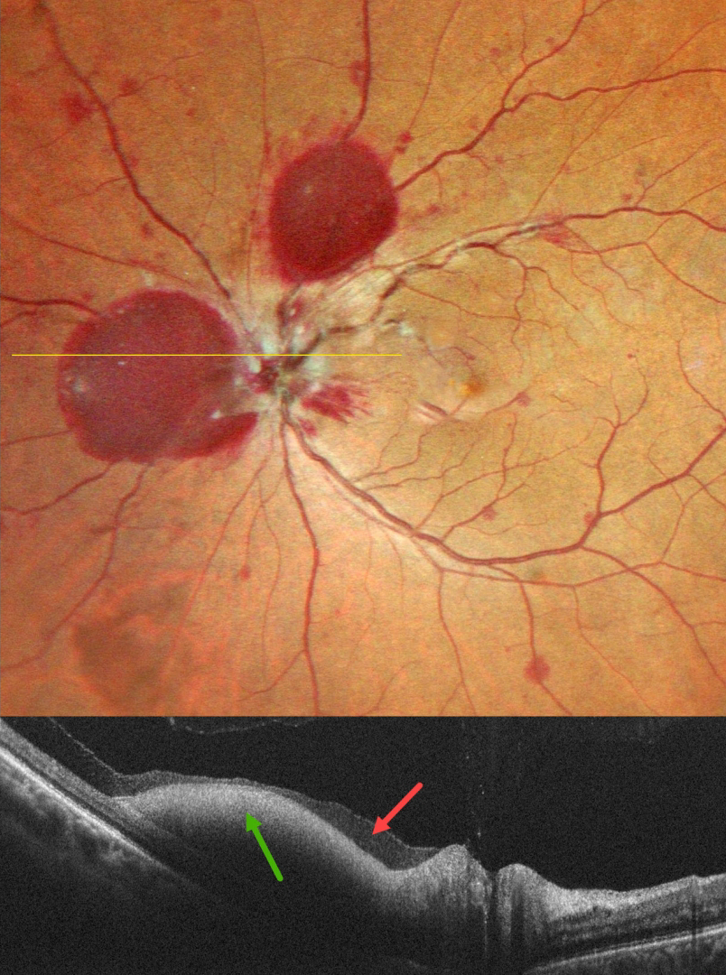
This 49YO male presented with 4 days of vision loss in his left eye. He denied any Valsalva maneuver. Vision was 20/25 in his normal OD and 20/20 OS.
Pseudocolor SLO imaging shows a large sub-internal limiting membrane (ILM) hemorrhage, nasal and superior to the nerve, along with scattered mostly deep, white-centered retinal hemorrhages. OCT scanning confirms the sub-ILM blood but also reveals shallow subhyaloid blood. Unfortunately, he was immediately lost to follow up. He then presented 1 month later with a breakthrough vitreous hemorrhage. Vitrectomy was advised due to his prior non-compliance. Vision 1 month postoperatively was 20/20, with complete resolution of all hemorrhages (not shown).
Learning Points:
Valsalva retinopathy, first described by Thomas Duane (Trans Am Ophthalmol Soc 1972; 70:298-313), is characterized by unilateral retinal and preretinal hemorrhages caused by elevated intrathoracic or intra-abdominal pressure. Various causes include coughing, heavy lifting, and vomiting. However, as in our case, patients sometimes present with these findings while denying any Valsalva maneuver. These hemorrhages virtually always resolve without sequelae, although the preretinal blood in severe cases can be drained into the inferior vitreous by creating a small ILM opening using either a thermal or Nd:YAG laser. Non -clearing break through vitreous hemorrhage is treated with vitrectomy.
VALSALVA RETINOPATHY
Nilesh Kumar
Originally posted on @retina.rocks September 26, 2024
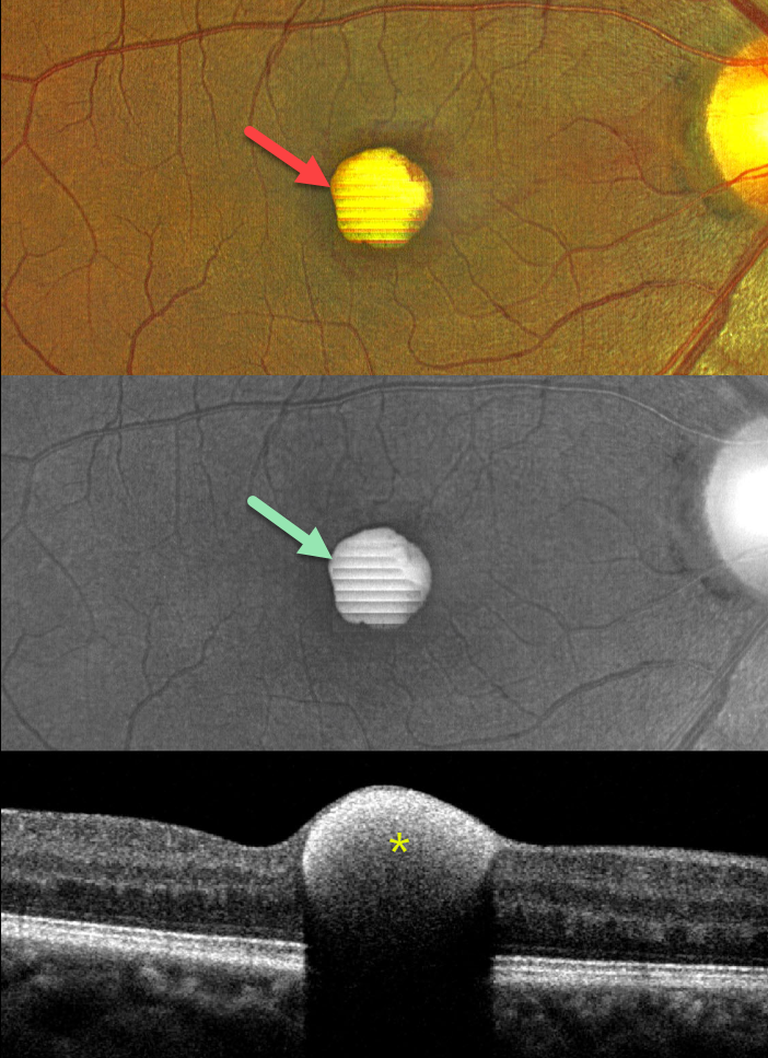
This 43YO male presented with a 1-week history of sudden decreased vision in his right eye. He denied any Valsalva maneuver. Vision was 20/70 in his right eye and 20/30 in his left eye.
Color imaging shows a foveal yellow sub-internal limiting membrane (ILM) hemorrhage. Fundus autofluorescence (FAF) shows intense hyper-FAF. Spectral domain OCT shows a hyperreflective sub-ILM lesion that shadows posteriorly.
Our patient was treated with observation, and the blood levels were greatly decreased 3 weeks later (not shown).
Learning Points:
Valsalva retinopathy was first described by Thomas Duane (Trans Am Ophthalmol Soc 1972; 70:298-313). It is characterized by usually unilateral retinal and preretinal blood caused by raised intrathoracic or intra-abdominal pressure. Various causes include coughing, heavy lifting, and vomiting. However, as in our case, sometimes patients present with these findings, denying any Valsalva maneuver (Forshaw et al, Acta Ophthalmologica 2024;102:122-123)
These hemorrhages virtually always resolve without sequelae, although the preretinal blood in severe cases can be drained into the inferior vitreous by creating a small ILM opening using either a thermal or Nd:YAG laser (Durukan et al, Eye 2008;22:214-218).
Porphyrins are organic molecules that are part of the heme molecule in hemoglobin. When these compounds break down, the devitalized blood appears yellow and becomes intensely hyperautofluorescent (Bloom and Spaide, Retinal Cases & Brief Reports 2022;16:401-402).
VALSALVA RETINOPATHY
Asma Samsudeen and Ashish Sharma
Originally posted on @retina.rocks December 7, 2023
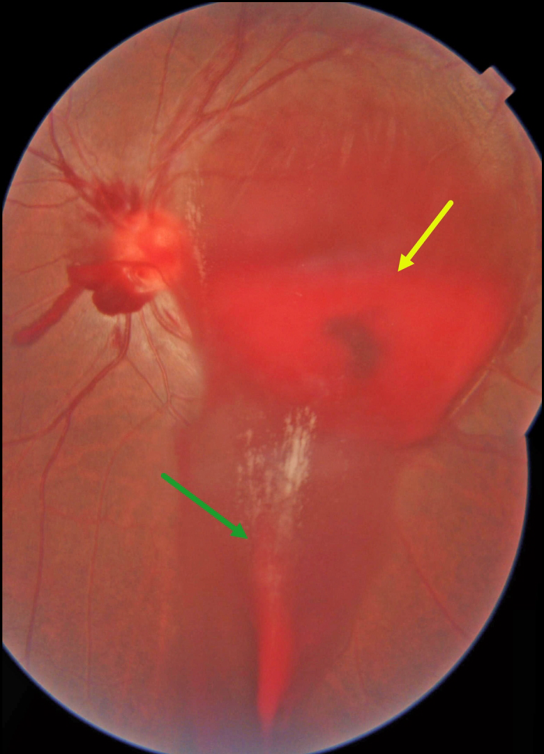
This 28YO male noted sudden vision loss in his left eye 1 day earlier, immediately following coughing. Vision was 20/20 in his normal right eye and counting fingers in his left eye.
Color imaging shows a large layered sub-internal limiting membrane (ILM) hemorrhage occupying the entire macula. The blood extends inferiorly in the subhyaloid space. Some peripapillary vitreous hemorrhage is also noted.
Our patient elected to be observed and, unfortunately, was subsequently lost to follow-up.
Learning Points:
Valsalva retinopathy is characterized by usually unilateral retinal and preretinal hemorrhages caused by raised intrathoracic or intra-abdominal pressure. Various causes include coughing, heavy lifting, and vomiting.
These hemorrhages virtually always resolve without sequelae, although the preretinal blood in severe cases can be drained into the inferior vitreous by creating a small ILM opening using either a thermal or Nd:YAG laser.
VALSALVA RETINOPATHY
Marianna Kavalaraki
Originally posted on @retina.rocks September 20, 2023
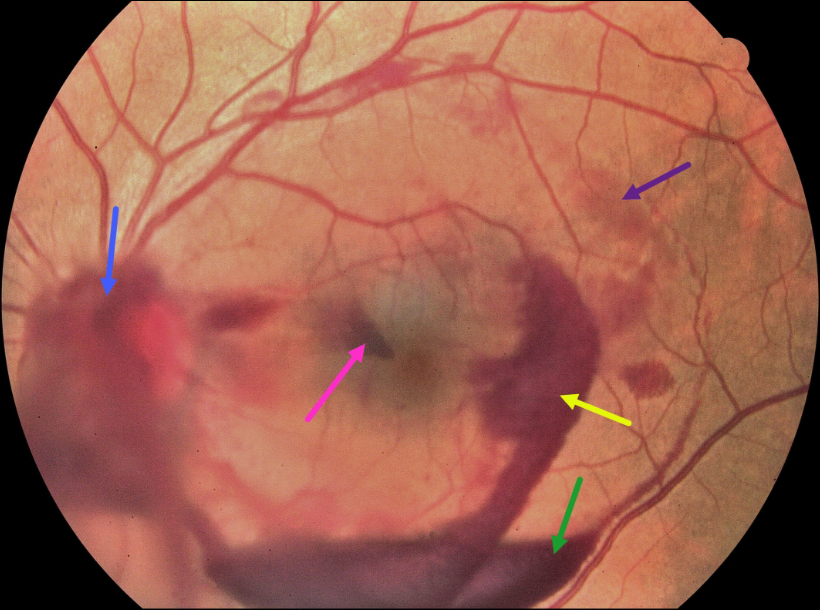
This 39YO male presented with a 3-day history of sudden floaters and central scotoma in his left eye following heavy lifting. Vision was 20/20 in his normal right eye and 20/20 OS.
Color fundus imaging shows multi-layered hemorrhages in the posterior pole, including vitreous, subhyaloid, sub-internal limiting membrane (ILM), Henle’s layer, and deep intraretinal or subretinal. The blood spontaneously resolved after 2 weeks of observation.
Learning Points:
Valsalva retinopathy is characterized by pre-retinal blood (classically sub-ILM but also subhyaloid and vitreous) following a Valsalva maneuver (heavy lifting, vomiting, coughing, sneezing, and compression injuries).
The hemorrhages virtually always resolve without sequelae, although the sub-ILM/hyaloid blood can be drained into the inferior vitreous by creating a small opening in the ILM/hyaloid using either a thermal or Nd:YAG laser.
The differential diagnosis includes other conditions causing multi-layered hemorrhages, including ruptured retinal arterial macroaneurysm, trauma, and anemic/leukemic retinopathy.
VALSALVA RETINOPATHY
Shivraj Tagare and Nishant Maindargi
Originally posted on @retina.rocks August 17, 2023
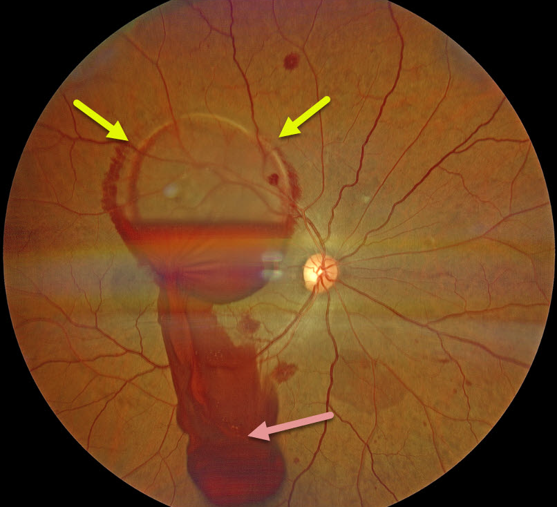
This 37YO male presented 1 day after sudden vision loss in his right eye following vigorous coughing. Vision was 20/400 in his right eye and 20/20 in his normal left eye.
Fundus examination of his right eye showed scattered posterior pole retinal hemorrhages and a large sub-internal limiting membrane (ILM) hemorrhage obscuring his right macula.
A Neodymium:YAG laser was used to create a small inferior opening in the ILM. Fundus photography shows that the blood immediately began draining into the inferior vitreous.
Learning Points:
Valsalva retinopathy is characterized by usually unilateral retinal and preretinal hemorrhages caused by raised intrathoracic or intra-abdominal pressure. Various causes include coughing, heavy lifting, and vomiting.
These hemorrhages virtually always resolve without sequelae, although the sub-ILM blood in severe cases can be drained into the inferior vitreous by creating a small ILM opening using either a thermal or Nd:YAG laser, as was done for this patient.
VALSALVA-INDUCED CHOROIDAL HEMORRHAGE
Originally posted on @retina.rocks May 2, 2023
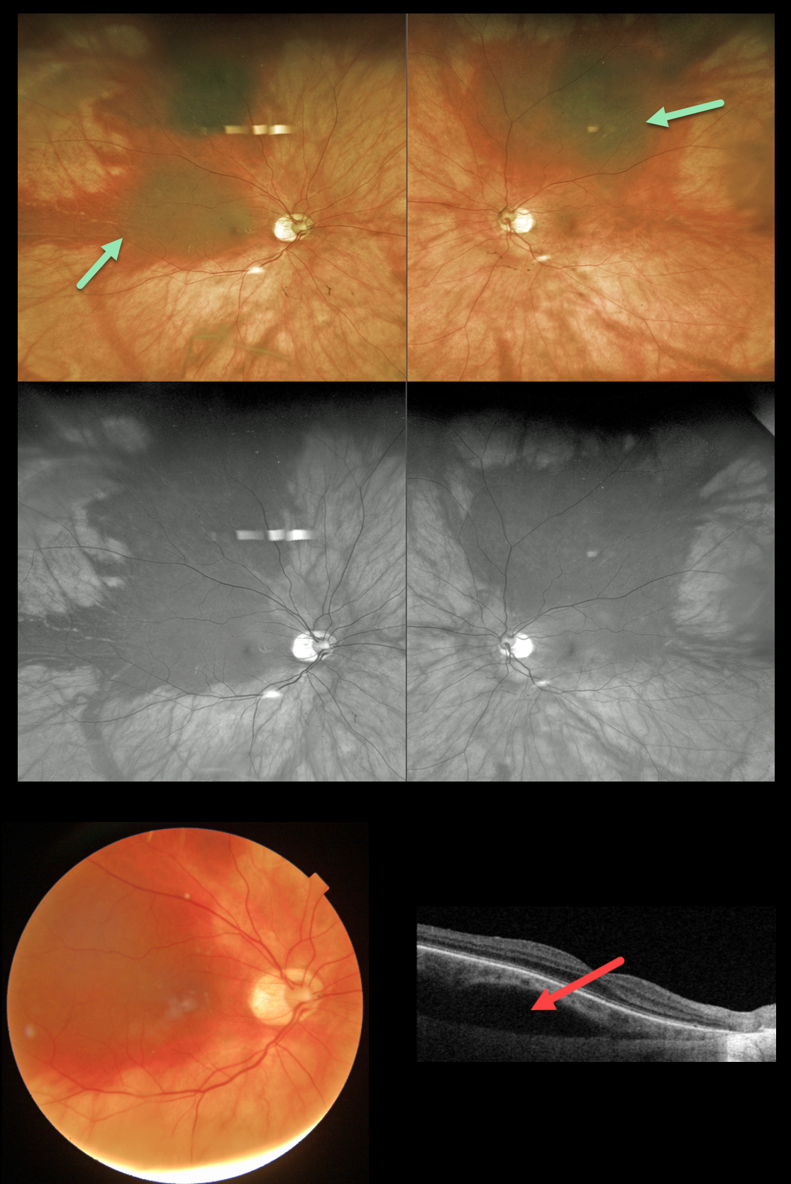
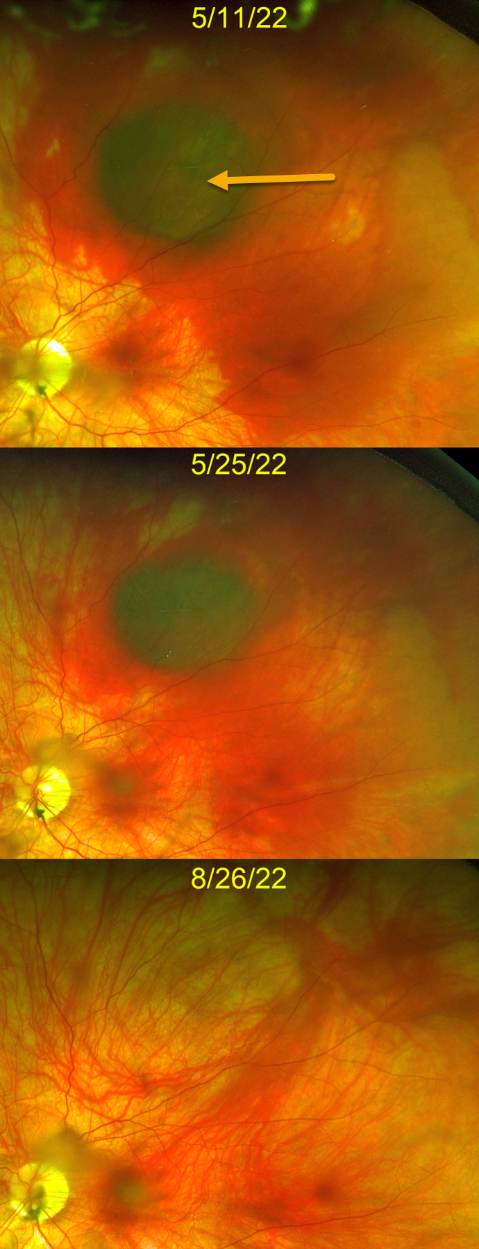
This 61YO healthy female presented with sudden blurred vision immediately following forceful blowing of her nose 3 days earlier. Vision was 20/25 OU.
Optos imaging shows bilateral superior suprachoroidal blood, which extends into the right macula and temporal periphery.
Swept-source OCT through the macula hemorrhage shows hyporeflective suprachoroidal elevation. Observation was recommended, and we expect the blood to absorb over the coming months.
Learning Points:
Valsalva-induced retinal findings almost always present with sub-internal-limiting-membrane blood. Suprachoroidal hemorrhage most commonly develops intra- or postoperatively, following trauma or with thrombolytic agents (Ghu and Green, Survey Ophthalmology 1999;43:471-486).
Valsalva-induced choroidal hemorrhage is exceedingly rare, with only a handful of cases reported in the literature, including a prior Retina Rocks case (see Image Gallery, Valsalva retinopathy folder, Valsalva UIV-20220511).
As with other localized suprachoroidal hemorrhages, these lesions may simulate uveal melanoma (Marous et al, Retina 2018;12:336-341).
VALSALVA RETINOPATHY
Akansha Sharma, Urmil Shah and Denish Patel
Originally posted on @retina.rocks May 1, 2023
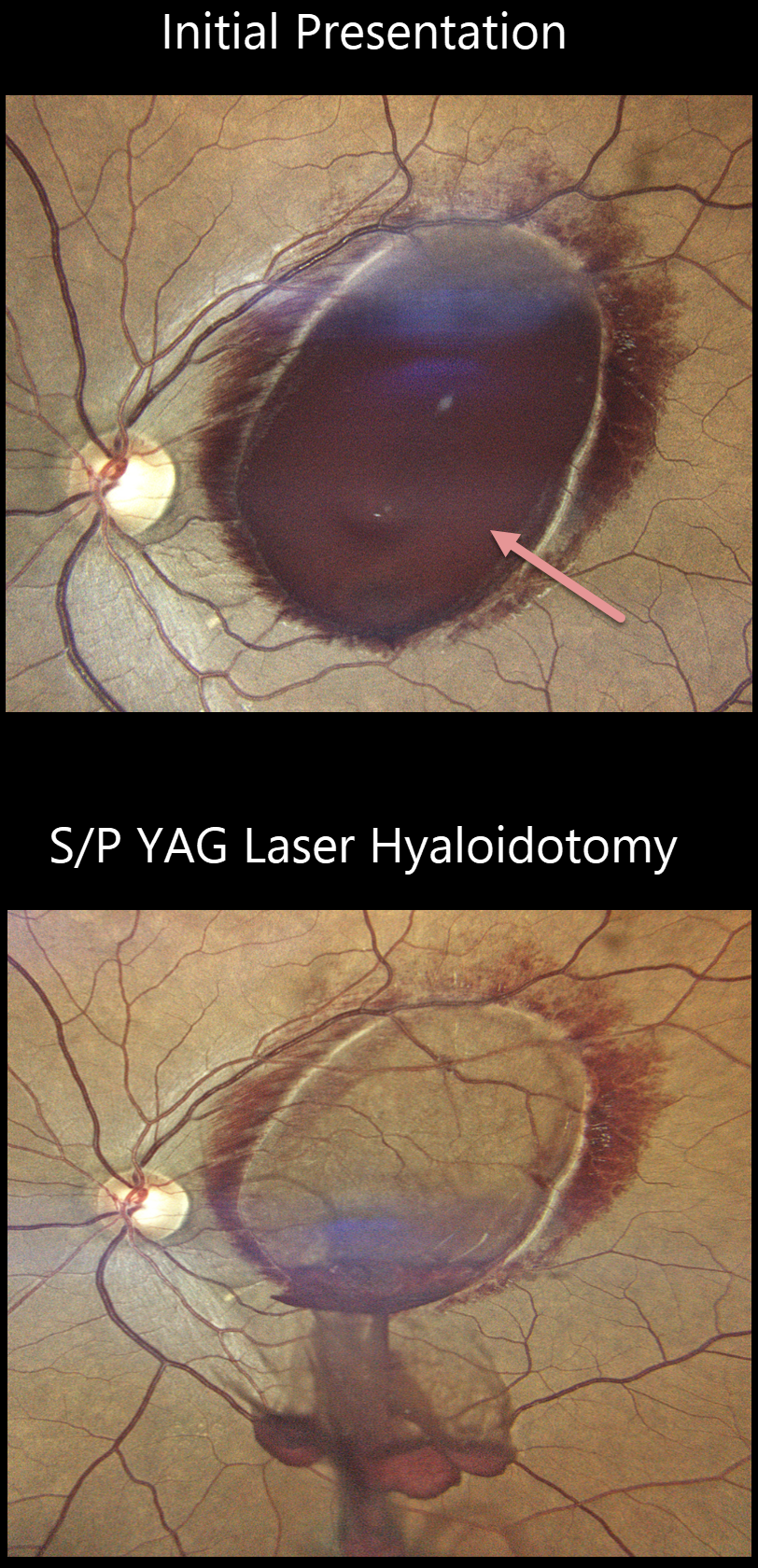
This 40YO male developed sudden loss of vision in his left eye after sneezing 4 days earlier. Vision was 20/1200 OS.
Fundus imaging shows a large sub-internal limiting membrane (ILM) hemorrhage covering the left macula. There is a rim of surrounding nerve fiber layer vs Henle’s blood.
He underwent a YAG laser hyaloidotomy on the same day with immediate improvement to 20/60. Final follow-up vision was 20/30 at 8 weeks.
Learning Points:
Valsalva hemorrhages are usually in the sub-ILM space. These hemorrhages virtually always resolve without sequelae, although the sub-ILM blood in severe cases can be drained into the inferior vitreous by creating a small ILM opening using either a thermal or Nd:YAG laser.
VALSALVA RETINOPATHY
Michael Wong and Jonathan Tsui
Originally posted on @retina.rocks March 9, 2023
This 38YO healthy female presented with one day of peripheral vision loss in her left eye after an episode of forceful vomiting from a migraine headache. Vision was 20/30 OS.
Optos color imaging shows a dark, round, localized suprachoroidal hemorrhage in the superior midperiphery surrounded by subretinal blood. The right fundus was normal (not shown).
The blood gradually improved with observation, completely resolving over the next 3-4 months with return of 20/20 vision.
Learning Points:
Valsalva-induced retinal findings almost always present with sub-internal-limiting-membrane blood. Suprachoroidal hemorrhage most commonly develops intra- or postoperatively, following trauma or with thrombolytic agents (Ghu and Green, Survey Ophthalmology 1999;43:471-486).
Valsalva-induced choroidal hemorrhage is exceedingly rare, with only a handful of cases reported in the literature. As with other localized suprachoroidal hemorrhages, these lesions may simulate uveal melanoma (Marous et al, Retina 2018;12:336-341).
VALSALVA RETINOPATHY
Originally posted on @retina.rocks January 17, 2023
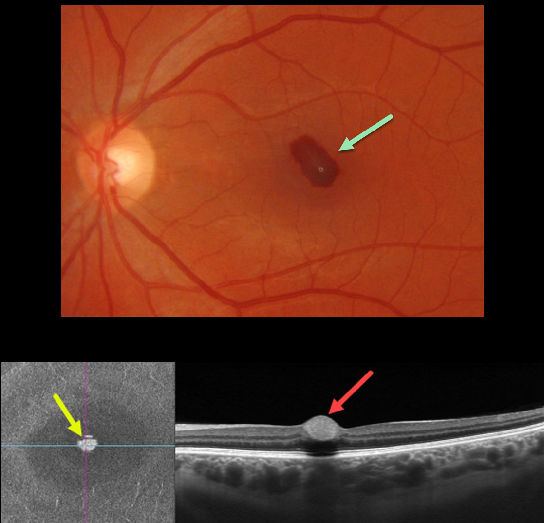
This 33YO female had a severe upper respiratory illness with intense coughing and sneezing. She woke one day earlier with a central scotoma in her left eye.
Optos color imaging shows a teardrop-shaped sub-internal limiting membrane (ILM) hemorrhage. Swept-source OCT B-scan shows a hyperreflective sub-ILM lesion with underlying shadowing. En face scanning of the inner retina provides a unique perspective on the blood.
Learning Points:
Valsalva hemorrhages are usually in the sub-ILM space. These hemorrhages virtually always resolve without sequelae, although the sub-ILM blood in severe cases can be drained into the inferior vitreous by creating a small ILM opening using either a thermal or Nd:YAG laser.
VALSALVA RETINOPATHY
Originally posted on @retina.rocks March 24, 2022
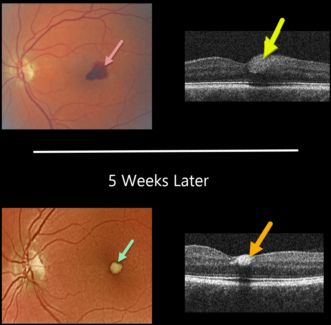
This 56YO female presented with acute vision loss in her left eye from Valsalva retinopathy. Vision was 20/100. A temporal foveal retinal hemorrhage is seen, located within the inner retina on OCT B-scan.
Five weeks later, the devitalized blood is contracting and turning yellow. The blood also became more hyperreflective on the OCT B-scan. All blood subsequently resolved with the return of normal vision.
Learning Points:
Valsalva hemorrhages are usually in the sub-internal limiting membrane (ILM) space, although this case was in the inner retina. These hemorrhages virtually always resolve without sequelae, although the sub-ILM blood can be drained into the inferior vitreous by creating a small opening in the ILM using either a thermal or Nd:YAG laser.
VALSALVA RETINOPATHY
Originally posted on @retina.rocks March 3, 2022
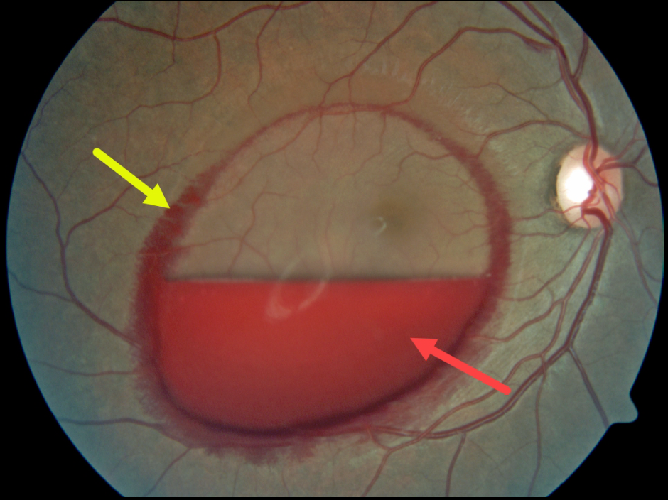
This patient presented with an acute layered sub-internal limiting membrane (ILM) hemorrhage due to a Valsalva maneuver. There is also a rim of blood along the circumference of the ILM detachment, which is most likely located in Henle’s layer, given its radiating deep retinal pattern.
Learning Points:
Sub-ILM hemorrhages most commonly follow a Valsalva maneuver or trauma, but can also happen spontaneously, as with anemia.
These hemorrhages virtually always resolve without sequelae, although the blood can be drained into the inferior vitreous by creating a small opening in the ILM using either a thermal or Nd:YAG laser.
VALSALVA RETINOPATHY
Originally posted on @retina.rocks November 16, 2021
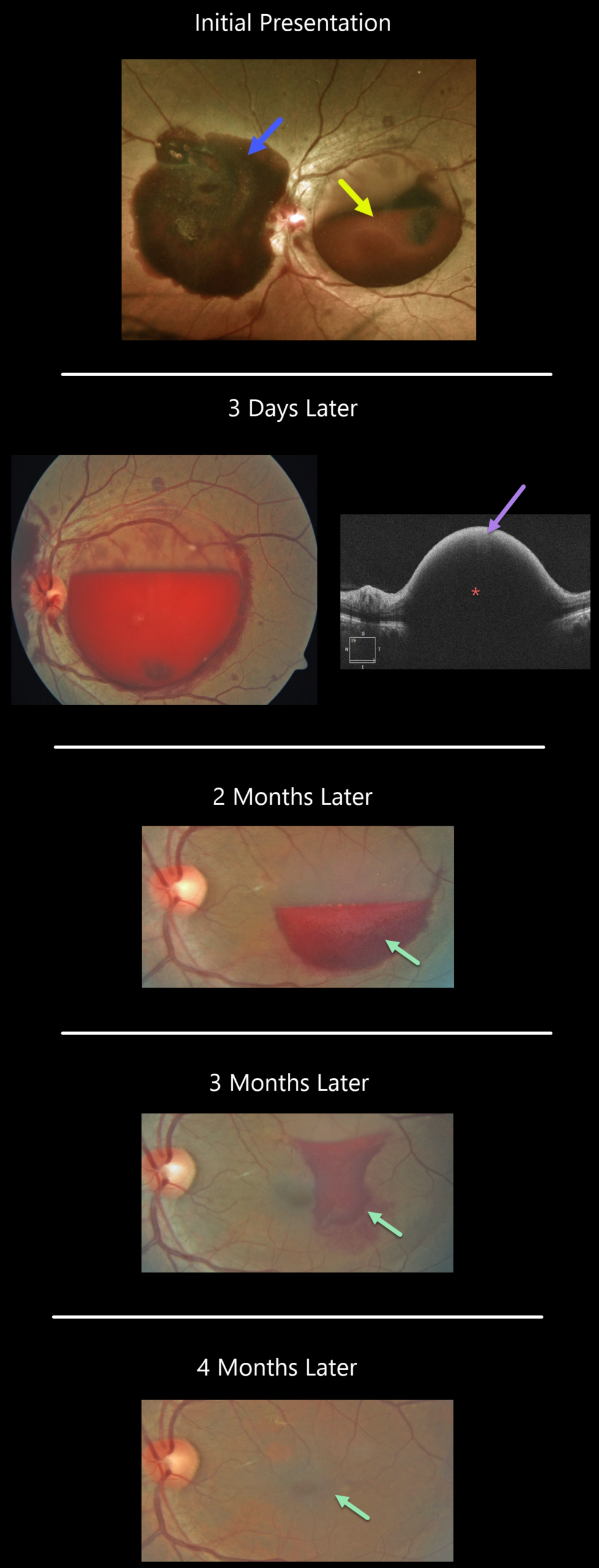
This patient developed sudden vision loss in their left eye while on the toilet.
A large sub-internal limiting membrane (ILM) hemorrhage occupies the macula, while a large subhyaloid hemorrhage extends from the nerve nasally. Scattered retinal hemorrhages are also seen.
Blood pressure was 191/141, and they were sent to the emergency room for hypertensive control.
When examined 3 days later, the macular blood had already begun to decrease. OCT scanning shows marked hyperreflectivity from the blood with posterior shadowing.
The blood gradually resolved over the next four months, and vision improved to 20/30.
Learning Points:
Acute sub-internal limiting membrane (ILM) hemorrhage most commonly is a result of a Valsalva maneuver or trauma, but can also happen spontaneously, as with anemia. The presence of subhyaloid hemorrhage, as well as the association with severe hypertension as occurred in our patient, is quite unusual.
VALSALVA RETINOPATHY
Originally posted on @retina.rocks March 15, 2021
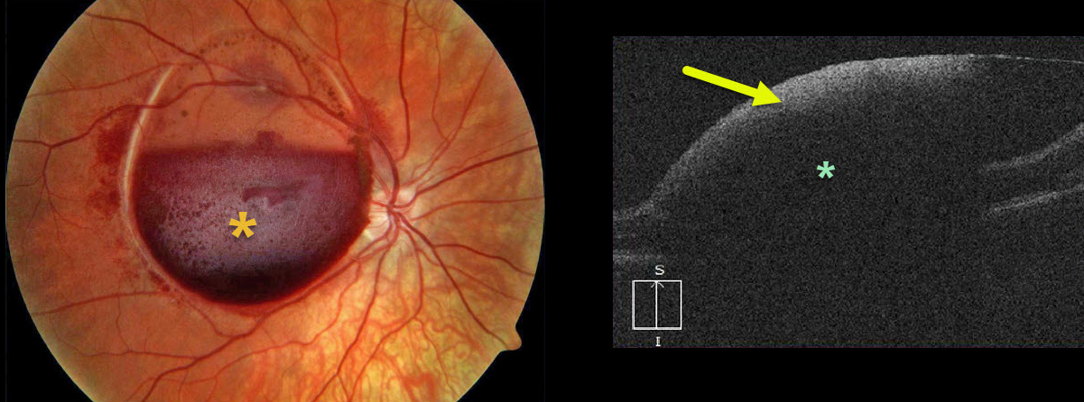
This is a beautiful photograph of an acute sub-internal limiting membrane (ILM) hemorrhage, most commonly found following a Valsalva maneuver or trauma, but can also happen spontaneously, as with anemia.
OCT scanning shows marked hyperreflectivity from the sub-ILM blood with posterior shadowing.
Learning Points:
Sub-ILM hemorrhages virtually always resolve without sequelae, although the blood can be drained into the inferior vitreous by creating a small opening in the ILM using either a thermal or Nd:YAG laser.
TERSON SYNDROME
Originally posted on @retina.rocks January 17, 2020
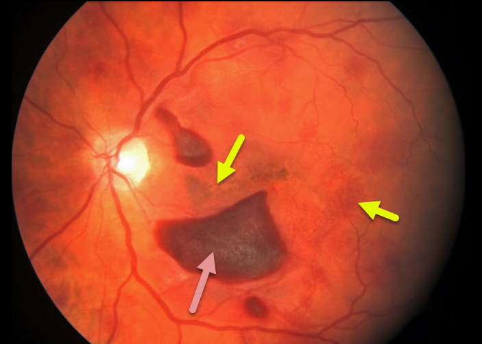
This patient presented with these retinal findings following a ruptured brain aneurysm.
The fundus photo of the left eye shows subhayloid and deep, radiating retinal hemorrhages in the outer plexiform layer.
Learning Points:
Terson’s syndrome is a condition in which retinal and/or vitreous hemorrhages are associated with spontaneous or traumatic intracranial hemorrhage. These deeper retinal hemorrhages are somewhat similar to those seen in hemorrhagic unilateral retinopathy.
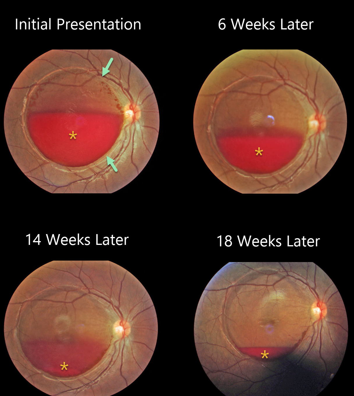
VALSALVA RETINOPATHY
Originally posted on @retina.rocks January 9, 2020
This pregnant female developed sudden vision loss in her right eye following delivery of her baby.
There is a large, round detachment of the internal limiting membrane (ILM) throughout the entire macula with layered blood initially blocking the central fovea.
The blood gradually resolved 6 weeks, 14 weeks, and 18 weeks thereafter.
Learning Points:
Valsava retinopathy is characterized by preretinal hemorrhages, which virtually always resolve without sequelae over time. The blood can also be drained into the inferior vitreous by creating a small opening in the ILM using either a thermal or Nd:YAG laser.


