

Vitamin A Deficiency
<< Back to Cases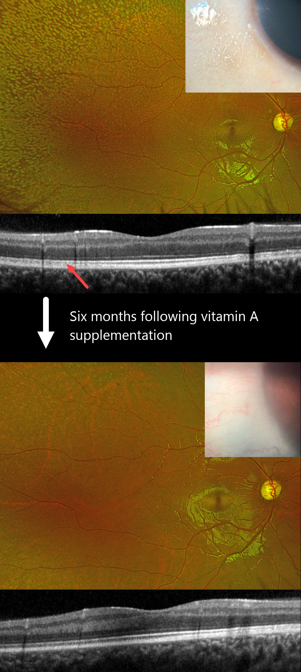
VITAMIN A DEFICIENCY
Mariano Cotic, Ariel Schlaen, Saint Martin Madeleine, and Enzo Dilascio
Originally posted on @retina.rocks January 14, 2025
This 10YO boy presented with bilateral nyctalopia and vision loss. Vision was counting fingers OD and 20/100 OS. Identical findings were noted bilaterally, so only the images for the right eye are shown.
Bitot’s spots were observed on the temporal conjunctiva. Innumerable yellow-white subretinal deposits extend from the peripheral macula into the periphery. OCT scanning shows blurred detail of the outer segment bands due to diffuse deposition of this hyperreflective material.
On further questioning, our patient comes from a low socioeconomic environment with a diet consisting of french fries and pasta. Fasting serum vitamin A was severely low at <0.11 mg/L (normal 0.13-0.81), and supplemental oral vitamin A therapy was begun.
Six months later, vision improved to 20/70 OD and 20/30 OS with complete resolution of his nyctalopia. The conjunctival lesions and subretinal deposits had dramatically improved.
Learning Points:
Vitamin A deficiency is rare in developed countries and is sometimes related to malabsorption in chronic pancreatitis or inflammatory bowel disease. It leads to several well-documented ocular complications, including nyctalopia, Bitot spots, and xanthopsia. Outer nuclear layer thinning with disruption of the outer retinal bands can be visualized on OCT (Berkenstock et al, Int J Retin Vitr 2020;6:23). The outer retinal white dots colocalize to OCT outer retinal hyperreflective lesions (Aleman et al, Doc Ophthalmol 2013;127:239-243). Oral or intramuscular vitamin A replacement can reverse the retinal changes if initiated early enough.
VITAMIN A DEFICIENCY
Mattie Adams
Originally posted on @retina.rocks November 4, 2024
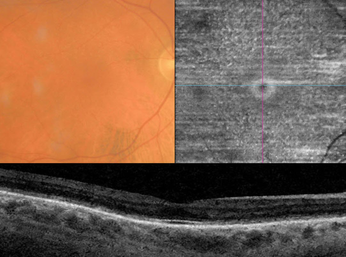
This 70YO male presented with six months of vision loss. He has a history of severe Crohn’s disease. He has undergone 9 bowel surgeries resulting in short bowel syndrome being treated with total parenteral nutrition. Vision was 20/50 OD and 20/40 OS.
Triton color imaging shows numerous smaller subretinal yellow-white dots. These dots are well-visualized on en face swept-source OCT through the outer retina. OCT B scan shows outer retinal thinning and loss of the hyperreflective bands detail.
Fasting serum vitamin A was severely low at 2.5 (normal 20-60), and he was started on parenteral vitamin A therapy.
Learning Points:
Vitamin A deficiency is rare in the United States and is sometimes related to malabsorption in chronic pancreatitis or inflammatory bowel disease. It leads to several well-documented ocular complications including nyctalopia, Bitot spots and xanthopsia. Outer nuclear layer thinning with disruption of the outer retinal bands can be visualized on OCT (Berkenstock et al, Int J Retin Vitr 2020;6:23). The outer retinal white dots colocalize to OCT outer retinal hyperreflective lesions (Aleman et al, Doc Ophthalmol 2013;127:239-243). Oral or intramuscular vitamin A replacement has been shown to reverse the retinal changes if initiated early enough. We are hoping that our patient’s vision and retinal findings will improve with continued treatment.
VITAMIN A DEFICIENCY
Originally posted on @retina.rocks March 30, 2022
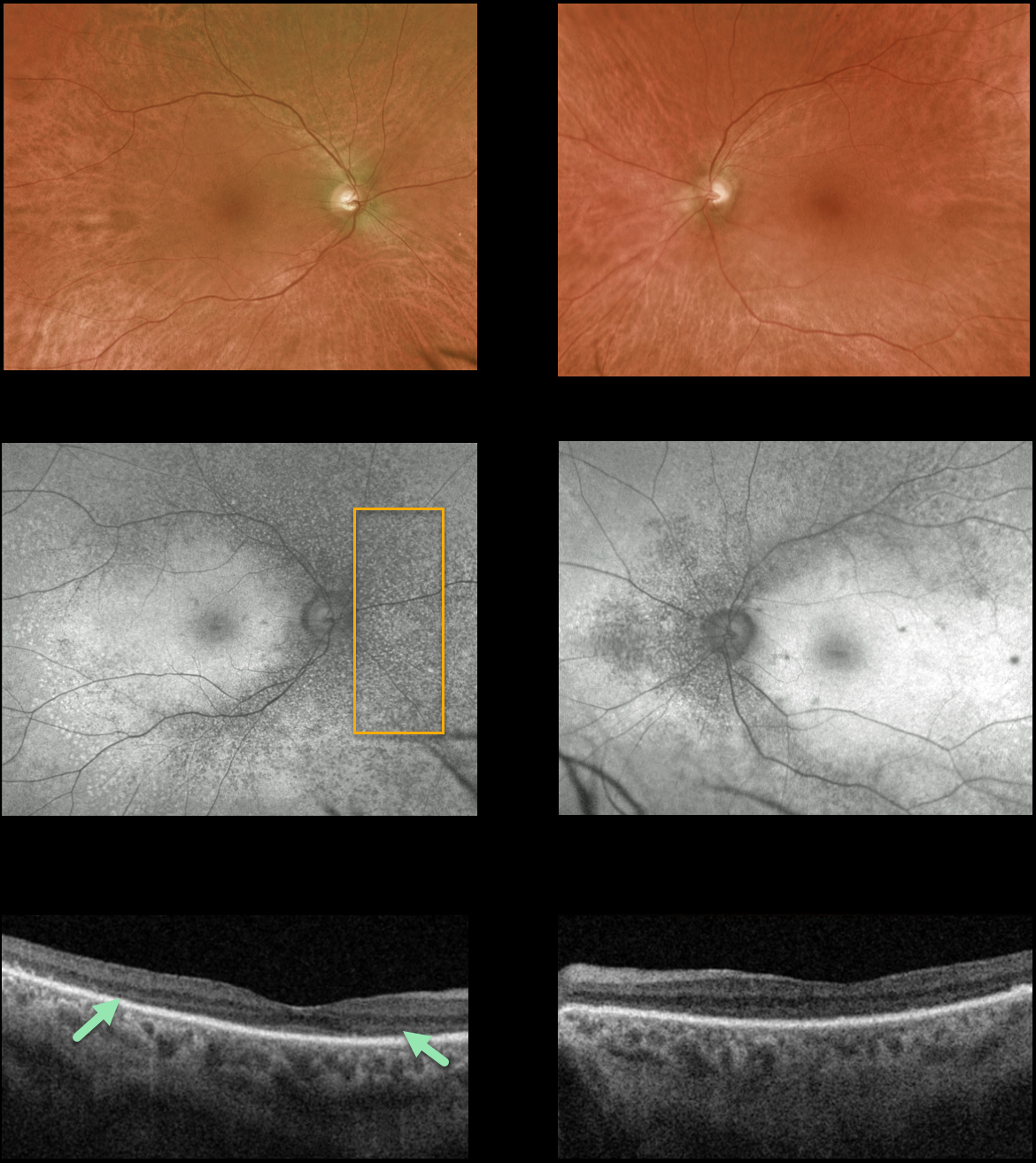
This 64YO man presented with 1 year of gradual severe bilateral vision loss. Vision was counting fingers at 5 feet OU.
Retinal examinations showed fine subretinal pigmentary changes with tiny whitish dots throughout the extramacular retina, best visualized as hypoautofluorescent spots on Optos ultra-widefield autofluorescence.
Triton swept-source OCT shows profound loss of the outer segment layers.
The patient denied any systemic diseases or current medications, though he admitted he hadn’t been to a physician in years. He appeared in poor health and malnourished, so we ordered nutritional testing, including folate, B12, vitamin A, antiretinal antibodies, and Invitae genetic testing.
All tests were negative except for a decreased Vitamin A level. He subsequently started daily vitamin A supplementation at 20,000 IU, and after a few weeks, his vision improved to 20/400 OD and 20/200 OS without funduscopic or OCT changes.
Learning Points:
Vitamin A deficiency is rare in the United States and is sometimes related to malabsorption in chronic pancreatitis or inflammatory bowel disease. It leads to several well-documented ocular complications, including nyctalopia, Bitot spots, and xanthopsia.
Outer nuclear layer thinning with disruption of the outer retinal bands can be visualized on OCT (Berkenstock et al, Int J Retin Vitr 2020;6:23).
Oral or intramuscular vitamin A replacement has been shown to reverse the retinal changes if initiated early enough. We hope our patient’s vision and retinal findings will improve with continued treatment.


