

Vitreomacular Traction (VMT)
<< Back to Cases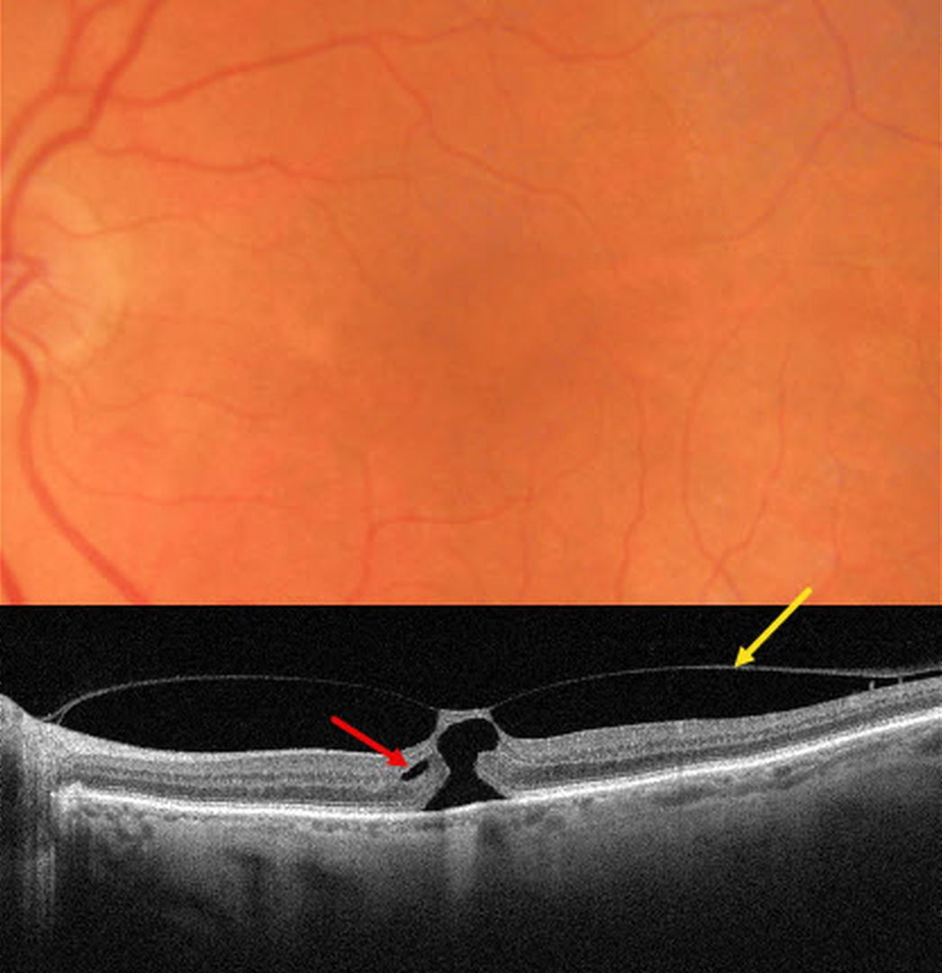
FOCAL VITREOMACULAR TRACTION
Originally posted on @retina.rocks February 9, 2026
This 82YO female was referred for an asymptomatic macular finding noted on screening OCT scanning. Vision was 20/200.
Triton color imaging shows a poorly defined foveal reflex. Swept-source OCT shows a partial macular posterior vitreous detachment (PVD) that is adherent to the foveal center. A thin strip of intact inner tissue is adherent to the overlying vitreous with an underlying outer macular hole. A tiny area of split outer plexiform layer is noted nasally.
Learning Points:
The International Vitreomacular Traction Study Group classified vitreomacular traction by size (focal <= 1500 microns or broad >1500 microns) and etiology (isolated or concurrent, Ophthalmology 2013;120:2611-2619). The prevalence of vitreomacular traction (VMT) increases with age and is present in about 1% of people over 40YO. The natural history is still being defined, but most patients have stable traction and vision. VMT can also resolve spontaneously in about 20% (Errera et al, Ophthalmology 2018;125:701-707).
Management includes observation, pneumatic vitreolysis, or vitrectomy. In our experience, most patients with VMT remain completely asymptomatic despite impressive OCT findings. We therefore almost always observe VMT unless the patient has significant symptoms. Our patient elected to continue observation since she was asymptomatic bilaterally.
OUTER FOVEAL MICRODEFECT
Originally posted on @retina.rocks December 13, 2023
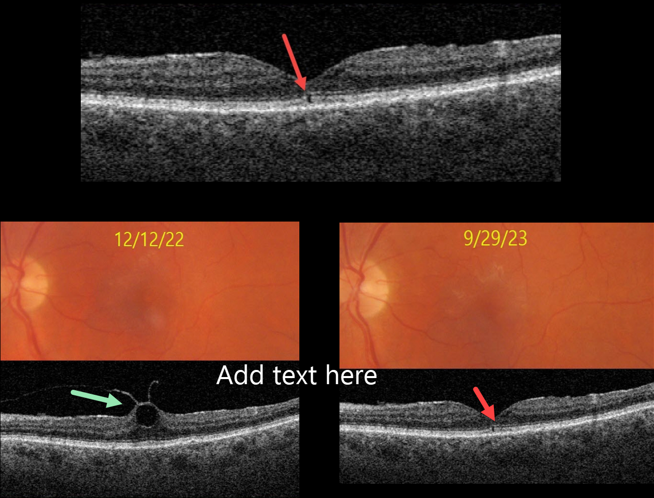
This 76YO female presented with no visual symptoms, 20/40 vision, and an abnormal OCT in her left eye. What caused this subtle OCT finding?
She was previously being followed for asymptomatic focal vitreomacular traction (VMT) in this eye. On 12/12/22, Triton color imaging showed a subtle foveal cyst with focal VMT on swept-source OCT.
We recommended observation due to the absence of symptoms, and when she returned on 9/29/23, these findings spontaneously resolved as the vitreous separated, leaving a tiny outer foveal microdefect.
Learning Points:
Outer foveal microdefects (Cohen et al, Ophthalmology Retina 2021;5:553-561) have been described in numerous conditions, including macular telangiectasia, tamoxifen use, ABCA4 disorders (cone-rod dystrophies, Stargardt disease, and fundus flavimaculatus), phototoxicity, trauma, and vitreomacular traction disorders.
The prevalence of VMT increases with age, and is present in about 1% of people over 40YO. The natural history is still being defined, but most patients have stable traction and vision. VMT can also resolve spontaneously in about 20% (Errera et al, Ophthalmology 2018;125:701-707).
Many patients with VMT remain completely asymptomatic despite impressive OCT findings. We therefore almost always observe VMT, as in this case, unless the patient has significant symptoms.
VITREOMACULAR TRACTION
Originally posted on @retina.rocks January 5, 2023
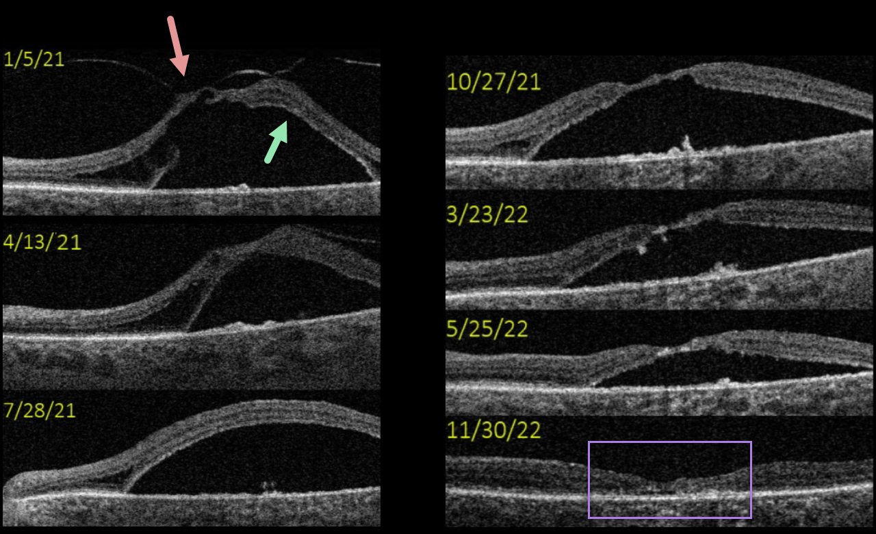
This 77YO female presented with vision of 20/400 in her left eye from severe vitreomacular traction with a secondary tractional macular detachment. The patient underwent pars plana vitrectomy in January 2021.
A few months post-vitrectomy, all traction was relieved, and the detachment was slightly decreased.
The detachment gradually resolved over the next year and a half, ultimately flattening completely with secondary foveal atrophy.
Although her final vision remained at 20/400, she was subjectively happy with her surgical results.
VITREOMACULAR TRACTION + LAMELLAR MACULAR HOLE
Erdem Dinç
Originally posted on @retina.rocks March 29, 2022
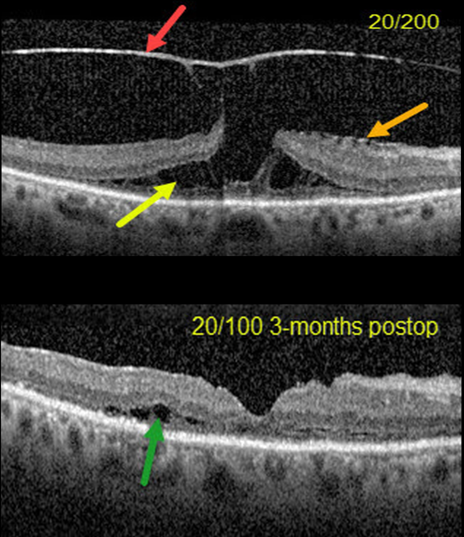
This 70YO female presented with 20/200 vision in her right eye from severe vitreomacular traction with a tractional lamellar macular hole (LMH).
OCT scanning shows a highly elevated posterior hyaloid adherent to the underlying fovea, with tractional splitting of the outer plexiform layer (OPL). There is also a faint nasal epiretinal membrane.
Combined cataract surgery, 23-gauge pars plana vitrectomy, membrane peeling, temporal half-moon inverted ILM flap, and fluid-air-C3F8 exchange was performed.
Three months postoperatively, the tractional LMH was closed with 20/100 acuity. There is still some temporal OPL splitting along with disorganization and thinning of the outer macular layers.
Learning Points:
LMH can be defined as tractional or degenerative (see Govetto et al, AJO 2016;164:99-109). Tractional LMH shows OPL splitting, an intact ellipsoid zone, and is associated with vitreomacular traction and tractional epiretinal membranes.
OCT features of a degenerative LMH include loss of outer retinal tissue below the OPL and an outer retinal ‘bump’. Non-tractional epimacular proliferation is often present and will often extend around the posterior edge of the degenerative LMH.
VITREOMACULAR TRACTION
Originally posted on @retina.rocks May 26, 2021
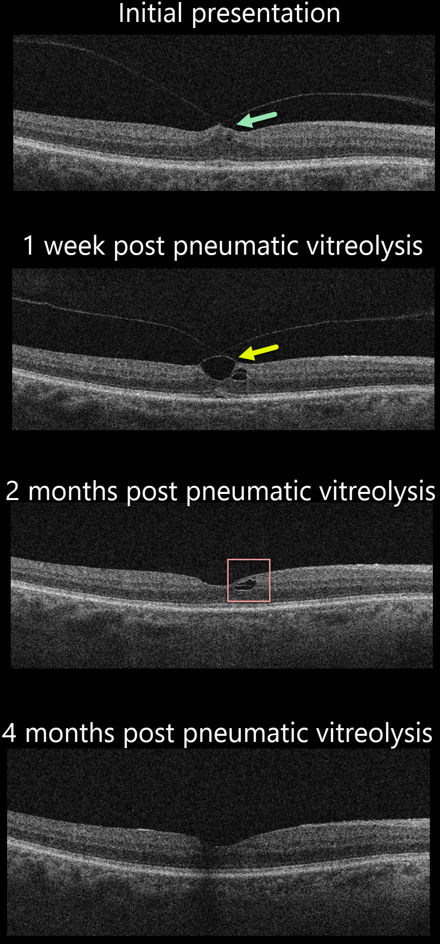
This 72YO male presented with symptomatic vitreomacular traction (VMT) and 20/40 vision. Various treatment options were discussed, including observation, ocriplasmin (Jetrea), pneumatic vitreolysis, and vitrectomy. He decided to proceed with pneumatic vitreolysis.
One week later, OCT showed that the vitreous remained focally attached to the central fovea, with increased traction.
Two months following the procedure, there was a complete vitreous detachment with normalization of the foveal contour and a small residual splitting of the outer plexiform layer nasally.
Four months following the procedure, OCT was virtually normal, and vision was 20/30. The patient was completely symptom-free.
Learning Points:
The prevalence of VMT increases with age and is present in about 1% of people over 40. The natural history is still being defined, but most patients have stable traction and vision. VMT can also resolve spontaneously in about 20% (Errera et al, Ophthalmology 2018;125:701-707).
Many patients with VMT remain completely asymptomatic despite impressive OCT findings. We therefore almost always observe VMT unless the patient has significant symptoms, as in this case.
Shortly after we treated this patient, the results of the DRCR Retina Network Protocol AG, which evaluated pneumatic vitreolysis with C3F8 for VMT, were published ahead of print (https://doi.org/10.1016/j.ophtha.2021.05.005). Gas successfully created a complete PVD in 78% of eyes. However, the study was terminated early due to safety concerns related to retinal detachment (8% of eyes). Based on these safety issues, we personally would no longer recommend this as a viable option unless medical issues make vitrectomy impossible.
VITREOMACULAR TRACTION + MACULAR HOLE
Originally posted on @retina.rocks February 11, 2020
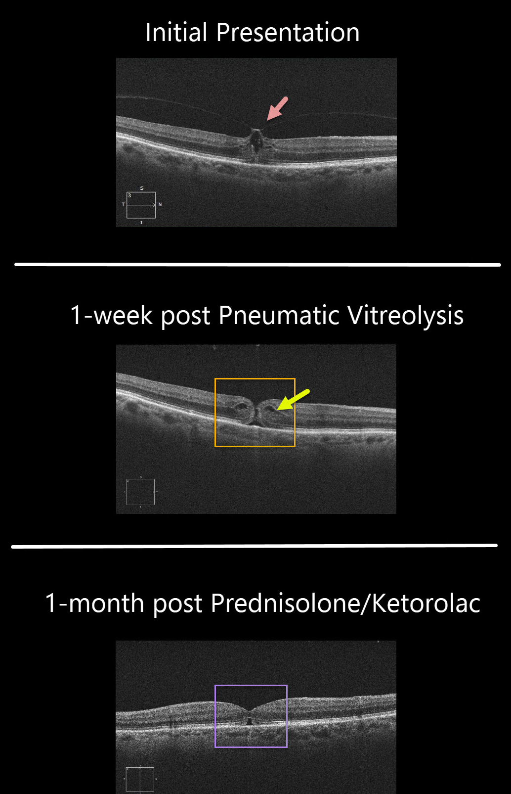
This patient’s symptomatic 20/80 vitreomacular traction was treated with pneumatic vitreolysis, where a small gas bubble was injected into the vitreous cavity to induce a posterior vitreous detachment. The patient wished to try this less invasive option instead of vitrectomy.
Although the traction successfully released 1 week postop, the patient developed an immediate macular hole with 20/100 vision.
Since the hole was tiny with some cystoid edema (CME), a trial of topical prednisolone acetate 1% and ketorolac was given.
One month later, the CME resolved and the hole closed with improvement to 20/60.
Learning Points:
Topical prednisolone acetate 1% and ketorolac is a newly described treatment that can occasionally be tried for macular holes without traction.


