

Vitreopapillary Traction
<< Back to Cases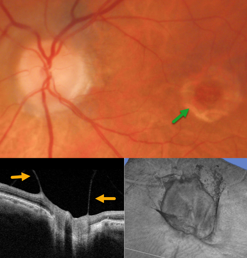
VITREOPAPILLARY TRACTION WITH CHRONIC MACULAR HOLE
Originally posted on @retina.rocks April 15, 2026
This 81YO male presented for his yearly examination without new complaints. Vision in his left eye was stable at 20/200 due to a chronic macular hole.
Triton color photography shows a full-thickness macular hole with a surrounding large rim of pigment loss. On slit lamp biomicroscopy, a partial PVD extended to the nerve with an elevated, partially attached Weiss ring. Vitreopapillary traction was confirmed on B-scan and 3D OCT. Continued observation was recommended.
Learning Points:
The normal vitreous undergoes a decades-long process of orchestrated degeneration, including liquefaction and gradual separation from the retinal surface. The posterior vitreous detachment (PVD) begins in the perifoveal retina early in life, followed by a foveal PVD, then a peripheral PVD, and then finally a complete PVD when the vitreous separates from the optic nerve (Johnson AJO 2010;149:371-382). The integrity of the internal limiting membrane (ILM) is integral to normal vitreoretinal adhesion, where a thinner ILM has a firmer adhesion than a thicker ILM. The ILM represents the basement membrane of the Müller cell. Hence, an area with fewer Müller cells reaching the inner retina will naturally have a firmer vitreoretinal adhesion. Since the optic nerve is completely devoid of Müller cells, this is the area of the posterior pole that has the firmest vitreoretinal adhesion, and this is the reason why the vitreous tends to remain so firmly attached to it.
Persistent vitreopapillary traction is a rarely reported phenomenon (Gabriel et al, Neuro-ophthalmology 2020;44:213-218), probably due to its lack of symptoms and often subclinical findings. In our patient’s case, the traction caused retinal thickening and hemorrhage, somewhat mimicking a peripapillary macular neovascularization. In our experience, these eyes usually remain asymptomatic and can be observed, although vitrectomy is very successful in relieving the traction and improving vision.
VITREOPAPILLARY TRACTION
Originally posted on @retina.rocks October 29, 2024
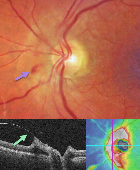
This 76YO female presented with asymptomatic optic nerve findings in her left eye. Color imaging shows somewhat opaque retina with some inner retinal hemorrhages along the nasal aspect of the nerve. OCT shows a partial PVD extending to the nerve with vitreopapillary traction and secondary thickening of the peripapillary nerve fiber layer. Observation was recommended.
Learning Points:
The normal vitreous goes through a decades long process of an orchestrated degeneration, including liquefaction and gradual separation from the retinal surface. The posterior vitreous detachment (PVD) begins in the perifoveal retina early in life, followed by a foveal PVD, then a peripheral PVD, and then finally a complete PVD when the vitreous separates from the optic nerve (Johnson AJO 2010;149:371-382). The integrity of the internal limiting membrane (ILM) is integral to normal vitreoretinal adhesion, where a thinner ILM has a firmer adhesion than a thicker ILM. The ILM represents the basement membrane of Muller cell. Hence an area that has a lower population of Muller cells reaching the inner retina will naturally have a firmer vitreoretinal adhesion. Since the optic nerve is completely devoid of Muller cells, this is the area of the posterior pole that has the firmest vitreoretinal adhesion, and this is the reason why the vitreous tends to remain so firmly attached to it.
Persistent vitreopapillary traction is a rarely reported phenomenon (Gabriel et al, Neuro-ophthalmology 2020;44:213-218), probably due to its lack of symptoms and often subclinical findings. In our patient’s case, the traction caused retinal thickening and hemorrhage, somewhat mimicking a peripapillary macular neovascularization. In our experience, these eyes usually remain asymptomatic and can be observed, although vitrectomy is very successful in relieving the traction and improving vision.
PROLIFERATIVE DIABETIC RETINOPATHY (PDR)
Originally posted on @retina.rocks January 29, 2024
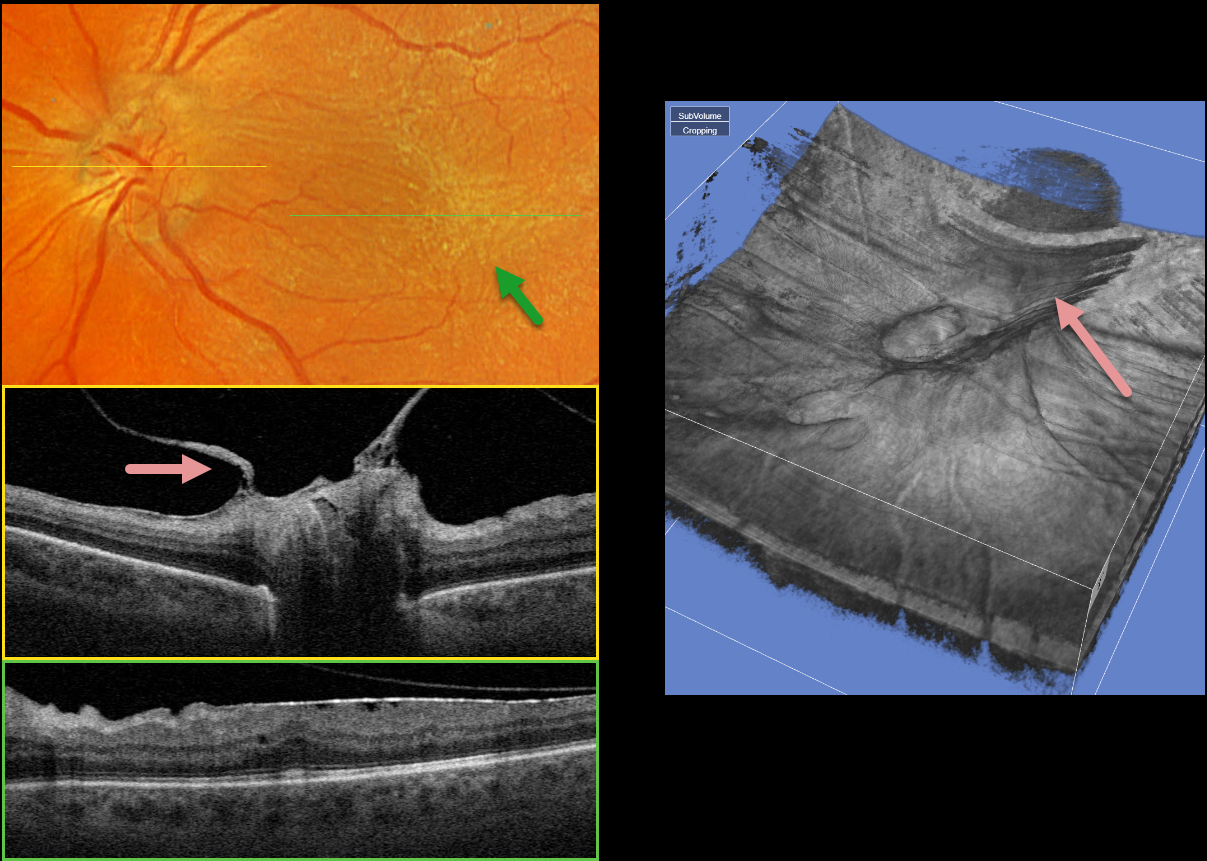
This 58YO male with type 2 diabetes has long-involuted proliferative diabetic retinopathy (PDR) following full panretinal photocoagulation (PRP). He has no visual complaints, and vision is 20/50 OS.
Optos color RGB imaging shows a diffuse macular pucker with striae extending nasally towards the nerve and temporally into the distal macula.
A condensation of elevated fibrosed tissue, representing a partially detached Weiss’ ring, surrounds the optic nerve. This traction is clearly evident in Triton swept-source OCT (image 1, middle) and in the 3D reconstruction (image 2). A moderate epiretinal membrane extends throughout the macula (image 1, bottom).
Observation was recommended, and vitrectomy with membrane peeling will be performed if the patient becomes more symptomatic.


