

Vitreous Cyst
<< Back to Cases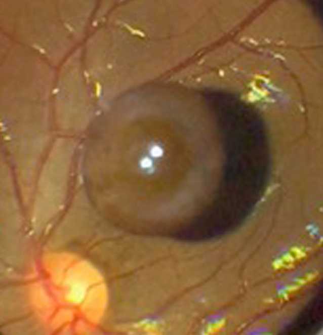
VITREOUS CYST
The European VitreoRetina Society (EVRS) and Prithvi Chandrakanth
Originally posted on @retina.rocks November 21, 2025
This healthy 20YO female presented with chronic floaters in her left eye. Vision was 20/20 in her healthy OD and 20/20 OS.
Funduscopy shows a large pigmented vitreous cyst floating over the superior macula. Observation was recommended.
Learning Points:
Vitreous cysts are extremely rare, idiopathic, and usually diagnosed incidentally. Likely originating from a ciliary body cyst (Dhull et al, Ophthalmology Retina 2020;4:1208), they can be either congenital or acquired. Acquired cysts have been associated with numerous ocular disorders, including trauma and retinitis pigmentosa.
VITREOUS CYST
Anand Temkar and Manish Nagpal
Originally posted on @retina.rocks November 11, 2024
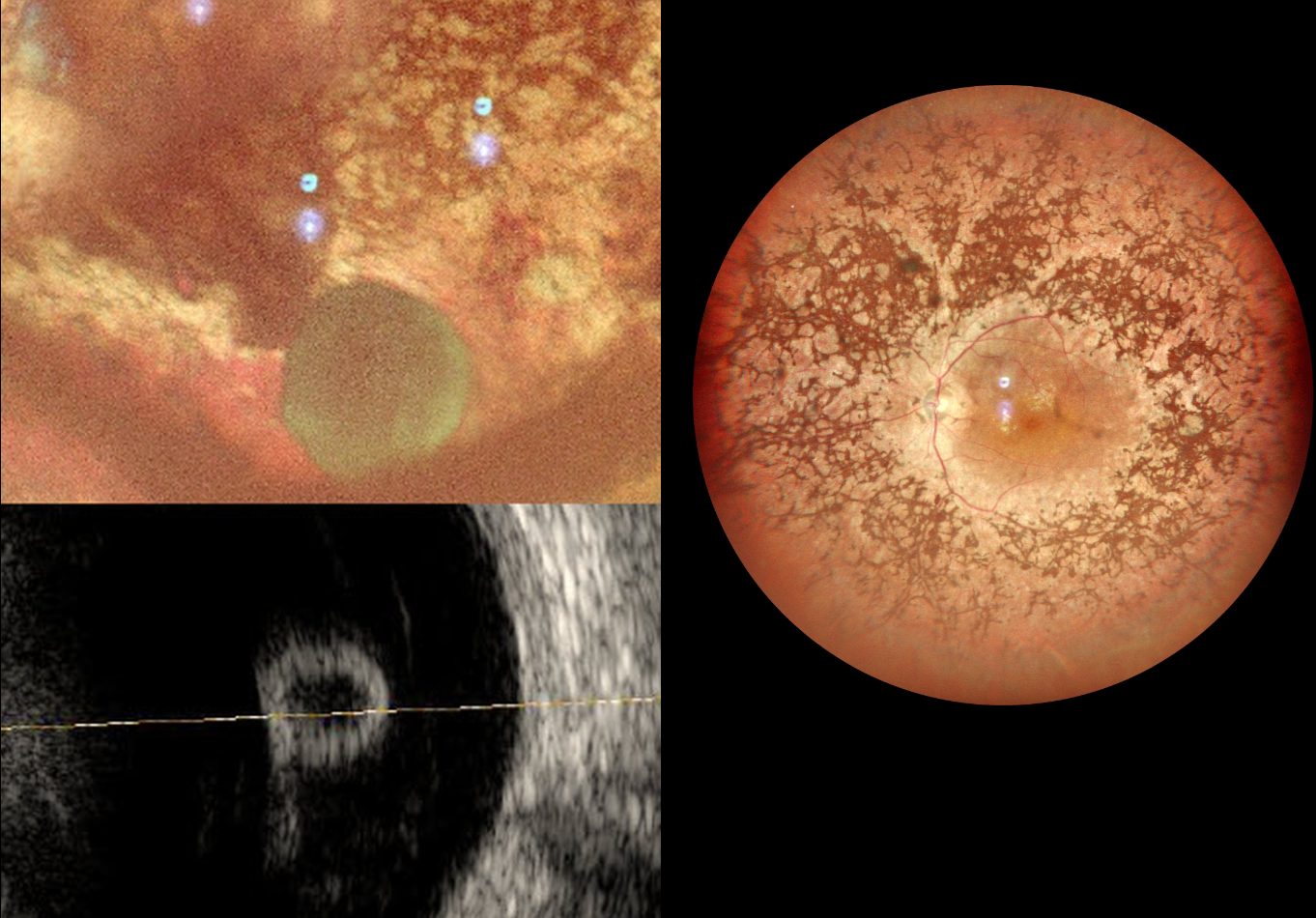
This 31YOF with a known history of retinitis pigmentosa (RP) presented with 2 days of decreased vision OD. There was a history of trauma to her right eye at 2 years of age. Vision was counting fingers OD and 20/120 OS. Slit lamp examination OD revealed a non-central corneal scar and anterior synechia consistent with the prior trauma.
MultiColor imaging shows bilateral RP intraretinal pigment migration and narrowed arterioles. A free-floating vitreous cyst is noted OD inferiorly.
Learning Points:
Vitreous cysts are extremely rare, idiopathic, and usually diagnosed incidentally. Likely originating from a ciliary body cyst (Dhull et al, Ophthalmology Retina 2020;4:1208), they can be either congenital or acquired. Acquired cysts have been associated with numerous ocular disorders, including trauma and retinitis pigmentosa, as in our patient.
VITREOUS CYST
Originally posted on @retina.rocks April 13, 2021
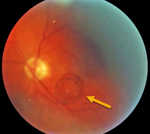
This patient was noted to have an asymptomatic vitreous cyst in the left eye.
Learning Points:
Vitreous cysts are extremely rare, idiopathic, and usually diagnosed as an incidental finding, as in this patient. If symptomatic, the cyst can literally be popped with a YAG laser.
These cysts likely originate from a ciliary body cyst (see Dhull et al, Ophthalmology Retina 2020;4:1208).
VITREOUS CYST
Originally posted on @retina.rocks March 20, 2020
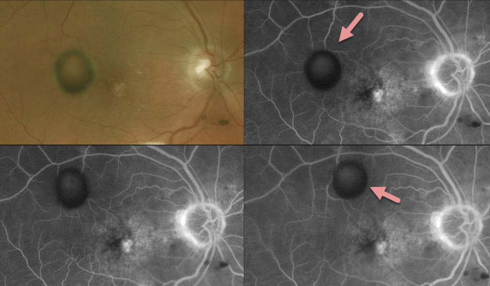
This patient has a vitreous cyst in the right eye, which can be seen moving around on fluoroscein angiography (FA) as it blocks different areas of the retina.
Learning Points:
Vitreous cysts are extremely rare, idiopathic asymptomatic and usually diagnosed as an incidental finding as in this patient.
If symptomatic, the cyst can be popped with a YAG laser.


