

Vogt-Koyanagi-Harada (VKH)
<< Back to Cases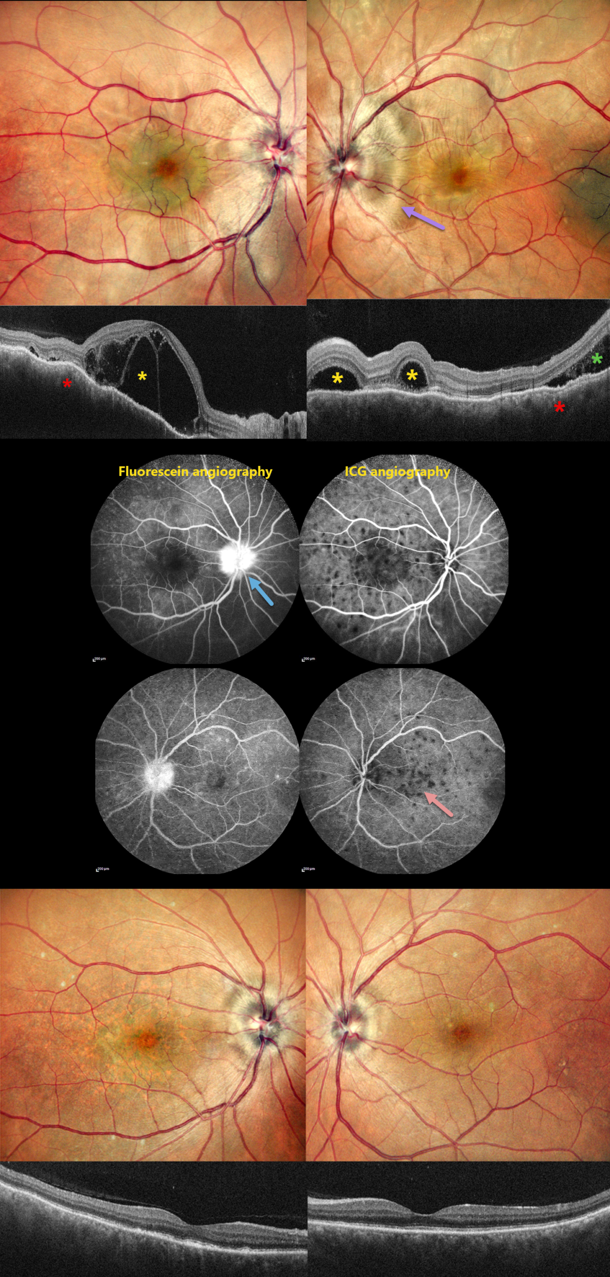
HARADA DISEASE
Rohan Jain and Manish Nagpal
Originally posted on @retina.rocks August 20, 2026
This 46YO male presented with 10 days of bilateral blurred vision with a left-sided headache. Vision was 20/200 OD and 20/20 OS. Anterior segments were normal.
Pseudocolor SLO imaging shows bilateral swollen nerves and irregular macular chorioretinal folds. OCT scanning shows multifocal bacillary layer detachments, subretinal fluid, and an undulating, thickened choroid. Fluorescein angiography shows bilateral multifocal areas of subretinal leakage and disc leakage, and indocyanine green angiography shows multiple hypofluorescent choroidal spots.
Based on the clinical and multimodal imaging findings, a diagnosis of acute Harada disease was established. He received a 3-day course of intravenous methylprednisolone followed by oral corticosteroids.
At the 2-month follow-up, the patient’s symptoms had completely resolved. Vision was 20/30 OD and 20/20 OS. The exudative findings are completely resolved on photography and OCT, and the choroidal thickening with overlying chorioretinal folds is also resolved.
Learning Points:
Harada disease is characterized by relatively acute bilateral panuveitis, optic nerve swelling, thickened choroid, chorioretinal folds, multifocal serous exudative retinal detachments, and bacillary layer detachments. Patients with only ocular findings have Harada disease (as in our patient), whereas those with additional systemic findings (including vitiligo, poliosis, headache, vertigo, and hearing loss) have Vogt-Koyanagi-Harada disease.
HARADA DISEASE
The European VitreoRetina Society (EVRS) and Ridham Nanda
Originally posted on @retina.rocks January 23, 2026
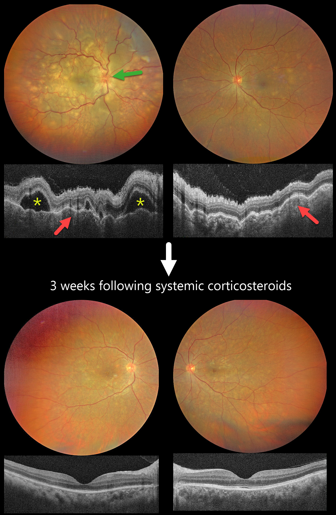
This previously healthy 40YO female presented with 2 days of blurred vision. She denied any systemic symptoms. Vision was 20/200 OD and 20/60 OS.
Color photography shows multifocal yellow-white subretinal lesions scattered throughout each posterior pole, with the right eye greater than the left, associated with overlying multifocal exudative retinal detachments. The right nerve is also edematous. OCT scanning shows a bilateral thickened choroid with overlying chorioretinal folds and multifocal serous detachments OD.
Intravenous methylprednisolone (1000mg/day) was administered for five days, followed by a 2-week course of oral prednisone (1mg/kg). Three weeks later, the funduscopic and OCT findings normalized. Vision improved to 20/30 OD and 20/20 OS.
Learning Points:
Harada disease is characterized by relatively acute bilateral panuveitis, optic nerve swelling, thickened choroid, chorioretinal folds, multifocal serous exudative retinal detachments, and bacillary layer detachments. Patients with just ocular findings have Harada disease (our patient), whereas those with additional systemic findings (including vitiligo, poliosis, headache, vertigo, and hearing loss) have Vogt-Koyanagi-Harada disease.
HARADA DISEASE
Originally posted on @retina.rocks December 16, 2025
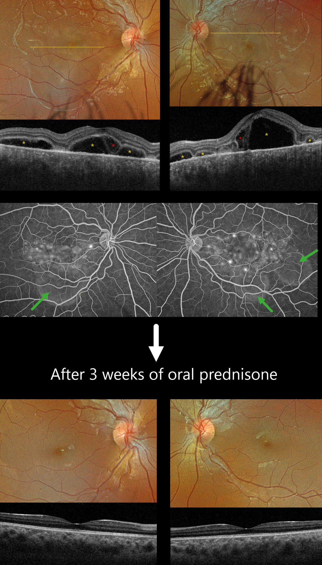
This previously healthy 13YO girl presented with several days of bilateral vision loss. Vision was 20/100 OD and 20/400 OS.
Optos color RGB imaging shows bilateral multifocal serous exudative retinal detachments. Triton swept-source OCT shows a complex pattern of multifocal pockets of subretinal fluid and bacillary layer detachments (BALAD). The choroid is markedly thickened. Fluorescein angiography shows multifocal pinpoint areas of subretinal leakage with pooling in the subretinal space and BALADs.
Learning Points:
Harada disease is characterized by relatively acute bilateral panuveitis, optic nerve swelling, thickened choroid, chorioretinal folds, multifocal serous exudative retinal detachments, and bacillary layer detachments. Patients with just ocular findings have Harada disease, whereas those with additional systemic findings (including vitiligo, poliosis, headache, vertigo, and hearing loss) have Vogt-Koyanagi-Harada disease.
Our patient was started on oral prednisone 40mg daily, and one week later, vision improved to 20/60 OU with resolution of all exudation (not shown). Three weeks later, the fundus appeared normal, and the choroid was approaching normal thickness. A slow steroid taper was started.
HARADA DISEASE
The European VitreoRetina Society (EVRS), Malvika Singh and Manish Nagpal
Originally posted on @retina.rocks October 3, 2025
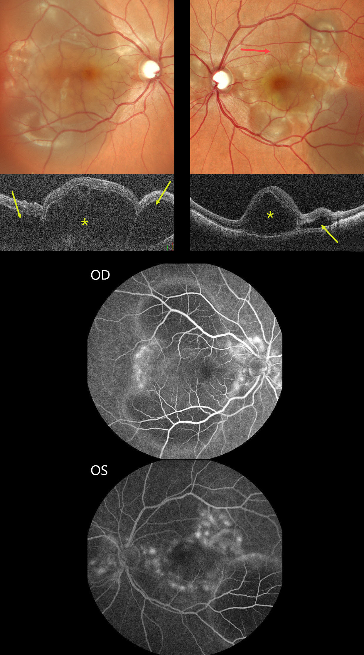
This previously healthy 30YO female presented with sudden bilateral vision loss for 5 days. Vision was counting fingers OU.
Color SLO imaging shows bilateral variably confluent serous detachments involving the posterior poles. Retinal folds are also noted in the left superior macula. OCT scanning shows a complex pattern of bacillary layer detachments (asterisks) and subretinal fluid (arrows). Fluorescein angiography shows multifocal subretinal leaks with pooling in the pockets of subretinal and intraretinal spaces.
She was started on oral steroids and immunomodulators in conjunction with a rheumatologist. One month later, vision returned to 20/20 OU with normalization of the funduscopic findings (not shown).
Learning Points:
Harada disease is characterized by relatively acute bilateral panuveitis, optic nerve swelling, thickened choroid, chorioretinal folds, multifocal serous exudative retinal detachments, and bacillary layer detachments. Patients with only ocular findings (as in our patient) have Harada disease, whereas those with additional systemic findings (including vitiligo, poliosis, headache, vertigo, and hearing loss) have Vogt-Koyanagi-Harada disease.
HARADA DISEASE
Nilesh Kumar
Originally posted on @retina.rocks August 20, 2025
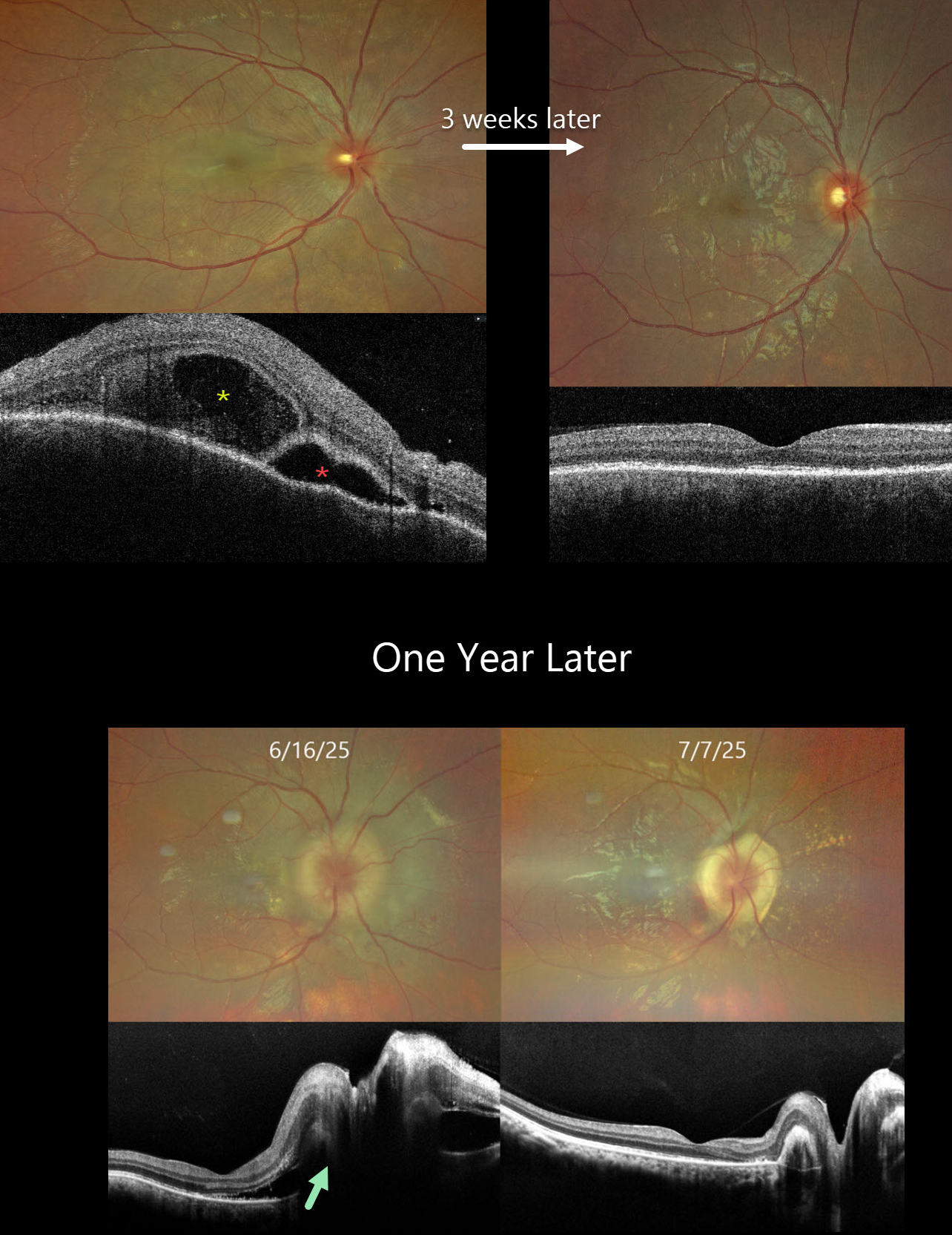
This previously healthy 24YO male presented with acute vision loss in his right eye. Vision was 20/200 OD and 20/20 in his normal OS. There were no neurologic or systemic symptoms.
Color photography shows an exudative retinal detachment involving the posterior pole. OCT shows a bacillary layer detachment, subretinal fluid with thickened retina and choroid. He was diagnosed with unilateral Harada disease and started on oral prednisone. Three weeks later, vision improved to 20/25 OD with marked clinical improvement. The oral prednisolone was tapered weekly over the next 6 weeks and stopped.
He returned one year later with recurrent vision loss. Vision was 20/200 OD. Exudative fluid with distal lipid flecks and a few small retinal hemorrhages surrounded the right nerve. OCT confirmed marked disk and peripapillary thickening with subretinal fluid. Oral prednisone was restarted, and oral azathioprine therapy was added. Three weeks later, the vision improved to 20/30. The fluid completely resolved with a ring of subretinal fibrosis surrounding the disc. The oral prednisone was tapered weekly over the next 6 weeks, and the oral azathioprine is planned to continue for the next year with regular monitoring.
Learning Points:
Harada disease is characterized by relatively acute bilateral panuveitis, optic nerve swelling, thickened choroid, chorioretinal folds, multifocal serous exudative retinal detachments, and bacillary layer detachments. Patients with just ocular findings have Harada disease, whereas those with additional systemic findings (including vitiligo, poliosis, headache, vertigo, and hearing loss) have Vogt-Koyanagi-Harada (VKH) disease. Although classically bilateral, early stages may present unilaterally before involving the fellow eye.
The disease generally progresses through four clinical stages: Prodromal, resembling a viral illness with systemic symptoms (fever, headache, meningismus); Uveitic, characterized by blurred vision, photophobia, optic disc edema, posterior choroidal thickening, and multifocal serous retinal detachments; Chronic/Convalescent, where depigmentation of the choroid leads to a “sunset-glow fundus” along with cutaneous signs like vitiligo, poliosis, and alopecia; and Recurrent, marked by repeated bouts of anterior uveitis and complications such as cataract, glaucoma, or macular neovascularization.
Management centers on high-dose systemic corticosteroids, often followed by steroid-sparing immunosuppressive agents to control inflammation and prevent recurrences. Early, aggressive therapy improves visual outcomes.
HARADA DISEASE
Navneet Mehrotra and Tamanna Patel
Originally posted on @retina.rocks January 23, 2025
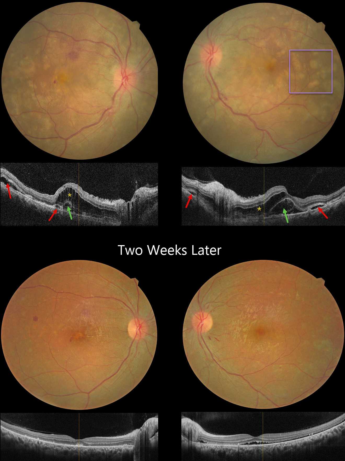
This 24YO female presented with several days of bilateral vision loss. She has a history of anti-glomerular basement membrane disease (anti-GBM) and is on hemodialysis. Her renal disease was quiescent, and there were no acute systemic complaints. There was no anterior segment inflammation, although mild vitreous cells were present bilaterally. Vision was 20/200 OD and 20/80 OS.
Fundus photography shows bilateral multifocal yellow subretinal lesions, inner retinal folds, some retinal hemorrhages, and mild disc edema. OCT scanning shows pockets of subretinal fluid, outer nuclear layer edema, and bacillary layer detachments. The choroid was thickened in both eyes on ultrasonography (not shown).
Oral prednisone 60mg/day was started in a tapering dose. Two weeks later, vision improved to 20/40 OD and 20/80 OS. The acute fundus findings were mostly resolved bilaterally.
Learning Points:
Harada disease is characterized by relatively acute bilateral panuveitis, optic nerve swelling, thickened choroid, chorioretinal folds, multifocal serous exudative retinal detachments, and bacillary layer detachments. Patients with just ocular findings have Harada disease, whereas those with additional systemic findings (including vitiligo, poliosis, headache, vertigo, and hearing loss) have Vogt-Koyanagi-Harada disease.
Anti-GBM disease is a rare autoimmune disorder characterized by autoantibodies that primarily target the basement membranes of the capillaries in the kidneys and lungs. Retinal findings include drusen occurring at an early age (Mansour et al, AJO Case Reports 2017;7:83-90). Acute fundus findings are rarely reported and consist of possibly steroid-induced central serous-like changes with multifocal retinal pigment epithelial detachments and exudative retinal detachments.
HARADA DISEASE
Anjana Mirajkar, Manish Nagpal and Navneet Mehrotra
Originally posted on @retina.rocks October 18, 2023
This 41YO female presented with headaches, redness, and pain OD for 1.5 months and OS for 2 weeks. She was diagnosed elsewhere with angle closure glaucoma, received a YAG peripheral iridotomy, and started on glaucoma drops. She then saw multiple other doctors who diagnosed her with papilledema.
Her CSF opening pressure was 21, and she was started on oral acetazolamide for a presumed diagnosis of idiopathic intracranial hypertension (IIH). In our office, vision was 20/100 OD and 20/60 OS. Intraocular pressure (IOP) was 30mmHG.
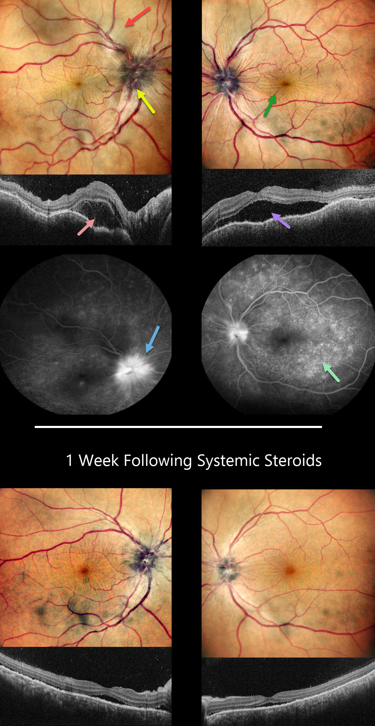
Anterior segments showed shallow anterior chambers with retrolental vitreous cells. Multicolor imaging shows bilateral irregular chorioretinal folds, multifocal serous detachments, and disc hyperemia.
OCT scanning shows a bacillary layer detachment OD, subretinal fluid OS, and a bilateral undulating thickened choroid. Fluorescein angiography shows bilateral optic nerve involvement and pinpoint subretinal leakage.
She was diagnosed with Harada’s disease and started on intravenous methylprednisolone for 3 days, followed by 60mg prednisone PO daily. She was also referred to rheumatology to start immunosuppressants. The acetazolamide was tapered and discontinued for the misdiagnosed IIH.
One week later, vision improved to 20/40 OU. IOP was 10mmHG OU with marked bilateral improvement in the chorioretinal folds, macular fluid, and choroidal thickening. A slow steroid taper was initiated pending initiation of immunosuppressants.
Learning Points:
This case has many classic findings for Harada’s disease, including bilateral panuveitis, optic nerve swelling, thickened choroid, chorioretinal folds, and multifocal serous exudative retinal detachments.
Patients with just ocular findings have Harada’s disease, whereas those with additional systemic findings (including vitiligo, poliosis, headache, vertigo, and hearing loss) have Vogt-Koyanagi-Harada (VKH) disease.
This case also reminds us that it is easy to misdiagnose these complex uveitis cases, especially when not all ocular findings are taken into full consideration in an attempt to give the patient a single, unifying diagnosis.
HARADA DISEASE
Originally posted on @retina.rocks February 7, 2022
This 35YO female presented with a 2-week history of severe bilateral vision loss, photophobia, and red eyes. There was no significant past medical history.
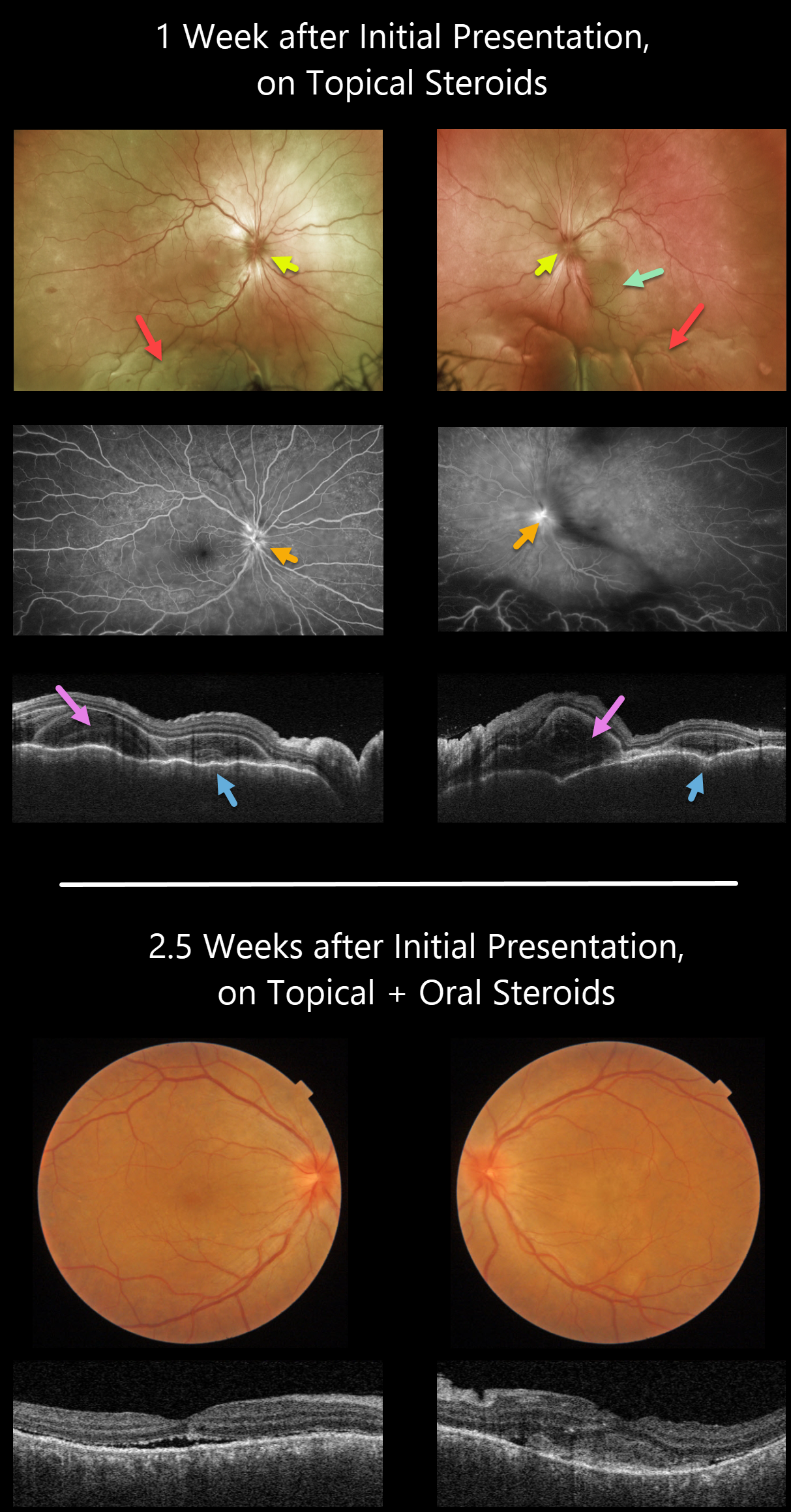
Vision was counting fingers OU. There was moderate bilateral conjunctival injection, severe cells with moderate flare, moderate fibrin, and 360-degree posterior synechiae. Topical cycloplegia and steroids were prescribed.
One week later, she was referred to our retinal service. There was marked improvement of her anterior uveitis and the posterior synechiae were mostly broken throughout. There were moderate bilateral vitreous cells, markedly swollen optic nerves, irregular diffuse macular exudative fluid with inferior exudative retinal detachments.
Fluorescein angiography showed bilateral optic nerve leakage. Triton swept-source OCT showed bilateral markedly thickened and rippled choroid with multiple bacillary pockets of outer retinal thickening and fluid. She started oral prednisone 80mg daily.
Around 10 days later, vision improved to 20/200 OU, with improvement in optic nerve edema, macular fluid, bacillary detachments, and choroidal thickening. The inferior exudative detachments were also resolving (not imaged).
Learning Points:
This case has many classic findings for Harada disease, including bilateral panuveitis, optic nerve swelling, thickened choroid, chorioretinal folds, and multifocal serous exudative retinal detachments.
Given the severity of the panuveitis and exudative subretinal fluid, we were surprised that angiography did not show the more classic multifocal subretinal leaks with pooling in the overlying serous detachments and bacillary cavities.
Patients with just ocular findings have Harada disease, whereas those with additional systemic findings (including vitiligo, poliosis, headache, vertigo, and hearing loss) have Vogt-Koyanagi-Harada disease.
HARADA DISEASE
Originally posted on @retina.rocks April 27, 2021
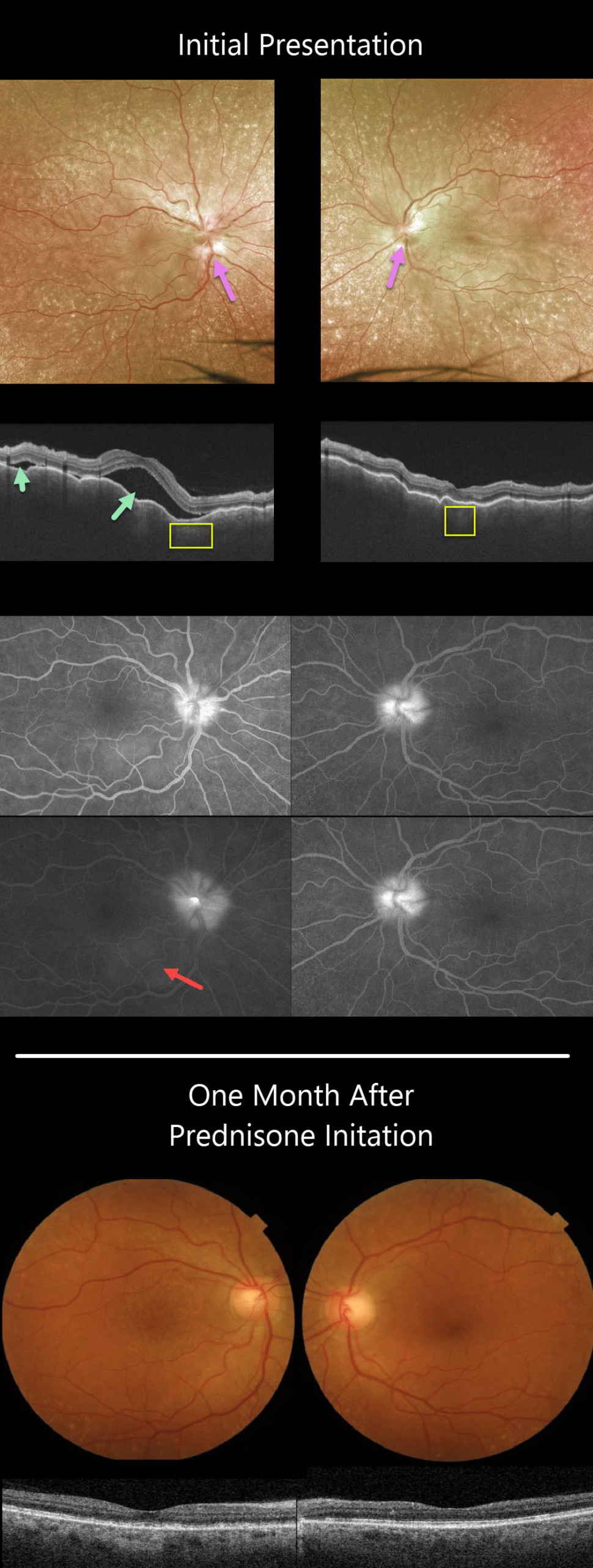
This 63YO female presented with a 3-week history of bilateral painful vision loss. Vision was 20/400 OD and 20/100 OS.
She had bilateral granulomatous anterior uveitis with a clear vitreous OU. Both nerves were swollen, and irregular chorioretinal folds were noted throughout each macula. Extensive incidental peripheral drusen were also present.
Triton swept-source OCT scanning shows a markedly irregularly thickened choroid OU with multifocal serous detachments in the right eye.
Optos fluorescein angiography (FA) shows bilateral optic nerve leakage and some faint subretinal leakage in the inferonasal right macula.
Oral prednisone was started, and there was near immediate improvement. One month later, vision improved to 20/40 OD and 20/60 OS, and all findings virtually normalized.
Learning Points:
This case has many classic findings for Harada’s disease, including bilateral panuveitis, optic nerve swelling, thickened choroid, chorioretinal folds, and multifocal serous retinal detachments.
We were surprised that the FA showed minimal subretinal leakage, since angiography typically shows extensive multifocal subretinal leaks with pooling in the overlying serous detachments and bacillary cavities.
Patients with just ocular findings have Harada’s disease, whereas those with additional systemic findings (including vitiligo, poliosis, headache, vertigo, and hearing loss) have Vogt-Koyanagi-Harada disease.
HARADA DISEASE
Originally posted on @retina.rocks September 23, 2020
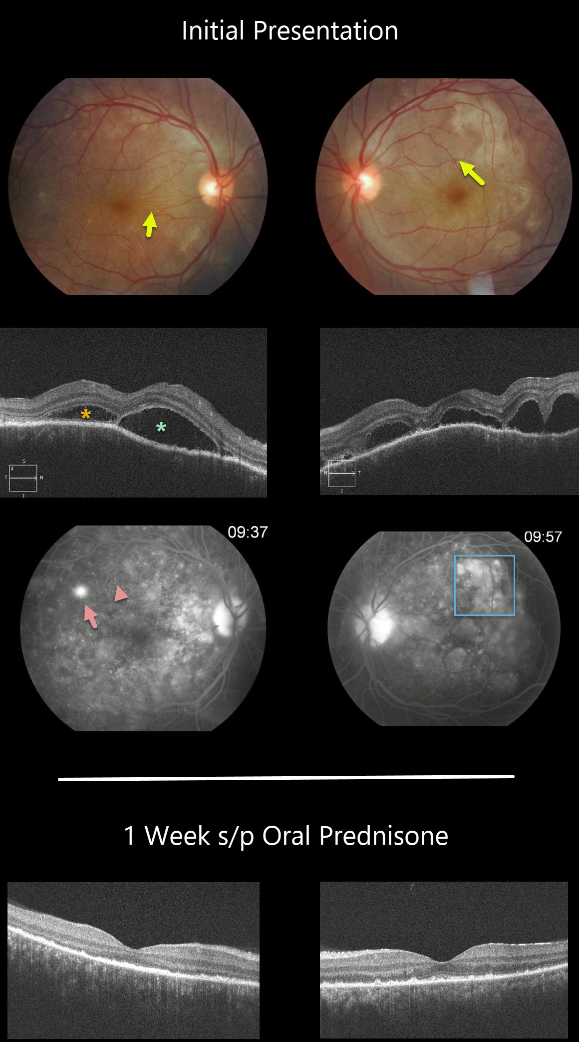
This patient has classic posterior segment findings of Harada disease, including multifocal exudative serous retinal detachments.
Choroidal inflammation produces a markedly thickened choroid with overlying chorioretinal folds (seen in both eyes as variably radiating outer retinal folds).
The OCT scans show multifocal serous detachments with bacillary detachments. Fluorescein angiography shows multiple subretinal leaks with pooling of fluorescein within the various subretinal and bacillary spaces.
The patient started 60mg of oral prednisone daily. One week later, there was a dramatic improvement, with complete resolution of all intra- and subretinal fluid and normalization of choroidal thickness.
Learning Points:
Harada disease is a bilateral granulomatous panuveitis. These patients often need extended treatment with oral prednisone. Most can be successfully weaned off of steroids, although chronic treatment is sometimes necessary.
Choroidal thickening is a good biomarker for disease activity and thins with controlled disease activity.
Remember that in Harada disease, there are only ocular findings. Vogt-Koyanagi-Harada disease occurs when there are associated auditory, integumentary or neurologic symptoms.
HARADA DISEASE
Originally posted on @retina.rocks May 19, 2020
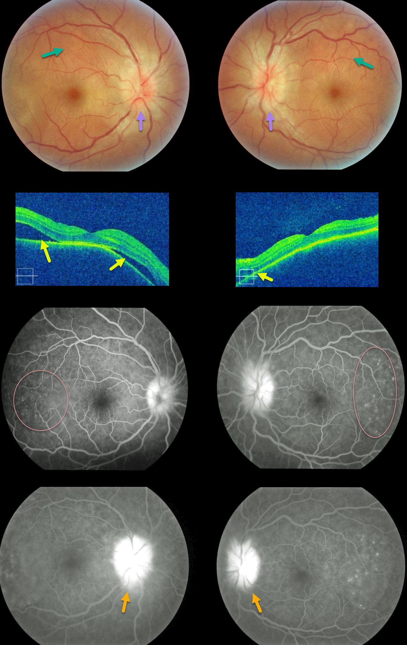
Our patient shows classic findings for acute Harada’s. The fundus photos and OCT images show bilateral multifocal subretinal exudative detachments with irregular, scattered overlying chorioretinal folds and bilateral optic disc swelling.
Fluorescein angiography (FA) shows multifocal subretinal leaks with pooling of fluid below the retina and within outer retinal cystoid spaces, along with leaking “hot” nerves.
These findings dramatically resolved after just 1 week of oral prednisone (not pictured).
Learning Points:
Bilateral, multifocal yellow-white, serous retinal detachments are the hallmark sign of acute Vogt-Koyanagi-Harada (VKH) disease.
The choroid is markedly thickened with overlying chorioretinal folds. The multiple sets and various orientations of these folds are quite different from those seen with idiopathic chorioretinal folds.
The optic nerves are usually inflamed and swollen. Patients often have a panuveitis with anterior chamber and vitreous cells.
Those with just ocular findings have Harada disease, whereas patients with additional systemic findings (including vitiligo, poliosis, headache, vertigo, and hearing loss) have Vogt-Koyanagi-Harada disease.


